E-ISSN:2456-1487
P-ISSN:2456-9887
RNI:MPENG/2017/70771

Case Report

Hemophagocytic Lymphohistiocytosis

Tropical Journal of Pathology and Microbiology

2023 Volume 9 Number 3 July-September
Publisherwww.medresearch.in

An Unusual Case of Residual Follicular Lymphoma with Hemophagocytic Lymphohistiocytosis

Tejesh M1, K Padma S2*, Apoorva K3, Shetty K J4
DOI: https://doi.org/10.17511/jopm.2023.i03.02

1 M Tejesh, Postgraduate, Department of Pathology, K S Hegde Medical Academy, Mangalore, Karnataka, India.

2* Shetty K Padma, Professor, Department of Pathology, K S Hegde Medical Academy, Mangalore, Karnataka, India.

3 K Apoorva, Assistant Professor, Department of Pathology, K S Hegde Medical Academy, Mangalore, Karnataka, India.

4 Jayaprakash Shetty K, Professor and Head, Department of Pathology, K S Hegde Medical Academy, Mangalore, Karnataka, India.

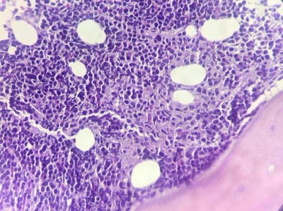
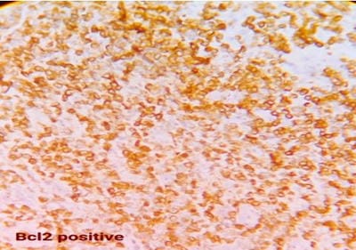
Follicular lymphoma is an indolent B-cell lymphoproliferative disorder of transformed follicular centre B cells characterised by diffuse lymphadenopathy, bone marrow involvement and splenomegaly. An uncommon, fatal clinical disease known as hemophagocytic lymphohistiocytosis is marked by hyperinflammation. HLH is brought on by abnormally activated macrophages and cytotoxic T cells, which produce acute organ dysfunction and cytokine storm. HLH caused by lymphoma is a rare but devastating condition. A rapid diagnosis will aid in effective treatment. This is a rare case report highlighting the diagnosis and treatment plan of a 60-year-old female patient with residual follicular lymphoma with hemophagocytic lymphohistiocytosis.

Keywords: Follicular, Lymphoma, Hemophagocytic Lymphohistiocytosis

Corresponding Author How to Cite this Article To Browse
Shetty K Padma, Professor, Department of Pathology, K S Hegde Medical Academy, Mangalore, Karnataka, India.
Email:
Tejesh M, K Padma S, Apoorva K, Shetty K J, An Unusual Case of Residual Follicular Lymphoma with Hemophagocytic Lymphohistiocytosis. Trop J Pathol Microbiol. 2023;9(3):28-31.
Available From
https://pathology.medresearch.in/index.php/jopm/article/view/625

Manuscript Received Review Round 1 Review Round 2 Review Round 3 Accepted
2023-09-07 2023-09-09 2023-09-16 2023-09-23 2023-09-30
Conflict of Interest Funding Ethical Approval Plagiarism X-checker Note
Nil Nil Yes 16%

© 2023by Tejesh M, K Padma S, Apoorva K, Shetty K Jand Published by Siddharth Health Research and Social Welfare Society. This is an Open Access article licensed under a Creative Commons Attribution 4.0 International License https://creativecommons.org/licenses/by/4.0/ unported [CC BY 4.0].

Introduction

Hemophagocytic lymphohistiocytosis (HLH) is a syndrome that results in organ infiltration by activated cytotoxic T cells and histiocytes as a result of an excessive inflammatory response and cytokine storm. Patients with HLH exhibit abnormally high levels of TNF-alpha, interleukins (IL) 4, 6, 8, 10, and 12, and soluble IL-2 receptor (sCD25), in addition to abnormally low or nonexistent NK cell activity.[1-3]. The aforementioned factors can help identify this condition from aetiologies that have similar symptoms.

Hemophagocytic lymphohistiocytosis (HLH) is a severe, potentially fatal immune activation condition. HLH is a sporadic or familial condition that can be brought on by several things that disturb immunological homeostasis [4]. This is a rare case report highlighting the diagnosis and treatment plan of a 60-year-old female patient with residual follicular lymphoma with hemophagocytic lymphohistiocytosis.

Case Report

A 60-year-old female patient presented with complaints of intermittent cough for 10 days accompanied by generalised lymphadenopathy. The patient also gave a history of COVID-19-positive status a year ago. The patient was diagnosed with stage IV NHL Follicular Grade I Lymphoma by USG-guided lymph node biopsy 2 years back. The patient was given 6 cycles of chemotherapy. The patient was also a known case of pulmonary tuberculosis for which she was on ATT for one year.

A year later, on follow-up examination, the patient was found to have multiple enlarged lymph nodes in the cervical region (anterior triangle), left axilla and inguinal region. PET-CT scan showed multiple enlarged lymph nodes in cervical, inguinal and retroperitoneal regions. Laboratory investigations included hemoglobin 8.3g/dl, platelet count 8000 cells/cumm, total WBC count 8800 cells/cumm, differential count neutrophil 01%, lymphocytes 54%, lymphoblasts 45%, triglycerides 578 mg/dl, total cholesterol 375 mg/dl, ferritin 20,250 ng/ml, LDH 11,780 U/L, SGOT 606 U/L, SGPT 147 U/L, Alkaline phosphatase 2126 U/L and Fibrinogen 150 mg/dl.Peripheral smear showed many atypical lymphoid cells with a high N: C ratio, and clumped chromatin. Some showed a cleft nucleus and a single prominent nucleolus.

Bone marrow aspiration showed sheets of uniform population of lymphoid cells suggestive of non-Hodgkin’s lymphoma infiltration of bone marrow. (Figure 1)

patho_625_01.JPG
Figure 1:
Bone marrow aspirate

Bone marrow biopsy revealed hypercellular marrow with complete effacement of architecture. Marrow was diffusely infiltrated by sheets of a uniform population of small to medium-sized lymphoid cells. Many of them showed cleaved nuclei and inconspicuous nucleolus with scant cytoplasm. (Figure 2). Bone marrow macrophages exhibiting haemophagocytosis were seen.

patho_625_02.JPG
Figure 2:
Bone marrow biopsy

Immunohistochemistry (IHC) was performed which showed positive for LCA, CD20, CD79a, Bcl2 (Figure 3), CD10 and Ki67 20% (Figure 4) - features were those of residual follicular lymphoma with HLH.


patho_625_03.JPG
Figure 3:
Bcl2

patho_625_04.JPG
Figure 4:
ki67 20%

Because of the histopathology and IHC report the patient was advised chemotherapy. But despite the prompt diagnosis and chemotherapy, the patient succumbed to the diseas.

Discussion

As a result of an excessive inflammatory response and cytokine insufficiency, hemophagocytic lymphohistiocytosis (HLH) is a disease that causes organ infiltration by activated cytotoxic T cells and histiocytes.[1,2] In a similar rare report by Devitt et al., HLH was connected to adult-onset Still's illness and an underlying autoimmune condition with T-cell/histiocyte-rich large B cell lymphoma.[5] According to a retrospective study by Shabbir et al, adult patients with HLH-associated malignancies had a poor prognosis with a 67% death rate.[6]

Among the clinical symptoms are persistent fever, hepatosplenomegaly, lymphadenopathy, pancytopenia, rash, and neurological problems.[7] Typical laboratory findings in our patient included high triglycerides, notably raised ferritin levels, hypofibrinogenemia, and abnormal liver function tests.

A bone marrow examination commonly reveals separate hemophagocytes, or histocytes containing absorbed hematopoietic components such as nucleated erythroid progenitors, white blood cell precursors, and platelets, similar to the outcomes in our case. Immune dysregulation has been associated with the coexistence of HLH with lymphoid malignancy, and the ongoing activation of T-cells by the neoplastic B-SLL clone is a phenomenon that has drawn a lot of attention in the literature.[8]

Our patient underwent immunohistochemistry (IHC), which revealed 20% positive results for LCA, CD20, CD79a, Bcl2, CD10, and Ki67. The diagnosis of HLH was made by correlating morphology and other biochemical findings. Our case is exceptional because it involves HLH, an immunological dysregulation condition that the presence of clonal and oligoclonal T-cells can cause. Chemotherapy may make a secondary HLH more likely to form as a result of clonal phenomenon.

Lehmberg K et al observed that between 40% and 70% of HLH instances in adults are assumed to be caused by immunosuppression, infection, or the tumour itself at presentation or when treatment is started.[9]

The latter may even occur in individuals who are in remission. Patients must thus have a complete oncologic examination, paying specific attention to lymphomas. Adults still have significant rates of HLH-related death, particularly those who have underlying malignancies. As has been the case with children, it is predicted that more knowledge of HLH, a quicker diagnostic process, and innovative therapy strategies would improve the prognosis of HLH in adults.

Although HLH is a rare condition, it is probably underdiagnosed. The overall mortality rate is consistently high, making an early diagnosis crucial. Infections frequently cause HLH, whether it is inherited or acquired. Recent discoveries on the pathophysiology of hereditary HLH have led to the development of genetic diagnostics and therapy regimens that have been found to improve prognosis. With a deeper knowledge of the aetiology of this disease subtype, more specialised diagnostics and cutting-edge targeted treatments may be made available.


Conclusion

Due to its rarity, cryptic presentation, and abnormal test results, HLH is a difficult diagnosis. Given the importance of early treatment to provide the best outcome, prompt recognition and diagnosis are crucial. The fundamental objective of treatment, particularly when HLH is linked to malignancies, is to target the underlying issue while making an effort to manage the heightened inflammatory response.

References

1. Okamoto M, Yamaguchi H, Isobe Y, Yokose N, Mizuki T, Tajika K, Gomi S, Hamaguchi H, Inokuchi K, Oshimi K, Dan K. Analysis of triglyceride value in the diagnosis and treatment response of secondary hemophagocytic syndrome. Intern Med. 2009;48(10):775-81. doi: 10.2169/internalmedicine.48.1677. Epub 2009 May 15. PMID: 19443971 [Crossref][PubMed][Google Scholar]

2. Fukaya S, Yasuda S, Hashimoto T, Oku K, Kataoka H, Horita T, Atsumi T, Koike T. Clinical features of haemophagocytic syndrome in patients with systemic autoimmune diseases: analysis of 30 cases. Rheumatology (Oxford). 2008 Nov;47(11):1686-91. doi: 10.1093/rheumatology/ken342. Epub 2008 Sep 9. PMID: 18782855 [Crossref][PubMed][Google Scholar]

3. Mehta RS, Smith RE. Hemophagocytic lymphohistiocytosis (HLH): a review of literature. Med Oncol. 2013 Dec;30(4):740. doi: 10.1007/s12032-013-0740-3. Epub 2013 Oct 9. PMID: 24105023 [Crossref][PubMed][Google Scholar]

4. Janka G. Hemophagocytic lymphohistiocytosis: when the immune system runs amok. Klinische Padiatrie. 2009;221(5):278–285. [Crossref][PubMed][Google Scholar]

5. Devitt, K. , Cerny, J. , Switzer, B. , Ramanathan, M. , Nath, R., Yu, H., … Chen, B. J. (2014). Hemophagocytic lymphohistiocytosis secondary to T-cell/histiocyte-rich large B-cell lymphoma. Leukemia Research Reports, 3(2), 42–45. doi:10.1016/j.lrr.2014.05.004 [Crossref][PubMed][Google Scholar]

6. Shabbir M, Lucas J, Lazarchick J, Shirai K. Secondary hemophagocytic syndrome in adult series of 18 patients in a single institution and a review of literature. Hematol Oncol. 2010;29:100–6. [Crossref][PubMed][Google Scholar]

7. Jordan MB, Allen CE, Weitzman S, Filipovich AH, McClain KL. How I treat hemophagocytic lymphohistiocytosis. Blood. 2011;118(15):4041-4052. doi: 10.1182/ blood-2011-03-278127 [Crossref][PubMed][Google Scholar]

8. Martinez A, Pittaluga S, Villamor N, Colomer D, Rozman M, Raffeld M, Montserrat E, Campo E, Jaffe ES. Clonal T-cell populations and increased risk for cytotoxic T-cell lymphomas in B-CLL patients: clinicopathologic observations and molecular analysis. Am J Surg Pathol. 2004 Jul;28(7):849-58. doi: 10.1097/00000478-200407000-00002. PMID: 15223953 [Crossref][PubMed][Google Scholar]

9. Lehmberg K, Nichols KE, Henter JI, Girschikofsky M, Greenwood T, Jordan M, Kumar A, Minkov M, La Rosée P, Weitzman S. Study Group on Hemophagocytic Lymphohistiocytosis Subtypes of the Histiocyte Society. Consensus recommendations for the diagnosis and management of hemophagocytic lymphohistiocytosis associated with malignancies. Haematologica. 2015 Aug;100(8):997-1004. doi: 10.3324/haematol.2015.123562. PMID: 26314082; PMCID: PMC5004414 [Crossref][PubMed][Google Scholar]