E-ISSN:2456-1487
P-ISSN:2456-9887
RNI:MPENG/2017/70771

Research Article

Spinal Cord Tumours

Tropical Journal of Pathology and Microbiology

2022 Volume 8 Number 4 July-August
Publisherwww.medresearch.in

The spectrum of Spinal Cord Tumours in a Tertiary Care Centre with Emphasis on Rare Tumours: An Observational Study

Sharma M.1*, Tyagi S.2, Pandey J.3, Sharma B.4, Choudhary A.5
DOI: https://doi.org/10.17511/jopm.2022.i04.01

1* Mudita Sharma, M.D. Pathology, Junior resident, Department of Pathology, Mahatma Gandhi Medical College, Jaipur, Rajasthan, India.

2 Surabhi Tyagi, Professor of Pathology, Department of Pathology, Mahatma Gandhi Medical College, Jaipur, Rajasthan, India.

3 Jyoti Pandey, M.D. Pathology (JR III), Department of Pathology, Mahatma Gandhi Medical College, Jaipur, Rajasthan, India.

4 B. S. Sharma, Professor & HOD Neurosurgery Director Neurosciences, Mahatma Gandhi Medical College, Jaipur, Rajasthan, India.

5 Anubha Choudhary, M.D Pathology, Junior resident, Department of Pathology, Mahatma Gandhi Medical College, Jaipur, Rajasthan, India.

Introduction: Spinal cord tumour is an abnormal mass of tissue within/or surrounding spinal cord &/or spinal column. They are referred to according to vertebral levels and area in which they are located within the spine - Extradural & Intradural (Extramedullary & Intramedullary). Aims and Objectives: To integrate histopathological spectrum of spinal cord tumours with their relevant immunohistochemistry, their incidence and spectrum about spinal levels, location within spine, age and sex. Materials and Methods: Hospital-based observational study in Department of Pathology, Mahatma Gandhi Medical College, Jaipur, for two years & includes 70 cases of spinal cord tumours. Results: In our study, 70 cases of spinal cord tumours showed 14 tumour types. Peak incidence was seen in 61-70 years followed by 41-50 years and lowest in children ˂ 10 years. Male preponderance was seen. The major histological type was meningiomas 17 cases (24.3%) followed by schwannomas 12 cases (17.14 %) and metastasis in 8 cases (11.4%). In relation to anatomical site in spinal cord, tumours were most common in the thoracic spine (48.6%) followed by the cervical spine (15.7 %). Conclusion: In our study, the spectrum of spinal cord tumours is exhibited in 14 tumour types. Peak incidence was seen in sixth decade with male preponderance and propensity for the thoracic segment of the spine. The most common tumour type was meningiomas followed by schwannomas. IHC plays a vital role in accurate diagnosis that helps in recognizing tumour histogenesis, clinical & radiological correlation, its pathological course, treatment & prognosis. MIB-1 assesses grade & aggressiveness of tumour thus helping in evaluating its chances of recurrence.

Keywords: Spinal cord tumours, extradural, intradural extramedullary, intramedullary, immunohistochemistry

Corresponding Author How to Cite this Article To Browse
Mudita Sharma, M.D. Pathology, Junior resident, Department of Pathology, Mahatma Gandhi Medical College, Jaipur, Rajasthan, India.
Email:
Mudita Sharma, Surabhi Tyagi, Jyoti Pandey, B. S. Sharma, Anubha Choudhary, The spectrum of Spinal Cord Tumours in a Tertiary Care Centre with Emphasis on Rare Tumours: An Observational Study. Trop J Pathol Microbiol. 2022;8(4):52-59.
Available From
https://pathology.medresearch.in/index.php/jopm/article/view/607

Manuscript Received Review Round 1 Review Round 2 Review Round 3 Accepted
2022-07-27 2022-07-29 2022-08-05 2022-08-12 2022-08-19
Conflict of Interest Funding Ethical Approval Plagiarism X-checker Note
Nil Nil Yes 17%

© 2022by Mudita Sharma, Surabhi Tyagi, Jyoti Pandey, B. S. Sharma, Anubha Choudharyand Published by Siddharth Health Research and Social Welfare Society. This is an Open Access article licensed under a Creative Commons Attribution 4.0 International License https://creativecommons.org/licenses/by/4.0/ unported [CC BY 4.0].

Introduction

A spinal cord tumour is an abnormal mass of tissue within/or surrounding the spinal cord &/or spinal column. They are referred to according to vertebral levels and the area in which they are located within the spine - Extradural & Intradural (Extramedullary & Intramedullary) [1]. They occur predominantly in the third and fourth decades & are less common in childhood and elderly [2].


Spinal tumours are < 15 % of all CNS tumours. [3]. They are referred to in two ways (a) By the vertebral levels or anatomical locations ∙Cervical ∙Thoracic ∙Lumbar ∙Sacral (b) By the area of location within the spine-Extradural and Intradural. Intradural can further be divided into-Extramedullary and Intramedullary [4]. Most are extradural approximately 55-60 % and 40-45% are intradural of which, 40% are extramedullary and 5% are intramedullary. [5]. They account for 20% of all intraspinal tumours in adults and 35% of all intraspinal tumours in children. [6]. The objective of this study is to integrate immunohistochemistry in the diagnosis of various spinal cord tumours and to determine their incidence in a tertiary care hospital like ours. The objective is to determine the total number of cases of spinal cord tumours in this observational study throughout two years from January 2020 to June 2021 and to study them in relation to spinal levels and their location within the spine, age and sex. In this study enumeration of a few rare cases encountered is also done

Classification of spinal cord tumours according to the location within the spinal cord

A. Extradural

B. Intradural -Extramedullary

C. Intramedullary

patho_607_01.PNG

Intramedullary spinal cord tumours grow inside the spinal cord. They are derived from glial or ependymal cells. The frequency of occurrence is approximately 5%. Astrocytomas and ependymomas are the most common. Astrocytomas are commonly seen in children and occupy the thoracic region followed by the cervical spine [7].

Ependymomas occur in the filum terminale and are common in adults. [8]. Although intracranial ependymoma is associated with an aggressive clinical course and poor prognosis, spinal ependymoma is linked with an indolent clinical course and good prognosis [9,10,11].

Intramedullary lipomas are rare congenital tumours, most commonly located in the cervicothoracic spine. Intradural-extramedullary spinal tumours are situated inside the dura, but outside the spinal cord. Spinal intradural extramedullary (IDEM) tumours account for two-thirds of all primary intraspinal neoplasms. These lesions are uncommon [12]. Most common are Meningiomas, which develop in the meninges of the brain and spinal cord. [13].

Meningiomas are often benign, recurrence is common. Schwannomas and neurofibromas arising from the nerve roots extending from the spinal cord or filum terminale and ependymomas occur at the spinal cord base and are usually benign. [14]. Nerve root tumours are also benign but neurofibromas may become malignant over time. Ependymomas, if large, make removal challenging. [15].

Extradural spinal cord tumours are located outside the dura. The incidence is approximately 55%. Occasionally, an extradural tumour extends through the intervertebral foramina, lying partially within and partially outside of the spinal canal. The spinal column is the most common site for bony metastasis. The most common primary-vertebral haemangiomas- are benign and rarely cause symptoms.[16].

Common primary cancers that spread to the spine are lung, breast and prostate. [17]. Lung carcinomas commonly in males and carcinoma breasts in females. Other cancers that can spread to the spine are multiple myeloma, lymphomas, melanomas, sarcomas and cancers of the G.I. tract, kidney and thyroid.

Diagnosis of spinal tumours is done by H & E, supported by ancillary aids- immunohistochemical and ultrastructural tests.


IHC plays an important role in the diagnosis of spinal cord tumours, in identifying the differentiation or origin of the tumour and to determine the proliferative index and expression of growth factor receptors and oncoproteins reflecting the malignant potential of the tumour.

The ultimate prognosis depends upon the histopathological type and grade of the tumour.

Aims and Objectives

  • To integrate the histopathological spectrum of spinal cord tumours with their relevant immunohistochemistry, their incidence and spectrum concerning spinal levels, location within the spine, age and sex.
  • Special reference to the rare spinal tumours encountered in our study.

Materials and Methods

  • Hospital-based observational study in Department of Pathology, Mahatma Gandhi Medical College, Jaipur, for two years & includes 70 cases of spinal cord tumours.
  • All neuropathological specimens of spinal cord tumours recieved were fixed & stained routinely with

Haematoxylin and Eosin (H & E). IHC was done depending on the diagnostic need of the individual case such as Epithelial Membrane Antigen (EMA), Vimentin, S-100, Cytokeratin(Pan CK), MIB-1, Neurofilament, G-FAP, R132H-IDH1

Table 1: Age-associated distribution of study subjects.patho_607_02.PNG

The most common age of presentation for spinal cord tumours was 61-70 years in 24.3% of the cases followed by 41-50 years. The Mean age was found to be 48.71 ± 18.47 years.

Table 2: Gender-based distribution of study subjects.patho_607_03.PNG
The occurrence of spinal cord tumours showed male preponderance. There were 43 males (61.4%) and 27 females (38.6%).

Table 3: Distribution of the study subjects according to the type of spinal cord tumour.patho_607_04.PNG
Out of 70 cases of spinal cord tumours, 17 were Meningiomas (24.3%) followed by schwannomas ( 17.14 %),

8 Rare cases - 2 Neuroendocrine tumours (2.9%), 2 Glioblastomas (2.9%), 1 giant cell tumour (1.4%), 1 melanotic cell neoplasm (1.4%), 1 aneurysmal bone cyst (1.4%) and 1 solitary fibrous tumour.

Table 4: Distribution of the study subjects according to the region of spinal cord tumour.patho_607_05.PNG
34 cases occupied the thoracic spine (48.6%), 11 in the cervical spine (15.7 %), 7 in the lumbar (10%), 6 in cervico-dorsal ( 8.6%), 5 in dorso-lumbar (7.1%), 3 in lumbo- sacral (4.3%) and none in the coccyx.


Table 5: Type of spinal tumours in relation to their location within the spinepatho_607_06.PNG

Table 6. Compartmental distribution of spinal cord tumours. 

Type of spinal tumour Extradur al (No.) Extradur al (%) Intradural extramedullary(N o.) Intradural extramedulla ry (%) Intradural intramedulla ry (No.) Intradural intramedulla ry (%)
Aneurysmal bone cyst 1 3.4 0 0 0 0
Meningioma 1 3.4 15 50 1 9.1
Neuroendocri ne tumour 1 3.4 1 3.3 0 0
Chordoma 2 6.9 0 0 0 0
Astrocytoma 0 0 0 0 3 27.3
Ependymoma 0 0 1 3.3 5 45.5
Giant cell tumour 1 3.4 0 0 0 0
Glioblastoma 0 0 1 3.3 1 9.1
Lymphoma 7 24.1 0 0 0 0
Melanotic neoplasm 1 3.4 0 0 0 0
Metastasis 7 24.1 0 0 1 9.1
Plasma cell neoplasm 6 20.7 1 3.3 0 0
Schwannoma 1 3.4 11 36.7 0 0
Solitary fibrous tumour 1 3.4 0 0 0 0
TOTAL 29 100 30 100 11 100

The compartmental distribution of lesions within the spinal cord was 30/70 cases as Intradural-extramedullary (IDEM) 42.90 % followed closely by extradural 29 /70 cases - 41.40% and 11/70 cases as intramedullary (15.70 %).

Results

  • In our study, 70 cases of spinal cord tumours showed 14 tumour types diagnosed on routine H & E and IHC and evaluated for their occurrence at different spinal levels and their location within the spinal cord.
  • Peak incidence was seen in 61-70 years followed by 41-50 years and lowest in children ˂ 10 years. Male preponderance was seen.
  • Major histological type was meningiomas - 17 cases (24.3%) followed by schwannomas - 12 cases (17.14 %) and metastasis - 8 cases (11.4%) lymphomas (10%), 7 plasma cell neoplasms (10%), 6 ependymomas (8.6%), 3 astrocytomas (4.3%) were seen.The rare cases were-

2 Glioblastomas (2.9%),1 Solitary fibrous tumour (1.4%),1 Melanotic cell neoplasm (1.4%),1 Aneurysmal bone

cyst (1.4%),1 Giant cell tumour (1.4%),2 Neuroendocrine tumours (2.9%)

Rare Spinal Tumors in Our Study

1. Glioblastoma Multiforme(GBM): In our study, 2 cases of glioblastoma multiforme were seen. A 17 year/M was diagnosed with GBM. Tumour was cervical & intradural extramedullary. It was immunopositivity for Olig 2, p53 and 3K27M. MIB1 was 60-70%.

A second case was of a 28-year-old/M diagnosed with GBM in the cervicothoracic region, which showed GFAP and p53 positivity. GBM represent approximately 7.5% of all intramedullary gliomas and has a predilection for the cervical spine or cervicothoracic region.[17]

patho_607_07.PNG
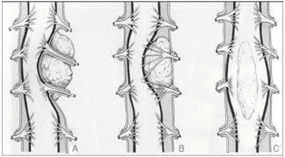
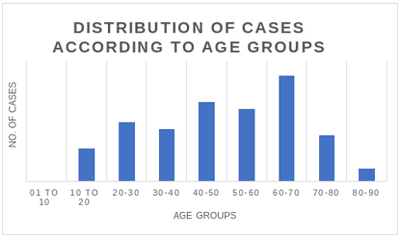
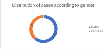
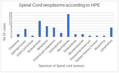
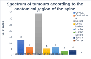
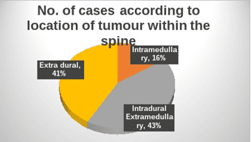
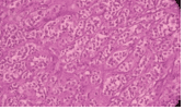
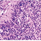
Fig 1: Glioblastoma multiforme H & E 10x

2. Solitary Fibrous tumour: A 48 year/F was diagnosed a case of solitary fibrous tumour in the thoracic spine, extradural in position.


Immunopositive for Vimentin, Cd-99, CD-34(Patchy), STAT-6 and negative for S-100. MIB was 30-35%. Based on histological features such as uniform spindle cells arranged in interlacing fascicles with intermingled collagen and dense reticulin fibre network and IHC profile, it was diagnosed as a Solitary Fibrous tumour.

patho_607_08.PNG
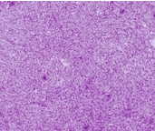
Fig 2: Solitary fibrous tumour -H & E 10x

3. Melanoma: A 50-year/F was diagnosed as case of melanoma occupying an extradural position in the thoracic spine. Diffusely positive for S-100, SOX 10 and patchy positive for HMB 45.MIB index was 10-12%.

These lesions usually occur as diffuse melanomatosis. Solitary lesions are rare. [19].

patho_607_09.PNG
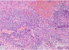
Fig 3: Melanoma- H & E 10x

4. Aneurysmal Bone cyst: A 45 year/F has been diagnosed with a case of an aneurysmal bone cyst, occupying the cervical segment. On H & E, the cystic spaces of variable sizes lined by fibrous septa containing fibroblasts and myofibroblasts. Areas of spindled stroma were seen.

Aneurysmal bone cyst represents 1.4 % of primary bone tumours. The vertebral column is involved in 3-20% of cases. According to the location in the spine, cervical and thoracic spines are involved more than the lumbar spine.

The lesion occurs in the first two decades of life with slight female preponderance. [20].

patho_607_10.PNG
Fig 4A: Aneurysmal bone cyst H & E 10x

patho_607_11.PNG

Fig 4B: Aneurysmal bone cyst H & E 10x

5. Giant Cell Tumour: A single case of Giant cell tumour diagnosed in 33 year/M occupying the sacrum. It showed numerous osteoclast giant cells and spindle cells arranged in fascicles with vascular stroma with areas of hemorrhage and necrosis with significant mitotic activity.

IHC profile was- p 63,SALL4 ,SATB2,CD68(Positive) H3K36M(Negative).MIB-1 was 55-60% and the tumour was extradural.


Spinal giant cell tumours, especially above the sacrum are relatively infrequent. The incidence is 2.9% in vertebrae above the sacrum and 2.5% in the sacrum in all GCTs of the bone and presents with spinal cord compression due to extension into the spinal canal. Age ≥55 years and location in the sacrum or coccyx are associated with worsened survival.[21]

patho_607_12.PNG
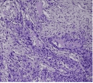
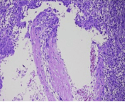
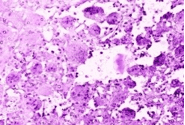
Fig 5: Giant cell tumour H & E 10x

6. Neuroendoocrine tumours

Two cases of neuroendocrine tumours were seen.

A 55 year/M was diagnosed as a case of neuroendocrine tumour eroding the D4 lamina with its spinous process with intra thoracic extension and IDEM in location.IHC profile- Focal positive for NF1, diffuse positivity for chromogranin A & synaptophysin; CK negative.MIB1 index was 18-20%.

A 62 year/M was diagnosed with a case of Neuroendocrine Tumour occupying the lumbar spine and ED position.On IHC, positivity for EMA and Pan CK, Synaptophysin and chromogranin A was seen;CK7,CK20,p40,TTF-1,CD10,HEPAR-1,PSA,CA19.9,NSE were negative.

patho_607_13.PNG

Fig 6: Neuroendocrine tumour H & E 10x

Conclusion

In our study, the spectrum of spinal cord tumours is exhibited in 14 tumour types. Peak incidence was seen in the sixth decade with male preponderance and propensity for a thoracic segment of the spine. Most common tumour types were meningiomas followed by schwannomas. Few very rare cases of the aneurysmal bone cyst, giant cell tumour, glioblastoma, melanoma, solitary fibrous tumour and atypical carcinoid were also seen.

The 2021 classification of CNS and spinal cord divides it into an array of lesions, both -benign and malignant. The histopathological diagnosis remains the main stray for further workup. It is conjugal to thorough history, detailed clinical examination with relevant state-of-the-art investigations.

We have witnessed that IHC serves as an advantageous tool for an accurate diagnosis- being rapid and practical. It helps in recognizing the nature of the lesion and thus assessing its history and its natural, pathological course.MIB-1 helps in assessing the tumour grade & aggression helping in evaluating the chances of recurrence and prognosis. It, therefore, improves the chances of compromise-free survival, procuring targeted therapy for the patient & improving their quality of life. Prompt & precise diagnosis is utmost as spinal pathologies pose considerable morbidity, a compromised state of survival, a permanent neurological deficit &/or ambulatory dependence for life.

Author contribution: Photographs- Dr Surabhi Tyagi. Articles and references - Dr Jyoti Pandey. Editing – Dr Anubha Choudhary. Proof reading- Dr. Jyoti Pandey, Dr.Anubha Choudhary.

Our series represents an observational study of 70 cases coming to the department of Pathology in a tertiary care hospital in Jaipur. The histology, location, extent of invasion, and metastasis affect the surgical resection of the tumour. Intradural extramedullary tumours are amendable to surgical resection usually.

Reference

01. Schellinger KA, Propp JM, Villano JL, McCarthy BJ. Descriptive epidemiology of primary spinal cord tumors. J Neurooncol. 2008 Apr;87(2):173-9. doi: 10.1007/s11060-007-9507-z [Crossref][PubMed][Google Scholar]


02. Gadgil, Nitin M. , et al. A clinicopathological study of lesions of spinal cord and its coverings: A tertiary care hospital experience. " Annals of Pathology and Laboratory Medicine 3. 3 (2016): A148-156 [Crossref][PubMed][Google Scholar]

03. M Das J, Hoang S, Mesfin FB. Intramedullary Spinal Cord Tumors. [Updated 2022 Apr 28]. In: Stat Pearls [Internet]. Treasure Island (FL): Stat Pearls Publishing; 2022 Jan-. Available from: [Article][Crossref][PubMed][Google Scholar]

04. Karsy M, Guan J, Sivakumar W, Neil JA, Schmidt MH, Mahan MA. The genetic basis of intradural spinal tumors and its impact on clinical treatment. Neurosurg Focus. 2015 Aug;39(2):E3. doi: 10.3171/2015.5.FOCUS15143 [Crossref][PubMed][Google Scholar]

05. Arnautovic, Kenan, and Aska Arnautovic. Extramedullary intradural spinal tumors: a review of modern diagnostic and treatment options and a report of a series. " Bosnian journal of basic medical sciences 9. Suppl 1 (2009): S40. [Crossref][PubMed][Google Scholar]

06. Abul-Kasim K, Thurnher MM, McKeever P, Sundgren PC. Intradural spinal tumors: current classification and MRI features. Neuroradiology. 2008 Apr;50(4):301-14. doi: 10.1007/s00234-007-0345-7 [Crossref][PubMed][Google Scholar]

07. Frühwald MC, Rutkowski S. Tumors of the central nervous system in children and adolescents. Dtsch Arztebl Int. 2011 Jun;108(22):390-7. doi: 10.3238/arztebl.2011.0390 [Crossref][PubMed][Google Scholar]

08. Cachia D, Johnson DR, Kaufmann TJ, Lowe S, Andersen S, Olar A, Cooper SL, Frankel BM, Gilbert MR. Case-based review: ependymomas in adults. Neurooncol Pract. 2018 Aug;5(3):142-153. doi: 10.1093/nop/npy026 [Crossref][PubMed][Google Scholar]

09. Singh, Pratima K. , et al. Differential involvement of protein 4. 1 family members DAL-1 and NF2 in intracranial and intraspinal ependymomas. " Modern pathology 15.5 (2002): 526-531 [Crossref][PubMed][Google Scholar]

10. Teo, Charles, et al. Ependymoma. " Child's Nervous System 19. 5 (2003): 270-285. [Crossref][PubMed][Google Scholar]

11. Vera-Bolanos, Elizabeth, et al. Clinical course

and progression-free survival of adult intracranial and spinal ependymoma patients. " Neuro-oncology 17. 3 (2015): 440-447. [Crossref][PubMed][Google Scholar]

12. Narayan S, Rege SV, Gupta R. Clinicopathological Study of Intradural Extramedullary Spinal Tumors and Its Correlation With Functional Outcome. Cureus. 2021 Jun 18;13(6):e15733. doi: 10.7759/cureus.15733 [Crossref][PubMed][Google Scholar]

13. Alruwaili AA, De Jesus O. Meningioma. [Updated 2022 May 8]. In: StatPearls [Internet]. Treasure Island (FL): StatPearls Publishing; 2022 Jan-. Available from: [Article][Crossref][PubMed][Google Scholar]

14. Marchetti, Marcello, Elena De Martin, and Laura Fariselli. Schwannomas and Neurofibromas. " Adult CNS Radiation Oncology. Springer, Cham, 2018. 105-116 [Crossref][PubMed][Google Scholar]

15. Celano E, Salehani A, Malcolm JG, Reinertsen E, Hadjipanayis CG. Spinal cord ependymoma: a review of the literature and case series of ten patients. J Neurooncol. 2016 Jul;128(3):377-86. doi: 10.1007/s11060-016-2135-8 [Crossref][PubMed][Google Scholar]

16. . . J Neurooncol. 2016 Jul;128(3):377-86. doi: 10.1007/s11060-016-2135-8 [Crossref][PubMed][Google Scholar] [Crossref][PubMed][Google Scholar]

17. Ecker RD, Endo T, Wetjen NM, Krauss WE. Diagnosis and treatment of vertebral column metastases. Mayo Clin Proc. 2005 Sep;80(9):1177-86. doi: 10.4065/80.9.1177 [Crossref][PubMed][Google Scholar]

18. Ziu E, Viswanathan VK, Mesfin FB. Spinal Metastasis. [Updated 2022 Mar 3]. In: StatPearls [Internet]. Treasure Island (FL): StatPearls Publishing; 2022 Jan-. Available from: [Article][Crossref][PubMed][Google Scholar]

19. Shen CX, Wu JF, Zhao W, Cai ZW, Cai RZ, Chen CM. Primary spinal glioblastoma multiforme: A case report and review of the literature. Medicine (Baltimore). 2017 Apr;96(16):e6634. doi: 10.1097/MD.0000000000006634 [Crossref][PubMed][Google Scholar]

20. Yadav YR, Tandon JK, Kriplani TC, Kapoor JP, Arora BM, Chandrakar S. Primary extradural spinal


melanoma. Neurol India. 1995 Jul-Sep;43(3):161-164. [Crossref][PubMed][Google Scholar]

21. Zileli M, Isik HS, Ogut FE, Is M, Cagli S, Calli C. Aneurysmal bone cysts of the spine. Eur Spine J. 2013 Mar;22(3):593-601. doi: 10.1007/s00586-012-2510-x [Crossref][PubMed][Google Scholar]

22. Bhojraj SY, Nene A, Mohite S, Varma R. Giant cell tumor of the spine: A review of 9 surgical interventions in 6 cases. Indian J Orthop. 2007 Apr;41(2):146-50. doi: 10.4103/0019-5413.32047 [Crossref][PubMed][Google Scholar]