E-ISSN:2456-1487
P-ISSN:2456-9887
RNI:MPENG/2017/70771

Research Article

Laryngeal Lesions

Tropical Journal of Pathology and Microbiology

2022 Volume 8 Number 2 March April
Publisherwww.medresearch.in

Clinico-Pathological Spectrum of Laryngeal Lesions Over Seven years in a Tertiary Hospital

Fatima S.1*, Nasir Mohamed Badri R.2, Francis Valere D’sa B.3, Ahmed Siddiqui W.4, Ibrahim Mirza N.5
DOI: https://doi.org/10.17511/jopm.2022.i02.02

1* Sohaila Fatima, Assistant Professor, Department of Pathology, King Khalid University, Abha, , KSA.

2 Rabab Nasir Mohamed Badri, , Department of Laboratory Medicine, Aseer Central Hospital, Abha, , KSA.

3 Bouvier Francis Valere D’sa, , Department of Laboratory Medicine, Aseer Central Hospital, Abha, , KSA.

4 Wajih Ahmed Siddiqui, , Department of Hemato-Oncology, Aseer Central Hospital, Abha, , KSA.

5 Nihal Ibrahim Mirza, , Department of Laboratory Medicine, Aseer Central Hospital, Abha, , KSA.

Objectives: To study the frequency of different pathological lesions in the larynx and their histopathological features. Methods: This is a seven years study conducted in the histopathology section of the department of laboratory medicine, ACH from January 2014 to December 2020 on tissue specimens from the larynx. Light microscopy was used for making a diagnosis and immunohistochemistry was done for confirmation of diagnosis as required. Results: A total of 49 cases were received during this period of which 36 were males and 13 were females. The age ranged from 4 years to 90 years with the highest incidence found between 31 to 40 years (12 cases). The most common site of involvement was glottis (42 cases), followed by supra-glottis (4) and sub-glottis (3). The majority of lesions were benign (35), dysplasia (3), and malignant (11). There were 19 polyps, 3 nodules, 3 contact ulcers, 3 pyogenic granulomas, 2 cysts, 2 papillomatosis, 1 inflammatory myofibroblastic tumor, 1 stenosis, 1 Wegener’s granulomatosis, 10 squamous cell carcinoma and 1 lymphoma. All cases of squamous cell carcinomas involved glottis with a mean age of 68.1 years with an M: F of 9:1. Lymphoma involved supra-glottis in a young 24-year-old male and it was B-cell non-Hodgkin lymphoma. Conclusions: Our study revealed that laryngeal lesions have a predilection for males and predominantly involve glottis (vocal cord). The majority of lesions were benign and non-neoplastic with polyps constituting 38.7% of all cases. Malignancies formed 22.9% of the total with squamous cell carcinoma making up 90.9% of all malignant cases.

Keywords: Clinico-Pathological Spectrum, Laryngeal Lesions, Laryngeal pathology

Corresponding Author How to Cite this Article To Browse
Sohaila Fatima, Assistant Professor, Department of Pathology, King Khalid University, Abha, , KSA.
Email:
Sohaila Fatima, Rabab Nasir Mohamed Badri, Bouvier Francis Valere D’sa, Wajih Ahmed Siddiqui, Nihal Ibrahim Mirza, Clinico-Pathological Spectrum of Laryngeal Lesions Over Seven years in a Tertiary Hospital. Trop J Pathol Microbiol. 2022;8(2):41-46.
Available From
https://pathology.medresearch.in/index.php/jopm/article/view/605

Manuscript Received Review Round 1 Review Round 2 Review Round 3 Accepted
2022-03-01 2022-03-03 2022-03-10 2022-03-17 2022-03-24
Conflict of Interest Funding Ethical Approval Plagiarism X-checker Note
Nil Nil Yes 18%

© 2022by Sohaila Fatima, Rabab Nasir Mohamed Badri, Bouvier Francis Valere D’sa, Wajih Ahmed Siddiqui, Nihal Ibrahim Mirzaand Published by Siddharth Health Research and Social Welfare Society. This is an Open Access article licensed under a Creative Commons Attribution 4.0 International License https://creativecommons.org/licenses/by/4.0/ unported [CC BY 4.0].

Introduction

The human larynx plays a pivotal role in airway protection, respiration, and phonation. The larynx lies in front of the hypopharynx opposite the third to sixth cervical vertebrae. It moves vertically and in an anteroposterior direction during swallowing and phonation. A laryngeal cavity starts at the laryngeal inlet where it communicates with the pharynx and ends at the lower border of the cricoid cartilage where it is continuous with the lumen of the trachea [1]. The larynx is a complex organ composed of various stromal and epithelial tissues. It is divided into three major compartments:  supraglottic, glottic and subglottic compartments [2].

Benign lesions are more common as compared to malignancies in the larynx. The diseases affecting the larynx are polyps and nodules, cysts and laryngoceles, contact ulcers and tumors. Most patients with benign laryngeal disorders present with dysphonia [3]. Laryngeal carcinoma is the most common site of malignancy in the head and neck worldwide. Worldwide, laryngeal tumors account for 156,000 new cases per year and 83,000 deaths. The incidence is higher in men worldwide, with an estimated 138,000 men being diagnosed each year compared to 18,000 women. There are signi­ficant variations in incidence across the world [4]. According to the Saudi Cancer Registry 2017 report, laryngeal carcinoma accounted for 0.48 percent of all newly diagnosed cancer cases in adults [5]. and as per the Global Cancer Observatory, 2020 report incidence of laryngeal malignancies was 0.71%, mortality 0.8% with a 5-year prevalence rate of 1.75 /per 100,000 population in Saudi Arabia [6]. Laryngeal tumours are most common after the age of 60 [4].

Material and Methods

Setting: Aseer Central Hospital, Abha which is a tertiary centre in the southwest Kingdom of Saudi Arabia.

Duration: Seven-year period from January 2014 to December 2020.

Type of study: A retrospective observational study.

Sampling methods and Sample size calculation: Laryngeal biopsies and specimens were received by the histopathology section of the Department of laboratory medicine during this period.

Inclusion criteria: All laryngeal specimens/biopsies.

Exclusion criteria: Lesions involving other head and neck structures are not included.

Data collection procedure: This retrospective study consisted of 49 laryngeal biopsies and specimens with available histopathological data of patients from records of the department of laboratory medicine. during these 7 years. The histopathological diagnosis was made based on light microscopy followed by special stains and immunohistochemistry wherever indicated.

Data analysis: The basic statistical analysis of cases and findings was done manually which has been represented by percentages.

Ethical consideration & permission: The data are collected from the histopathological section of the department of laboratory medicine. For this necessary permission was obtained from the concerned authorities. The identity of the patient and the treating doctor is not recorded.

Results

A total of 49 cases were received during this period of which 36 were males and 13 were females with M: F =2.7:1. The age ranged from 4 years to 90 years with the highest incidence found between 31 to 40 years (12 cases) followed by 41-50 years (10 cases) (Chart 1). The most common site of involvement was glottis - 42 cases (85.7%), followed by supra-glottis-4 cases (8.2%) and sub-glottis-3 cases (6.1%). The majority of lesions were benign -35 (71.4%) followed by malignant-11(22.4%) and dysplasias-3(6.1%). Benign lesions were 19 polyps, 3 nodules, 3 contact ulcers, 3 pyogenic granulomas, 2 cysts, 2 papillomatosis, 1 inflammatory myo-fibroblastic tumor, 1 stenosis, 1 Wegener’s granulomatosis. Malignant tumors were 11 with 10 squamous cell carcinoma and 1 lymphoma (Chart 2). All cases of squamous cell carcinomas (Figure2a, b) involved glottis with a mean age of 68.1 years with M: F of 9:1, one case had associated papilloma with high-grade dysplasia. Seven patients presented with hoarseness of voice with associated dysphonia and difficulty in breathing in one, and three with difficulty in breathing. Lymphoma involved supra-glottis in a young 24-year-old male and it was high-grade B-cell lymphoma (Figure2c, d).


patho_605_1.JPG
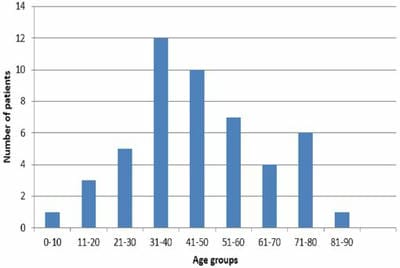
Chart 1: Distribution of laryngeal lesions according to age groups.

patho_605_02.JPG
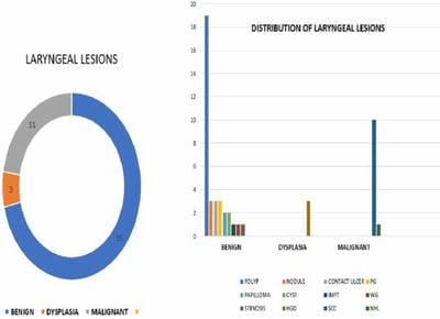
Chart 2: Distribution of laryngeal lesions according to numbers.

Discussion

The larynx is the passageway for air between the pharynx above and the trachea below. Many diseases affect the larynx, benign and malignant with the predominance of the former. In our study, the most common site of involvement was glottis (42 cases), followed by supra-glottis and sub-glottis. The majority of lesions were benign- 35(71.4%) followed by malignant cases-11(22.4%). Benign lesions could be non-neoplastic or neoplastic. The vocal cord (laryngeal) nodule represents a peculiar noninflammatory reaction to injury-causing hoarseness, which is seen more commonly in people who misuse their voices. In the early stages, there is the myxoid stage and later the fibrous stage [2].

They appear symmetrically on the free edge of the vocal cord, at the junction of the anterior one-third, with the posterior two-thirds, as this is the area of maximum vibration of the cord and thus subject

to maximum trauma. A vocal polyp is unilaterally arising from the same position. Mostly, it affects men in the age group of 30–50 years [1].

We had 19 polyps which made up 54.2% of all benign lesions and 3 laryngeal nodules. Contact granulomas are caused by severe phonotrauma, gastroesophageal reflux disease, and intubation. It is almost always found at the level of the posterior commissure, in the area of the vocal process of the arytenoid cartilage Microscopically, it has the appearance of exuberant granulation tissue [2].

We had 3 cases of contact granuloma. The two most common types of cyst of the larynx have been divided by DeSanto et al. according to their mechanism of formation into saccular (24%) and ductal (75%). The former arises from cystic distention of the laryngeal saccule. They are usually large and deep and are often found inside the ventricle [7].

There were 2 cysts seen in our study. One case of Wegener’s granulomatosis was associated with nasal septum involvement.

Pyogenic granulomas are capillary hemangiomas composed of thin-walled capillaries with scant stroma in a lobular arrangement [8]. We had 3 cases of pyogenic granuloma. Laryngeal papillomas constitute about 80% of benign neoplasms of the larynx [1] The papillomas usually involve the vocal cords and ventricles, followed by transmission to the false cords, epiglottis, subglottic area, hypopharynx, and nasopharynx [9].

Juvenile laryngeal papillomas are present in children or adolescents with multiple papillary tumours on the true cords. When extensive, papillomatosis may cause extreme respiratory difficulty and even death. Adult laryngeal papillomas have a male predominance and are most commonly solitary. As for the juvenile form, the involved viral types are usually HPV-6 and HPV-11 [9].

We had 2 cases of papillomatosis in a 4-year-old male and a 19-year-old female.  an inflammatory myofibroblastic tumour (IMFT) is a distinctive neoplasm composed of myofibroblastic and fibroblastic spindle cells accompanied by an inflammatory infiltrate of plasma cells, lymphocytes, and/or eosinophils. Laryngeal IMFT primarily arise in the glottic region [9]. We had 1 case of IMFT in the subglottic region (Figure 1c, d).


patho_605_03.JPG
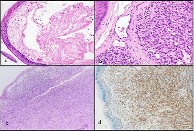
Figure 1a: Vocal cord polyp b. Pyogenic granuloma c. Inflammatory myo-fibroblastic tumour (a, b, c x Hematoxylin and Eosin) d. Smooth muscle actin positivity in tumour cells.

Dysplasia is seen mostly in adults and affects men more often than women, with a male-to-female ratio as high as 4.6:1. Dysplasia can occur anywhere in the larynx, but it occurs most frequently along one vocal cord and less frequently along with both vocal cords [9]. We had 3 cases of high-grade dysplasia, all males with a mean age of 63.3 years.

The majority of laryngeal cancers are squamous cell cancers, other malignancies are rare and may include adenocarcinomas, sarcomas, lymphomas, and neuroendocrine tumours. About 90-95% of laryngeal malignancies are squamous cell carcinoma with various grades of differentiation [1].

In our study malignancies formed 22.9% of the total with squamous cell carcinoma making up 90.9% (Figure 2 a,b). Carcinoma of the larynx accounts for 2.2% of all cancers in men and 0.4% in women [2]. Cigarette smoking has been established as the principal risk factor in laryngeal carcinogenesis, especially in combination with alcohol abuse [9]. The most common early symptoms of laryngeal SCC are hoarseness (with glottic and supraglottic SCC) and dyspnea and stridor (with subglottic SCC). Other symptoms include dysphagia, change in the quality of voice, sensation of a foreign body in the throat, hemoptysis, and odynophagia [9]. In our study 10 cases of SCC were found in a glottic region with a male: female ratio of 9:1 and a mean age of 68.1 years. Two patients gave a history of smoking, seven patients presented with hoarseness of voice with associated dysphonia and difficulty in breathing in one, and three with difficulty in breathing.

patho_605_04.JPG
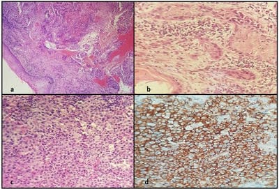
Figure 2 a, b: Squamous cell carcinoma (Hematoxylin and Eosin) c. Non-Hodgkin lymphoma d. CD20 positivity in malignant cells of lymphoma.

Hoarseness is often an early presenting symptom of glottic cancers due to vocal cord immobility or fixation, with pain on swallowing and referred ear pain indicating advanced disease. In contrast, pain with swallowing is the most common early symptom of supraglottic cancer, with hoarseness indicating advanced disease extending into the glottis.

Early-stage laryngeal cancers are treated with a locally-directed treatment modality, local radiation therapy or surgery [10]. Locally-advanced cancers are more difficult to treat and involve combination therapy. These cancers, if surgically resectable are not amenable to laryngeal preservation surgery, while definitive radiation concurrent with cisplatin chemotherapy remains an option for laryngeal preservation. In contrast to early-stage disease, the therapeutic approach to locally-advanced disease is combined chemotherapy and radiation demonstrating both improved locoregional control and larynx preservation [11-14].

Lymphomas arising in the larynx and trachea are rare, accounting for < 1% of neoplasms at these sites. Approximately 4% of head and neck lymphomas arise in the larynx; Lymphoma involves the supraglottic larynx more often than the subglottic larynx. Patients present with cough, dyspnea, and hoarseness. The most common primary lymphoma at this body site is mucosa-associated lymphoid tissue (MALT) lymphoma [9].

We had 1 case of non-Hodgkin lymphoma in supra-glottis in a 24-year-old male who presented with breathing difficulty and was diagnosed with high-grade B-cell lymphoma (Figure 2c, d).


Conclusions

Our study showed that laryngeal lesions have a predilection for the glottis (vocal cord) and affect males. The majority of laryngeal masses were benign and non-neoplastic with the predominant malignancy being squamous cell carcinoma. Histopathology being the mainstay of diagnosis contributes to the accurate management of these patients.

What does this study adds to existing knowledge?

This study enables us to know the relative frequency of laryngeal lesions in patients presenting to a tertiary hospital in southwest KSA, a region relatively unexplored in these terms. Histopathology being the mainstay of diagnosis contributes to the accurate management of these patients.

Author’s contribution: Dr. Sohaila Fatima: Concepts, design, definition of intellectual content, literature search, clinical studies, data acquisition, data analysis, statistical analysis, manuscript preparation, manuscript editing, manuscript review, Dr. Rabab Nasir Mohamed Badri: Concepts, definition of intellectual content, clinical studies, data acquisition, statistical analysis, manuscript preparation, manuscript editing, manuscript review, Dr. Bouvier Francis Valere D’sa: Concepts, definition of intellectual content, clinical studies, data acquisition, statistical analysis, manuscript preparation, manuscript editing, manuscript review, Dr. Wajih Ahmed Siddiqui: Concepts, definition of intellectual content, literature search, clinical studies, data analysis, statistical analysis, manuscript preparation, manuscript editing, manuscript review, Dr. Nihal Ibrahim Mirza: Concepts, Literature search, Clinical studies, Data acquisition, Manuscript preparation, Manuscript review.

Reference

01. Dhingra PS, Dhingra S. Diseases of ear, nose and throat & head and neck surgery. seventh edition. Elsevier, 2018. ISBN: 978-81-312-4884-3 e-Book ISBN: 978-81-312-4939-0 [Crossref][PubMed][Google Scholar]

02. Rosai and Ackerman’s Surgical Pathology. 11th ed. vol 2. Mosby St. Louis, Missouri; 2017 [Crossref][PubMed][Google Scholar]

03. Wareing, M. , R. Millard, and S. Yalamanchili. Benign laryngeal lesions. Current Diagnosis and Treatment: Otolaryngology Head and Neck Surgery. Third ed. New York: Mc Graw Hill Companies (2012): 449-55 [Crossref][PubMed][Google Scholar]

04. Clarke, Ray. Scott-Brown's Otorhinolaryngology and Head and Neck Surgery: 3 volume set. (2018). [Crossref][PubMed][Google Scholar]

05. National Health Information Center Saudi Cancer Registry. Cancer Incidence Report Saudi Arabia 2017. https://nhic. gov. sa/eServices/Documents/2017 [Crossref][PubMed][Google Scholar]

06. International Agency for Research on Cancer. Global cancer observatory. 2020. Cancer today. Disponível em: iarc. fr/today/home, acedido em 15 (2021) [Article][Crossref][PubMed][Google Scholar]

07. Mitchell RN, Schoen FJ. Blood Vessels. In: Kumar V, Abbas AK, Aster JC editors. Robbins and Cotran pathologic basis of disease, 9th edition. Philadelphia, PA: Elsevier/Saunders; 2015.p.483-522 [Crossref][PubMed][Google Scholar]

08. DeSanto LW, Devine KD, Weiland LH. Cysts of the larynx--classification. Laryngoscope. 1970 Jan;80(1):145-76. doi: 10.1288/00005537-197001000-00013 [Crossref][PubMed][Google Scholar]

09. Tumours of the hypopharynx, larynx, trachea and parapharyngeal space. In: EI-Naggar AK, Chan JKC, Grandis JR, Takata T, Slootweg PJ. World Health Organization Classification of Tumours. Pathology and genetics of head and neck tumours. Lyon: IARC Press; 2017.p. 81-104 [Crossref][PubMed][Google Scholar]

10. Koroulakis, A. , and M. Agarwal. Laryngeal Cancer. [Updated 2020 Aug. 10]." StatPearls [Internet]. Treasure Island (FL): Stat Pearls Publishing (2021): 205-11 [Crossref][PubMed][Google Scholar]

11. Aaltonen, Leena-Maija, et al. Voice quality after treatment of early vocal cord cancer: a randomized trial comparing laser surgery with radiation therapy. International Journal of Radiation Oncology* Biology* Physics 90. 2 (2014): 255-260. [Crossref][PubMed][Google Scholar]

12. Forastiere, Arlene


A. , et al. Use of larynx-preservation strategies in the treatment of laryngeal cancer: American Society of Clinical Oncology clinical practice guideline update. " Journal of Clinical Oncology 36. 11 (2018): 1143-1169 [Crossref][PubMed][Google Scholar]

13. Forastiere AA, Ismaila N, Lewin JS, Nathan CA, Adelstein DJ, Eisbruch A, et al. Use of Larynx-Preservation Strategies in the Treatment of Laryngeal Cancer: American Society of Clinical Oncology Clinical Practice Guideline Update. J Clin Oncol. 2018 Apr 10;36(11):1143-1169. doi: 10.1200/JCO.2017.75.7385 [Crossref][PubMed][Google Scholar]

14. Rosenthal DI, Mohamed AS, Weber RS, Garden AS, Sevak PR, Kies MS, et al. Long-term outcomes after surgical or nonsurgical initial therapy for patients with T4 squamous cell carcinoma of the larynx: A 3-decade survey. Cancer. 2015 May 15;121(10):1608-19. doi: 10.1002/cncr.29241 [Crossref][PubMed][Google Scholar]

15. Ruytenberg, Thomas, et al. Improvements in high resolution laryngeal magnetic resonance imaging for preoperative transoral laser microsurgery and radiotherapy considerations in early lesions. " Frontiers in oncology 8 (2018): 216. [Crossref][PubMed][Google Scholar]