E-ISSN:2456-1487
P-ISSN:2456-9887
RNI:MPENG/2017/70771

Research Article

Pre-Eclampsia

Tropical Journal of Pathology and Microbiology

2022 Volume 8 Number 2 March April
Publisherwww.medresearch.in

Histopathological Study of Placenta in Association with deranged Thyroid Profile, in Known Cases of pre-Eclampsia/ Eclampsia

Hudda S.1, Choudhary A.2*
DOI: https://doi.org/10.17511/jopm.2022.i02.01

1 Sangeeta Hudda, MD, Department of Pathology, Mahatma Gandhi University of Medical Sciences and Technology, Jaipur, Rajasthan, India.

2* Anubha Choudhary, MD, Department of Pathology, Mahatma Gandhi University of Medical Sciences and Technology, Jaipur, Rajasthan, India.

Objectives: To assess the histo-morphology of the placenta in association with abnormal thyroid hormone levels in patients with Pre-eclampsia/Eclampsia, concerning the birth weight of the baby. Methods: The study was performed in the department of Pathology over 3 months. Cases and controls were evaluated based on the 3rd trimester TSH level of the Antenatal patients. Cases were considered as patients who had levels of TSH outside the normal range in the 3rd trimester (0.3-3.1mIU/L). Out of 50 patients,15 cases and 33 controls were defined. A histopathological report of the resected placenta specimens was received. Clinical details of the mother and the birth weight of the baby were collected. Finally, the correlation between placental histo-morphology, mother's diagnosis and baby birth weight were correlated. Results: Among all the cases, 9 patients (60%) were diagnosed with pre-eclampsia, as compared to 4 (14.8%) among controls. Concerning histology, hemorrhage in the placenta was observed more among the cases (13/15; 86.6%) as compared to controls (51.8%). Calcification was more among the controls (55.5% vs 26.6%). Among other characteristics like placental weight, infarction, chorangiosis, syncytial knots and villitis, no significant difference was noted. 58.8% (10/17) of babies born to cases had Low birth weight (<2.5 kg) compared to 40.7% in controls. Conclusion: The occurrence of pre-eclampsia was high among mothers with abnormal thyroid hormone levels. Similar observations were seen in the increased incidence of low birth weight babies. So, we strongly recommend performing thyroid screening on all ANC patients.

Keywords: Histopathological Study, Placenta, Pre-Eclampsia, Eclampsia

Corresponding Author How to Cite this Article To Browse
Anubha Choudhary, MD, Department of Pathology, Mahatma Gandhi University of Medical Sciences and Technology, Jaipur, Rajasthan, India.
Email:
Sangeeta Hudda, Anubha Choudhary, Histopathological Study of Placenta in Association with deranged Thyroid Profile, in Known Cases of pre-Eclampsia/ Eclampsia. Trop J Pathol Microbiol. 2022;8(2):36-40.
Available From
https://pathology.medresearch.in/index.php/jopm/article/view/603

Manuscript Received Review Round 1 Review Round 2 Review Round 3 Accepted
2022-03-01 2022-03-03 2022-03-10 2022-03-17 2022-03-24
Conflict of Interest Funding Ethical Approval Plagiarism X-checker Note
Nil Nil Yes 16%

© 2022by Sangeeta Hudda, Anubha Choudharyand Published by Siddharth Health Research and Social Welfare Society. This is an Open Access article licensed under a Creative Commons Attribution 4.0 International License https://creativecommons.org/licenses/by/4.0/ unported [CC BY 4.0].

Introduction

Various endocrine, paracrine, and autocrine events within the fetoplacental unit affect fetal growth. Thyroid hormone levels have been proved to play an important role in the pathogenesis of morbidity in the fetus, especially concerning the growth and development of the central nervous system [1].

Trophoblast growth and development are controlled by thyroid hormones. T3 has a high binding capacity for trophoblast tissue and it has been suggested that the placenta is a thyroid hormone-dependent tissue. thyroid hormones stimulate trophoblast endocrine function and cause enhanced production of human placental lactogen and human chorionic gonadotrophin.  Studies have also proven that thyroid hormones enhance the production of epidermal growth factor, a strong trophoblast mitogen. Therefore villous development and placentation greatly depend upon the thyroid hormones [2].

Thyroid hormones also control the secretion of several cytokines and growth factors that are critical for Endovascular trophoblast (EVT) invasion and angiogenesis of maternal and fetal placental vessels, including angiogenin, angiopoietin 2 (Ang-2), vascular endothelial growth factor (VEGF), Interleukin 10 and TNF. Furthermore, it also attenuates trophoblast proliferation, motility and invasion [3].

Our purpose is to study histomorphological features of the placenta and correlate them with TSH levels, and the birth weight of the baby in 3rd-trimester mothers especially with disorders like eclampsia/pre-eclampsia.

Materials and Methods

Setting: The study was conducted in the Department of Pathology, Mahatma Gandhi Medical College & Hospital, Jaipur, Rajasthan, India.

Duration: Over three months.

Sample Collection: The study population was 3rd-trimester mothers undergoing labour. Placenta specimens were received in containers with 10% formalin. They were adequately examined and sectioned serially and fixed in a fresh preparation of 10% formalin. They were allowed to fix it for 24 hours. Grossing is done on the next day

wherein crucial parameters were noted down like weight and dimensions of the placental disc, length and diameter of the umbilical cord and distance of cord from disc margin. Then representative sections were obtained from the four quadrants of the placental disc, the central area and the area of cord insertion. Sections from the umbilical cord and placental membrane were also obtained. The sections are processed in an automatic tissue processor. The paraffin block was obtained at the end from which thin sections were cut using a microtome. The sections were then subjected to routine H&E staining.

Other relevant reports of the patients were received from the central laboratory. Thyroid function tests are performed on the VITROS 5600. As per guidelines of ICMR, TSH levels of 0.3-3.1 mIU/L were considered the normal range for 3rd-trimester mothers.

Clinical parameters like the baby's birth weight and clinical diagnosis of the mother were obtained from the master register in the labor room. The total sample size was 42 out of which cases and controls were divided based on TSH levels, yielding 27 controls and 15 cases. Cases were considered those with TSH levels outside the normal range. Correlation between the placental morphology, TSH levels, and birth weight of the baby was done, the results were tabulated and percentage values were obtained.

Results

Table 1: Distribution of controls & cases.

TSHLevel Controls (TSH=0.3- 3.1mIU/L) Cases (TSH>3.1)
No.n=42 27[65%] 15[35%]

Table 2: Final clinical diagnosis of the mother.

Final Diagnosis of Mother Controls Cases
Pre-eclampsia/Eclampsia 4[14.8%] 9[60%]
Oligohydramnios 7 2
IUGR 4 2
PreviousLSCS 4 4
ContractedPelvis/CPD 4 0
Anemia 3 3
PlacentaPrevia 0 1
HELLPSyndrome 1 0
Post-TermDelivery 5 0
IUD 1 0
Others 2 2

Among the cases, pre-eclampsia was diagnosed in 9 patients (60%) as compared to 4 (14.8%) among controls. Other common disorders among the controls were oligohydramnios, Intra-uterine growth retardation (IUGR), cephalopelvic disproportion (CPD) and post-term deliveries.

patho_603_01.JPG
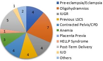
Figure 1: Final diagnoses among controls.

patho_603_02.JPG
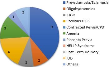
Figure 2: Final diagnoses among cases.

Table 3: Placental Weight.

Weight (gms) Controls Cases
<100 1 0
100-200 1 0
200-300 6 2
300-400 11(40.7%) 6 (40%)
400-500 5 (18.5%) 5 (33.3%)
500-600 3 (11.1%) 2 (13.3%)
Total 27 15

In relation to the placental weight, 86.6% of the cases had a placental weight of over 300gm. Whereas among the controls 70.3% weighted 300gm.

Concerning histology, hemorrhage in the placenta was found more among the cases (13/15;86.6%) as compared to controls (51.8%). Infarction was seen more among the cases (53.3%) as compared to the controls (33%). Calcification was found more among controls (55.5% vs 26.6%). No significant

differences were found among other characteristics like chorangiosis, syncytial knots and villitis.

Table 4: Histomorphology of placenta.

Feature   Controls Cases
Infarction   11 (33%) 8 (53.3%)
Syncytialknots Type-1 17 2
Type-2a 5 9
Type-2b 2 1
Chorangiosis   12 (44.4%) 6 (40%)
Haemorrhage   14 (51.8%) 13 (86.6%)
Calcification   15 (55.5%) 4 (26.6%)

Table 5: Birth Weight of Baby.

Birth Wt (Kg) Cases Controls
<1.5 1 3
1.5-2.5 9 8
2.5-3.5 4 14
>3.5 1 2
Total = 15 27

Low Birth Weight (<2.5 kg):  Cases- 10/17: 58.8% Controls- 11/27:  40.7%, 58.8 % (11/17) babies which were born to cases had Lowbirth weight (<2.5 kg) as compared to only 40.7 % among the controls.

Discussion

Our study segregates the cases and controls based on levels of TSH in their 3rd trimester. That yielded 15 cases and 27controls. Nine patients among the cases (60%) were having pre-eclampsia, as compared to only 4 (14.8%) among controls.

This finding correlates with the study conducted at the Regional Institute of Medical Sciences, Imphal, Manipur which found that severe preeclampsia was seen in 64.3% of the patients with thyroid dysfunction compared with39.6%in patients with normal thyroid hormone levels [4].

In a similar study at the Wenzhou Medical University, China, was compared in patients with euthyroid status, subclinical hypothyroidism (SCH) was associated with higher rates of preeclampsia (1.819%vs.3.504%, P=0.020) [5].

Amongst the other ailments that affected the cases, anaemia (18%) was the commonest, followed by Oligohydramnios, IUGR and placenta previa. Among the controls, there was a high incidence of oligohydramnios (25.9%), post-dated delivery (18.5%), cephalon-pelvic disproportion (14.8%)


and IUGR (14.8%). One baby which was born to a woman from control suffered from intrauterine death (IUD). History of previous LSCS was present in four patients from either side.

Placentalweightofover300gm was found in 86.6% of the cases Whereas among the controls it was seen in 70.3% of patients.

Concerning histology, the incidence of placental hemorrhage was significantly higher among the cases (86.6%) as compared to the controls (51.8%). Similar trend was seen for placental infarction as well. Infarction was seen in 53.3% of the cases as compared to only 33% among the controls.

In women with pre-eclampsia, the mean luminal diameter of uterine spiral arterioles is less than one-third of the diameter of similar vessels in an uncomplicated pregnancy. Consequently, there is reduced uteroplacental perfusion leading to ischemic changes and fetal hypoxia. Also, the villous surface area, the villi diameters and the density of fetal blood vessels in the terminal villi is reduced in a pre-eclamptic patient [1].

Calcification was more among the controls (55.5%vs26.6%). This is probably due to the increased incidence of post-dated pregnancies seen among the controls. Among other histologic criteria like syncytial knots and chorangiosis, significant differences were not seen between both groups. As far as the birth weight of the baby is concerned, 58.8% (10/17) of babies born to cases had Low birth weight (<2.5 kg) compared to only40.7 %( 11/27) in controls.

This is following the study conducted at the Wenzhou Medical University, China, where more LBW infants were delivered in the subclinical hypothyroidism (SCH) group than in the euthyroid group(4.582%vs.1.885%, P,0.001) [5]. In a study conducted at the Institute of Obstetrics & Gynecology, Chennai, the gestational age at birth was significantly decreased and the cesarian rate was significantly high in preeclampsia. Low birth weight was significant in preeclampsia [6].

A similar study done at the Regional Institute of Medical Sciences, Imphal, Manipur concluded that Complications like abruption, intrauterine fetal death (IUD), intrauterine growth restriction (IUGR) oligohydramnios, preterm deliveries, postpartum

haemorrhage (PPH), low birth weight babies, birth asphyxia in babies and subsequent neonatal intensive care unit (NICU) admissions were significantly higher (p<0.05 in the preeclampsia patients with thyroid dysfunction in comparison with euthyroid ones[4].

In another study conducted at Narayana medical college, Nellore, the morphology of stem and terminal villi (TV) was studied, and the surface area and diameter of TV and capillaries were measured. The gross placental morphometrical study revealed that the mean placental weight, thickness, diameter, and surface area were significantly lower in placentas with PE than in controls. The histomorphometric findings of the villous surface area and diameter were lower in placentas with PE, whereas the TVdensity was higher in placentas with PE than in controls, and the differences were significant (P<0.0001) [1].

In another study conducted at Navodaya medical college, Raichur on the maternal thyroid profile in pre-eclampsia, there was no significant difference in the thyroxine(T4) and tri-iodothyronine (T3) levels in the two groups, but there were a significant increase in thyroid-stimulating hormone (TSH) levels in pre-eclampsia patients(7.22± 1.3) compared to normal pregnancy(2.48±1.05)(p=0.0001)[7].

A study was done in the US by various contributors aimed to discern if stillbirth with preeclampsia and gestational hypertension (PE/GH) has a particular phenotype by comparing stillbirths with and without PE/GH. Among PE/GH pregnancies, stillbirths had increased maternal and fetal vascular lesions, including retroplacental hematoma, parenchymal infarction, fibrin deposition, fetal vascular thrombi, and avascular villi. Stillbirth pregnancies are overwhelmingly associated with placental lesions. Parenchymal infarctions are more common in PE/GH preterm stillbirths [8].

Another study conducted at University Kebangsaan Malaysia medical centre, KaulaLampur showed an increased syncytial knot formation In the placenta of hypertensive mothers. Vascular endothelial growth factor (VEGF) expression was seen in syncytiotrophoblasts of 14 of the hypertensive cases (14/15, 93.3%), while only two of the normotensive cases were positive (2/15,13.3%) [9].

This study showed that an increased number of


syncytial knots is a consistent histological finding in the placenta of a patient with hypertensive disorder of pregnancy (HDP). VEGF expression was significantly increased in syncytiotrophoblasts in the placenta of the hypertensive group, and it could be used as a biomarker for hypertension.

Conclusion

In our study, the incidence of pre-eclampsia was more among mothers with abnormal thyroid hormone levels. This can cause placental insufficiency in the form of increased hemorrhage, infarction and reduced placental weight. This is subsequently reflected in the increased incidence of low birth weight babies. So screening for hypothyroidism in pregnancy is recommended in all antenatal females and replacement of thyroid hormones in all indicated women will improve the obstetric outcome in future.

What does this study adds to existing knowledge?

In our study, we found that the incidence of pre-eclampsia was more among mothers with abnormal thyroid hormone levels.

Author’s contribution: Sangeeta Hudda: manuscript preparation, manuscript editing, and manuscript review. Anubha Chaudhary: Concepts, design, definition of intellectual content, literature search, clinical studies, data acquisition, data analysis, statistical analysis.

Reference

01. Sankar KD, Bhanu PS, Ramalingam K, Kiran S, Ramakrishna BA. Histomorphological and morphometrical changes of placental terminal villi of normotensive and preeclamptic mothers. Anat Cell Biol. 2013 Dec;46(4):285-90. doi: 10.5115/acb.2013.46.4.285 [Crossref][PubMed][Google Scholar]

02. Sardana D, Nanda S, Kharb S. Thyroid hormones in pregnancy and preeclampsia. J Turk Ger Gynecol Assoc. 2009 Sep 1;10(3):168-71. [Crossref][PubMed][Google Scholar]

03. Barjaktarovic, Mirjana, et al. The association of maternal thyroid function with placental hemodynamics. Human Reproduction 32. 3 (2017): 653-661. [Crossref][PubMed][Google Scholar]

04. Banik, Puja, et al. Thyroid dysfunction in preeclampsia and related fetomaternal outcomes. International Journal of Reproduction, Contraception, Obstetrics and Gynecology 8. 5 (2019): 1928-1934. [Crossref][PubMed][Google Scholar]

05. Chen LM, Du WJ, Dai J, Zhang Q, Si GX, Yang H, et al. Effects of subclinical hypothyroidism on maternal and perinatal outcomes during pregnancy: a single-center cohort study of a Chinese population. PLoS One. 2014 Oct 29;9(10):e109364. doi: 10.1371/journal.pone.0109364 [Crossref][PubMed][Google Scholar]

06. Rani S U, Arumaikannu J & Shanthi S. Hypothyroidism as a bio marker of preeclampsia: Our experience. Corpus ID: 86428148, Published 2018, Medicine, Biology. doi: 10.1371/journal.pone.0109364 [Crossref][PubMed][Google Scholar] [Crossref][PubMed][Google Scholar]

07. Satyanarayan, Anitha K. , et al. Maternal thyroid profile in pre-eclampsia. International Journal of Medical Science and Public Health 4. 10 (2015): 1401-1403 [Crossref][PubMed][Google Scholar]

08. Gibbins KJ, Silver RM, Pinar H, Reddy UM, Parker CB, Thorsten V, et al. Stillbirth, hypertensive disorders of pregnancy, and placental pathology. Placenta. 2016 Jul; 43:61-8. doi: 10.1016/j.placenta.2016.04.020 [Crossref][PubMed][Google Scholar]

09. Azliana AF, Zainul-Rashid MR, Chandramaya SF, Farouk WI, Nurwardah A, Wong YP, et al. Vascular endothelial growth factor expression in placenta of hypertensive disorder in pregnancy. Indian J Pathol Microbiol. 2017 Oct-Dec;60(4):515-520. doi: 10.4103/IJPM.IJPM_376_16 [Crossref][PubMed][Google Scholar]