E-ISSN:2456-1487
P-ISSN:2456-9887
RNI:MPENG/2017/70771

Case Report

Portal Hypertension

Tropical Journal of Pathology and Microbiology

2022 Volume 8 Number 2 March April
Publisherwww.medresearch.in

Unravelling the mystery behind an unusual case of Portal Hypertension

Unnikrishnan R.1*, Philip M.2, Yadav A.3, Mahadevan P.4
DOI: https://doi.org/10.17511/jopm.2022.i02.03

1* Rasmi Unnikrishnan, Senior Registrar, Department of Pathology, Lakeshore Hospital and Research Centre, Ernakulam, Kerala, India.

2 Mathew Philip, HOD - Senior Consultant, Department of Gastroenterology, Lisie hospital, Ernakulam, Kerala, India.

3 Abhishek Yadav, Senior Consultant, Lead Liver transplant surgeon, Lakeshore Hospital and Research Centre, Ernakulam, Kerala, India.

4 Pushpa Mahadevan, HOD - Senior consultant, Department of Pathology, Lakeshore Hospital and Research Centre, Ernakulam, Kerala, India.

A 38yr old male was incidentally detected to have chronic liver disease with portal hypertension. He gives a family history of liver disease. On complete workup, the arsenic levels in his family members were found to be high. His liver biopsy showed features of Hepatoportal sclerosis. Two years later, his liver imaging showed a suspicious nodule in the Right lobe, following which he underwent a live donor liver transplant. Histopathology of the explanted liver revealed, areas of cavernous hemangioma, epithelioid hemangioendothelioma with a focus of diffusely infiltrating Angiosarcoma, in a background of Hepatoportal sclerosis.

Keywords: Portal hypertension, Arsenic poisoning, Hepatoportal sclerosis, Angiosarcoma

Corresponding Author How to Cite this Article To Browse
Rasmi Unnikrishnan, Senior Registrar, Department of Pathology, Lakeshore Hospital and Research Centre, Ernakulam, Kerala, India.
Email:
Rasmi Unnikrishnan, Mathew Philip, Abhishek Yadav, Pushpa Mahadevan, Unravelling the mystery behind an unusual case of Portal Hypertension. Trop J Pathol Microbiol. 2022;8(2):47-51.
Available From
https://pathology.medresearch.in/index.php/jopm/article/view/600

Manuscript Received Review Round 1 Review Round 2 Review Round 3 Accepted
2022-03-19 2022-03-21 2022-03-28 2022-04-04 2022-04-10
Conflict of Interest Funding Ethical Approval Plagiarism X-checker Note
Nil Nil Yes 17%

© 2022by Rasmi Unnikrishnan, Mathew Philip, Abhishek Yadav, Pushpa Mahadevanand Published by Siddharth Health Research and Social Welfare Society. This is an Open Access article licensed under a Creative Commons Attribution 4.0 International License https://creativecommons.org/licenses/by/4.0/ unported [CC BY 4.0].

Introduction

Primary hepatic Angiosarcoma is a rare tumour that accounts for about 0.1%–2% of all primary malignant liver tumours. [1-3]. They are classified under mesenchymal tumours, of endothelial cell origin, generally appearing during the sixth or seventh decade of life. [3,4]. Most patients have a variable clinical presentation, from asymptomatic to nonspecific symptoms mimicking other chronic liver diseases including abdominal pain, fatigue, weight loss, jaundice, and anorexia or may present with liver failure.[4]. Various environmental carcinogens, such as vinyl chloride, thorium dioxide, and arsenic, are known to cause primary hepatic angiosarcoma. [5]. Exposure to these chemicals, especially arsenic, may be a part of medicinal, industrial or even occupational. Arsenic in drinking water sources has also been postulated. Among the spectrum of liver lesions attributed to chronic arseniasis, the significant ones include hepatocellular carcinoma, angiosarcoma, cirrhosis, and hepatoportal sclerosis. [5,6]. Hepatoportal sclerosis (HS) is a condition characterized by portal hypertension without evidence of liver cirrhosis. [6-8]. However, chronic arsenic exposure leading to Hepatoportal sclerosis followed by Angiosarcoma is very rare.

Case Report

patho_600_01.JPG
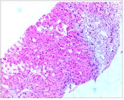
Figure 1: Enlarged portal areas showing inconspicuous portal veins, periportal mega sinusoids and herniation of veins into the lobules.

A 38yr old male presented with incidentally detected cirrhosis of the liver, grade 2 oesophagal varices, splenomegaly and a family

history of liver disease. The initial liver biopsy received in our department showed, cirrhosis with, dilated and engorged sinusoids (Fig: 1).

One year later, we received a review liver biopsy of his 10yr old daughter, showing similar pathology (Fig: 2).

patho_600_02.JPG
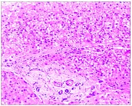
Figure 2: Daughter’s liver biopsy - Portal areas show fibrosis/sclerosis with inconspicuous portal veins. The sclerosis is seen extending into the lobule with atrophic hepatocyte trabeculae.

On complete workup of the case, we found that two months following his liver biopsy, our patient had an episode of GI bleeding and oesophagal variceal ligation was done. Around the same time, his daughter was being evaluated in an outside hospital for pyrexia of unknown origin and as a part of routine workup, her imaging done there showed, an enlarged left lobe and the caudate lobe of the liver with diffuse altered parenchymal echotexture, suspicious of acute / sub-acute granulomatous hepatitis. Her liver function test was normal. In this scenario, as a part of a complete workup, of the family, the nail and hair samples tested for arsenic surprisingly showed high levels. The following are the arsenic levels in his family members:

Patient    30.28 mcg/g (Hair) 6.12 μg/g (Nail)

Wife        1.52 mcg/g (Hair) 7.16 μg/g (Nail)

Daughter 8.2 mcg/g (Hair) 4.5 μg/g (Nail)

Son         9.8 mcg/g (Hair) 34.8 μg/g (Nail)

After acquiring this history, we reviewed the father's liver biopsy. Both the biopies showed inconspicuous portal veins, mega sinusoids,


dilated venules appearing to herniate into lobules with sclerosis and dilatation of efferent veins (Fig: 1, 2). Hence we gave a diagnosis in keeping with Hepatoportal Sclerosis.

Later, a repeat CT Abdomen of our patient showed Cirrhosis of the Liver, with multiple enhancing lesions in both lobes of the liver with a 1.6x1.5cm, subcapsular lesion in segment 6. He was classified as CHILD B; MELD 22. LFT done at this time showed total bilirubin of 7.1mg/dL (direct - 4.4) and an INR of 4.4. Following this, the patient underwent a live donor liver transplant at our hospital and we received the hepatectomy specimen.

Grossly, the liver weighed 1.57kg. The surface of the liver and cut surface showed multiple cirrhotic nodules of varying size, with many having a brownish hue. Also noted was a subcapsular, 1.5cm ill-circumscribed focus in the right lobe, having a brownish spongy appearance. Microscopy showed liver with a diffusely infiltrating vascular neoplasm, composed of papillary structures protruding into the vascular lumen with an epithelioid hemangioendothelioma morphology (Fig: 3).

patho_600_03.JPGFigure 3: Liver with diffusely infiltrating vascular neoplasm, composed of papillary structures protruding into the vascular lumen with an epithelioid hemangioendothelioma morphology.

The cells were seen lining vascular spaces with hobnailing of the nucleus. The tumour cells had a pleomorphic vesicular nucleus, prominent nucleoli and scanty cytoplasm with mitotic figures (0-2/HPF).

The spongy areas showed dilated vascular spaces filled with blood, reminiscent of a cavernous haemangioma (Fig: 4).

patho_600_04.JPG
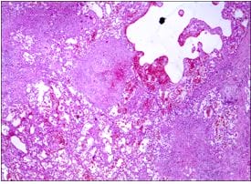
Figure 4: The spongy areas showing dilated vascular spaces filled with blood, reminiscent of a hemangioma.

patho_600_05.JPGFigure 5: One of the subcapsular nodules adjacent to the spongy area showing a poorly circumscribed neoplasm c/o lobules of cells with an epithelioid appearance and tumor emboli.

patho_600_06.JPG
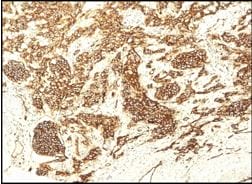
Figure 6: The tumor cells showed diffuse strong positivity for CD31.

Some of the vascular channels


showed foci of extramedullary hemopoiesis. The tumor cells showed diffuse strong positivity for CD31 (Fig: 6), and CD 34 and were negative for Hepar 1, CK, CK7 and CK20. Hence we offered a diagnosis of diffusely infiltrating Angiosarcoma in the case of Hepatoportal sclerosis.

Discussion

Angiosarcoma is an uncommon, highly malignant tumor that forms multiple or less often solitary haemorrhagic masses. Primary hepatic angiosarcoma is rare and accounts for about 0.1%–2% of all malignant primary liver tumours. [1-3]. The clinical presentation may vary, from asymptomatic to nonspecific symptoms mimicking other chronic liver diseases or may present as liver failure. [4]. The predisposing factors include treatment with arsenic, injection of radioactive contrast medium Thorotrast, industrial exposure to vinyl chloride and other postulates. [5].

Arsenic exposure can cause acute toxicity or chronic toxicity and has carcinogenic properties as well. It has a long history of use as an intentional poison, in medicines, pesticides, and even environmental exposure that stems from the pollution of drinking water in certain geographic areas. The significant liver lesions attributed to chronic arseniasis include cirrhosis, hepatocellular carcinoma, angiosarcoma and hepatoportal sclerosis. [6]. Hepatoportal sclerosis (also known as noncirrhotic portal fibrosis, idiopathic portal hypertension, and Banti syndrome) is an idiopathic noncirrhotic presinusoidal intrahepatic portal hypertension due to fibrosis of the intima of the portal vein and its branches. It is characterized by splenomegaly, hypersplenism and portal hypertension. [7-10].

In our case, the patient’s history of arsenic exposure and subsequent development of Hepatoportal Sclerosis were considered predisposing factors. The family resided abroad, and later the possible source of arsenic exposure was postulated to be their drinking water source.

Microscopy, in this case, revealed areas of cavernous hemangioma, epithelioid hemangioendothelioma and diffusely infiltrating Angiosarcoma.

Conclusion

In literature, cases have been reported regarding,

arsenic exposure predisposing to the development of Hepatoportal Sclerosis and cases with development of Hepatic Angiosarcoma, following Hepatoportal sclerosis. However, such an unusual association of arsenic exposure leading to Hepatoportal sclerosis and subsequent development of Angiosarcoma has not been reported. Hence our case is reported as an exceptional one.

Reference

01. Kim HR, Rha SY, Cheon SH, Roh JK, Park YN, Yoo NC. Clinical features and treatment outcomes of advanced stage primary hepatic angiosarcoma. Ann Oncol. 2009 Apr;20(4):780-7. doi: 10.1093/annonc/mdn702 [Crossref][PubMed][Google Scholar]

02. Chien CY, Hwang CC, Yeh CN, Chen HY, Wu JT, Cheung CS, et al. Liver angiosarcoma, a rare liver malignancy, presented with intraabdominal bleeding due to rupture--a case report. World J Surg Oncol. 2012 Jan 26;10:23. doi: 10.1186/1477-7819-10-23 [Crossref][PubMed][Google Scholar]

03. Millan M, Delgado A, Caicedo LA, Arrunategui AM, Meneses CA, Villegas JI, et al. Liver Angiosarcoma: Rare tumour associated with a poor prognosis, literature review and case report. Int J Surg Case Rep. 2016;28:165-168. doi: 10.1016/j.ijscr.2016.09.044 [Crossref][PubMed][Google Scholar]

04. Rujeerapaiboon N, Wetwittayakhlang P. Primary Hepatic Angiosarcoma: A Rare Liver Malignancy - Varying Manifestations but Grave Prognosis. Case Rep Gastroenterol. 2020 Apr 8;14(1):137-149. doi: 10.1159/000506928 [Crossref][PubMed][Google Scholar]

05. Popper H, Thomas LB, Telles NC, Falk H, Selikoff IJ. Development of hepatic angiosarcoma in man induced by vinyl chloride, thorotrast, and arsenic. Comparison with cases of unknown etiology. Am J Pathol. 1978 Aug;92(2):349-76 [Crossref][PubMed][Google Scholar]

06. Centeno JA, Mullick FG, Martinez L, Page NP, Gibb H, Longfellow D, et al. Pathology related to chronic arsenic exposure. Environ Health Perspect. 2002 Oct;110 Suppl 5(Suppl 5):883-6. doi: 10.1289/ehp.02110s5883 [Crossref][PubMed][Google Scholar]


07. Cuadrado Lavín A, Aresti Zárate S, Delgado Tapia A, Figols Ladrón De Guevara FJ, Salcines Caviedes JR. Hepatoportal sclerosis: a cause of portal hypertension to bear in mind. Gastroenterol Hepatol. 2008 Feb;31(2):104. doi: 10.1157/13116079 [Crossref][PubMed][Google Scholar]

08. Sanz-García C, Matilla-Peña A, Nogales-Rincón O, Núñez-Martínez O, Clemente-Ricote G. Esclerosis hepatoportal y angiosarcoma hepático: una asociación infrecuente y una posible etiología común [Hepatoportal sclerosis and liver angiosarcoma: an infrequent association with a possible common etiology]. Gastroenterol Hepatol. 2005 Nov;28(9):555-7. Spanish. doi: 10.1157/13080609 [Crossref][PubMed][Google Scholar]

09. Kleiner DE. Noncirrhotic portal hypertension: Pathology and nomenclature. Clin Liver Dis (Hoboken). 2015 May 27;5(5):123-126. doi: 10.1002/cld.459 [Crossref][PubMed][Google Scholar]

10. Rios FF, Rodrigues de Freitas LA, Codes L, Santos Junior GO, Schinoni MI, Paraná R. Hepatoportal sclerosis related to the use of herbals and nutritional supplements. Causality or coincidence? Ann Hepatol. 2016 Nov-Dec 2016;15(6):932-938. doi: 10.5604/16652681.1222113 [Crossref][PubMed][Google Scholar]