E-ISSN:2456-1487
P-ISSN:2456-9887
RNI:MPENG/2017/70771

Research Article

Lymph Node Lesions

Tropical Journal of Pathology and Microbiology

2021 Volume 7 Number 6 November December
Publisherwww.medresearch.in

A cytomorphological study of the spectrum of lymph node lesions in a tertiary care centre – a retrospective study

Saleem H.1, Fatima R.2, Shameem A.3, Chandra T.4, Shameem K.5*
DOI: https://doi.org/10.17511/jopm.2021.i06.03

1 Hafsa Saleem, Resident, Department of Pathology, Kamineni Institute of Medical Sciences, Narketpally, Telangana, A.P, India.

2 Rida Fatima, Undergraduate Medical student, , Ayaan Medical College, Moinabad, Telangana, A.P, India.

3 Afroze Shameem, , Department of Paediatrics, NMC Medical center, Dubai, Abu Dhabi, UAE.

4 T Jaya Chandra, Professor, Department of Microbiology, GSL Medical College, Rajahmundry, A.P, India.

5* Khatija Shameem, Associate Professor, Department of Pathology, Kamineni Institute of Medical Sciences, Narketpally, Telangana, A.P, India.

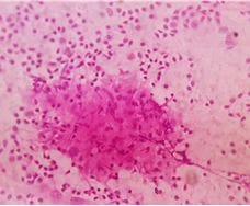
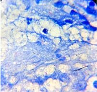
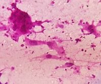
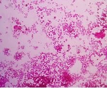
Introduction: Lymphadenopathy (LA) is a clinical condition where the lymph nodes (LNs) can be enlarged. Fine Needle Aspiration Cytology (FNAC) is a simple, inexpensive and rapid diagnostic technique practiced to find the cause for the superficial LA. With this, a study was conducted to find the cytomorphological features of the enlarged LNs and also to analyze the utility of FNAC in the suspected malignant lesions. Materials and methods: The study was conducted in the Department of Pathology, Kamineni Institute of Medical Sciences, Narketpally, from June 2018 to Dec 2020. Random sampling was considered. Individuals of all age groups with enlarged LNs were included. Inadequate or hemorrhagic samples, non-cooperative members were not considered. The participants were proceeded for FNAC as per the standard reports. Smear preparation was done at the bedside, fixed in 95% Ethyl alcohol. The smears were stained with H & E, Giemsa techniques. If required, Ziehl Neelsen (ZN) technique was also used to find acid-fast bacilli (AFB). After staining, the smears were air-dried and examined under the microscope. Results: A total of 161 (100%) participants were included, ages were ranged between 8 months to 85 years, and the mean age was 32.7 years. Gender wise, 89 (55.3%) were male, and 72 (44.7%) were female participants. The cervical region was identified to be the most common (108; 67%), followed by submandibular and inguinal. Among the TB LA, 8 (16%) were diagnosed to be group 1, 30 (60%) were group 2, and 12 (24%) were group 3. Out of the 22 (13.7%) malignant cases, metastasis was seen in 15 (10.55%) and lymphoma in 7 (4.34%). Conclusion: FNAC precludes the need for biopsy or surgery, saves time and patient resources. Hence FNAC can be considered an essential diagnostic tool in a clinical setup.

Keywords: Lymph nodes, lymphadenopathy, studies, lesions

Corresponding Author How to Cite this Article To Browse
Khatija Shameem, Associate Professor, Department of Pathology, Kamineni Institute of Medical Sciences, Narketpally, Telangana, A.P, India.
Email:
Hafsa Saleem, Rida Fatima, Afroze Shameem, T Jaya Chandra, Khatija Shameem, A cytomorphological study of the spectrum of lymph node lesions in a tertiary care centre – a retrospective study. Trop J Pathol Microbiol. 2021;7(6):286-291.
Available From
https://pathology.medresearch.in/index.php/jopm/article/view/590

Manuscript Received Review Round 1 Review Round 2 Review Round 3 Accepted
2021-11-27 2021-11-29 2021-12-06 2021-12-13 2021-12-20
Conflict of Interest Funding Ethical Approval Plagiarism X-checker Note
Nil Nil Yes 16%

© 2021by Hafsa Saleem, Rida Fatima, Afroze Shameem, T Jaya Chandra, Khatija Shameemand Published by Siddharth Health Research and Social Welfare Society. This is an Open Access article licensed under a Creative Commons Attribution 4.0 International License https://creativecommons.org/licenses/by/4.0/ unported [CC BY 4.0].

Introduction

Lymph nodes (LNs) are small oval-shaped organs in the lymphatic system, distributed throughout the body. [1, 2, 3]. These can form secondary lymphoid tissue, releasing B and T lymphocytes, the most important defence system in our body.  LNs collect and destroy various foreign materials and microbial populations from the lymph. [4]. Increased lymph flow causes swelling of LNs in response to debris, neutrophils, or macrophages to clear them. [3]. Lymphadenopathy (LA) is a clinical condition where the LNs can be enlarged, common in life-threatening malignancies or severe infections. [4]. However, this can be a common finding in outpatients in all age groups. [1]. Size of LN can be the common criteria to be considered along with age, gender, organomegaly and so on. These can give a clue to the pathologists for the differential diagnosis of benign and malignancy. [4]. Fine Needle Aspiration Cytology (FNAC) is a simple, inexpensive and rapid diagnostic technique. This is usually practiced in the outpatient ward to find the cause for the superficial LA. [5]. With this, a study was conducted to find the cytomorphological features of the enlarged LNs and also to analyze the utility of FNAC in the suspected malignant lesions.

Materials and Methods

Settings: The study was conducted in the Department of Pathology, Kamineni Institute of Medical Sciences, Narketpally.

Duration and type of study: This was a retrospective cum perspective and an observational study conducted over 30 months, from June 2018 to Dec 2020. 

Sampling method: Random sampling was considered in this study.

Sample size calculation: All the eligible members who satisfy the inclusion criteria were considered in this study. 

Inclusion criteria: Individuals of all age groups with enlarged LNs irrespective of their size and location in the body were included. 

Exclusion criteria:  Inadequate or hemorrhagic samples, non-cooperative members and those who did not submit the consent were not considered.

Data collection, procedure:

Initially, the data of the study members were collected and recorded. Then the clinical profile was recorded, followed by the examination. All these were documented in the study proforma. Then the participants were proceeded for FNAC as per the standard reports. [6] This was carried using a 22 to 24 gauge disposable ten cc syringe. Smear preparation was done at the bedside, fixed in 95% Ethyl alcohol. The sears were stained with H & E, Giemsa techniques. If required, Ziehl Neelsen (ZN) technique was also used to find acid-fast bacilli (AFB). After staining, the smears were air-dried and examined under a microscope.

The lesions were reported with the help of clinical history. Cellularity was assessed as high, moderate, and low. The cytomorphological diagnosis was categorized as reactive, tuberculous, acute suppurative, chronic nonspecific, metastatic and lymphoma. Tuberculous LNs were further divided into three groups; epithelioid granuloma without necrosis were included in group1, epithelioid granulomas with necrosis in group 2 and necrosis without epithelioid granulomas were categorized in group 3.

Ethical consideration and permission: The study protocol was approved by the institutional ethics committee. Informed consent was taken from all the participants.

Statistical analysis: SPSS 21.0 was used for the study of the data. Microsoft word and Excel were used to generate tables.

Results

A total of 161 (100%) participants were included in this research. The age of the participants was ranged between 8 months to 85 years, and the mean age was 32.7 years. Gender wise, 89 (55.3%) were male, 72 (44.7%) were female participants, and the male-female ratio was 1.23. The Maximum (26%) number of participants were in the 20 – 29 years group, followed by the 30 – 39, 40 – 49 years group (Table 1). 

Cervical region was identified to be the most common (108; 67%), followed by submandibular and inguinal, 16 (9.9%) each, respectively. In 01 (0.6%) individual, generalized LA was identified (Table 2). Sizes of LNs ranged between 1 to 8 cm; in 36.02% (58) members, LN was < 2 cm, varied between 2 to 3 cms in 42.2% (68) and > 3 cm


in 38 (23.6%) members.  Tuberculosis (TB) (29.8%; 50) was the most common etiological agent followed by reactive (24.8%; 40) and granulomatous (13.6%; 13) lymphadenitis (Table 3).

Table 1: Age-wise distribution of the study participants; n (%). 

Age Male Female Total
0 – 9 2 (1.2) 6 (3.7) 8 (4.9)
10 – 19 4 (2.4) 9 (5.6) 13 (8)
20 – 29 27 (16.8) 15 (9.3) 42 (26)
30 – 39 21 (13.1) 9 (5.6) 30 (18.6)
40 – 49 19 (11.8) 8 (4.9) 27 (16.8)
50 – 59 05 (3.1) 10 (6.2) 15 (9.3)
60 – 69 07 (4.3) 9 (5.6) 16 (9.9)
70 – 79 02 (1.2) 6 (3.7) 8 (4.9)
Total 89 (55.3) 72 (44.7) 161 (100)

The Maximum (26%) number of participants were in the 20 – 29 years group.

Table 2: Site of involvement among the study members; n (%). 

Site Right Left Bilateral Total
Cervical 43 (26.7) 31 (19.3) 34 (21.1) 108 (67)
Subterminal 0 4 (2.5) 0 4 (2.5)
Submandibular 10 (6) 5 (3.1) 1 (0.6) 16 (9.9)
Supraclavicular 3 (1.9) 6 (3.7) 0 9 (5.6)
Axillary 4 (2.5) 2 (1.2) 1 (0.6) 7 (4.3)
Inguinal 5 (12.4) 6 (3.7) 5 (3) 16 (9.9)
Generalized 0 0 1 (0.6) 1 (0.6)
Total 65 (40.3) 54 (33.5) 42 (26) 161 (100)

The cervical region was identified to be the most common.

Table 3: Gender wise etiology of LA among the study members; n (%). 

Diagnosis Male Female Total
TB 15 (9.3) 35 (21.7) 50 (31.1)
Reactive 20 (12.4) 20 (12.4) 40 (24.8)
Acute suppurative 8 (4.9) 7 (4.3) 15 (9.3)
Granulomatous 10 (6.2) 12 (7.4) 22 (13.7)
Chronic non specific 0 9 (5.6) 9 (5.6)
Metastatic 13 (8.1) 2 (1.2) 15 (9.3)
Lymphoma 4 (2.5) 3 (1.9) 7 (4.3)
Non diagnostic 2 (1.2) 1 (0.6) 3 (1.9)
Total  72 (44.7) 89 (55.3) 161 (100)

TB was diagnosed as the predominant cause for LA

Among the TB LA, 8 (16%)

were diagnosed to be group 1, 30 (60%) were group 2, and 12 (24%) were group 3 and grading was carried as per the standard guidelines (Fig 1 and 2). Out of the 22 (13.7%) malignant cases, metastasis was seen in 15 (10.55%) and lymphoma in 7 (4.34%). Among the lymphomas, there was 1 case was Hodgkin's lymphoma (HL), and the remaining were non-Hodgkins.

patho_590_01.JPG

Figure 1: TB well-formed granuloma with necrosis H&E stain (40 X).

patho_590_02.JPG

Figure 2: AFB in ZN stained smear (100X).

patho_590_03.JPG

Figure 3: Mets SCC dyscohesive cells with nuclear atypia H&E stain (40X).


patho_590_04.JPG

Figure 4: Mets malignant melanoma hyperchromatic nuclei, eosinophilic nucleoli, melanin pigment H&E (20X).

patho_590_05.JPG

Figure 5: Mets malignant melanoma hyperchromatic nuclei, eosinophilic nucleoli, melanin pigment H&E stain (40X).

Discussion

Infective LA is the most common and important health problem in developing countries like India. [1, 3]. With proper clinical history and ultrasound findings correlation, the etiology of LA can be established using FNAC, a cost-effective diagnostic tool. [7].

A total of 161 (100%) members were evaluated in this research. The study participants' age ranged between 9 months to 85 years, maximum (26%) were seen in the 20 – 29 years group. This was following the data published by Vimal S et al. [3].

Amita S et al. [8], Hemalatha A et al. [9] and Chaitra S et al. [10]. In this research, there was a decline in the infective etiology of LA, especially in the geriatric age group.  Hemalatha A et al. [9]. also reported a similar viewpoint. Female predominance was also observed in the literature. This was in contrast to the available literature. [9, 10]. But we were unable to report the cause for this. 

When the LN size was considered, it was <2cm in 36.02% (58) members, varied between 2 to 3 cms in 42.2% (68) and > 3 cm in 38 (23.6%) members.  This was similar to the data published by Patel A et al. [8]; these investigators reported that 10 out of 22 participants LNs were diagnosed to be > 3 cm.

In this report, the cervical region was identified to be the most common (108; 67%), followed by submandibular and inguinal, 16 (9.9%) each, respectively (Table 2) in a report by Ashwini H N et al. [11]. LA was reported to be 23.8% in the cervical area, 19.3% in the Axillary region, 12.3% in the submandibular area and 11.5% in the inguinal area. Vatsala S et al. [2]. reported that 63.48% of cervical LA followed by submandibular (4.78%), supraclavicular (4.78%), axillary (8.26%) and inguinal (3.48%). Similar findings were reported by Mehdi G et al. [1] Amita S Patel et al. [8] and Patro P et al. [12].

Lesion wise, TB (29.8%; 50) was found to be the most common etiological agent in this study, followed by reactive (24.8%; 40) and granulomatous (13.6%; 13) lymphadenitis (Table 3). India is one of the endemic countries of TB. [13]. Illiteracy, low socioeconomic status, overcrowding, and so on are the common causes. But in the available literature, there is a difference in the incidence of etiology for the LA. Vatsala S et al. [2], Vimal S et al. [3] and Chaitra et al. [10] reported that reactive LA is the common etiology. At the same time, Patel S A et al. [8], Ashwini et al. [11] reported that TB was the common cause for LA. 

Totally, in this research, 86.33% benign infective cytomorphological lesions were reported, whereas this was 96.9% as per Ashwini et al. [11] report. The lesions were classified as per Das et al. [14] research. The authors considered the cellular components as part of the spectrum of the natural history of disease progression based on the patient's immunity. Among the TB


LA, 8 (16%) were diagnosed to be group 1, 30 (60%) were group 2, and 12 (24%) were group 3.  The ZN stain smear positivity was 44% in group 3. This was due to the defect in the immune status [9]. There were 24% cases presented with necrosis alone. Clumps of amorphous cellular material, liquefied necrotic material with marked polymorphous infiltration can be confused with acute suppurative adenitis. Patel et al. [4]. also reported high (71%) AFB smear positivity in group 3 followed by group 2 (46.5%). Whereas Vimal et al. [3] reported that the AFB positivity was 42.59% in group 1 and 87.5% in group 3. Ashwini et al. [11] also reported AFB positivity, similar to our findings. Three cases were indecisive, having shown only hemorrhage despite repeated aspirations. Sellami M et al. [15] observed identical results, and it was reported that aspiration by a cytopathologist could provide an excellent clinical sample.

Neoplastic lesions were seen in 13.66%, and metastasis was the most common malignancy in this report. Metastases are the tumour deposits in the lymph node. The presence of metastasis in the LN is a significant independent prognostic factor that predicted cancer death. Metastasis in LN was 68% (15/ 22), the cervical region is the most common area and the 50 to 79 years group is most commonly affected.  Out of the 22 malignant lesions, Squamous cell carcinoma (SCC) was 45.5% (10); these showed clusters of polygonal cells with pleomorphic, enlarged, irregular, hyperchromatic nuclei and dense eosinophilic cytoplasm. Similar findings were reported by Mehdi G et al. [1] and Chaitra S et al. [10]; cervical LA was reported as the primary presentation. Cancer in the oropharynx was the most common site for SCC, and similar findings were reported in the literature. [1]. In this research, 2 cases of metastasis were from adenocarcinoma with cells arranged in a glandular pattern, ball-like clusters and acini and one case of malignant melanoma showed atypical cells with melanin pigment in the cytoplasm and pleomorphic hyperchromatic nuclei and eosinophilic nucleoli (Fig 4, 5). The aspirates showed highly atypical cells in sheets and clusters and singly scattered in the background as Mehdi et al. [1].

The diagnosis of lymphomas is controversial. [3]. Narang et al. reported 88.8% diagnostic accuracy, and in this study, seven non-hemorrhagic lymphomas (NHLs) and 1 HL were diagnosed (Fig 6). Flow cytometry of the excised LN was

done along with immune histochemistry to confirm these cases. Lymphoma was seen in the age group of more than 4th decade, in the cervical region. In the >50years age group, LA must be approached with more suspicion and clinical correlation and radiological evaluation. Chaitra et al. [10] recorded 14.6% lymphoma and argued that the diagnosis was based mainly on a finding of a monomorphic population of lymphoid cells and that FNAC was diagnostic more in HL than NHL. Our study correlated with that of the study reported by Prabhakar et al. [12] and Rajarikam et al. [16].

Conclusion

FNAC is a useful, inexpensive, and highly reliable diagnostic tool in diagnosing non-neoplastic and neoplastic lymphadenopathies. TB was the most common lesion, and the maximum number of AFB cases were seen in purulent cases due to high bacillary load. Malignant lesions of LNs with a primary metastatic tumour can be diagnosed easily by FNAC and give a clue to the diagnosis of the primary tumour of origin. FNAC precludes the need for biopsy or surgery, saves time and patient resources. Hence FNAC can be considered an essential diagnostic tool in a clinical setup.

Limitations: Small sample size and short duration are the limitations of this research.

What this study adds to the existing knowledge?

FNAC is a simple and inexpensive technique used to get the sample whenever LA is suspected.

Author's contribution: Hafsa Saleem: Benchwork, Literature search, Rida Fatima: Literature search, Afroze Shameem: Paper editing, T Jaya Chandra: Paper editing, Khatija Shameem: Research plan, Paper writing.

Reference

01. Mehdi G, Singh AK, Hasan M, Ansari HA, Rehman S, Mirza S, et al. Cytological evaluation of enlarged lymph nodes in metastatic disease: A hospital-based assessment. " Clinical Cancer Investigation Journal 4. 2 (2015): 152. [Crossref][PubMed][Google Scholar]

02. Vatsala Sharma, Parveen Shah, Vinod Raghava, Uma Sharma. Cytomorphological


Spectrum of Lymph Node Swellings: A Tertiary Care Hospital Study in Gurugram. . [Crossref][PubMed][Google Scholar] [Crossref][PubMed][Google Scholar]

03. Vimal S, Dharwadkar A, Chandanwale SS, Vishwanathan V, Kumar H. Cytomorphological study of lymph node lesions: A study of 187 cases. " Medical Journal of Dr. DY Patil University 9. 1 (2016): 43 [Crossref][PubMed][Google Scholar]

04. Altınboğa, Ayşegül Aksoy, and Gökhan Yüce. Fine-Needle Aspiration Cytology in the Diagnosis of Lymph Nodes: Correlation with Histopathological Diagnosis. " Southern Clinics of Istanbul Eurasia 30. 1 (2019). [Crossref][PubMed][Google Scholar]

05. Ahmad T, Naeem M, Ahmad S, Samad A, Nasir A. Fine needle aspiration cytology (FNAC) and neck swellings in the surgical outpatient. J Ayub Med Coll Abbottabad. 2008 Jul-Sep; 20(3):30-2. [Crossref][PubMed][Google Scholar]

06. Iacob A, Zazgyva A, Ormenişan A, Mezei T, Sin A, Tilinca M. Effectiveness of fine-needle aspiration cytology in the diagnosis of lateral cervical nonthyroid tumors. Medicine (Baltimore). 2016 Aug; 95(31):e4448. doi: 10.1097/MD.0000000000004448 [Crossref][PubMed][Google Scholar]

07. Sohail Anwar, Amna Rehman, Asifa Karamat, Huma Batool, Ali Afzal. FNAC: FNAC: A Simple & Cost-Effective Diagnostic Tool for Benign & Malignant Pathologies Associated with Cervical Lymphadenopathy. " Proceedings. Vol. 35. No. 4. 2021 [Crossref][PubMed][Google Scholar]

08. Amita S Patel, Gunvanti B Rathod, Kamlesh J Shah. Cytomorphological Study of Cervical Lymph Node Lesions. A Study of 173 cases. Ind J Pathol Res Pract. 2019; 8 (3): 255 – 62 [Crossref][PubMed][Google Scholar]

09. Hemalatha A, Shruti P, Kumar MU, Bhaskaran A. Cytomorphological patterns of tubercular lymphadenitis revisited. Ann Med Health Sci Res. 2014 May; 4(3):393-6. doi: 10.4103/2141-9248.133466 [Crossref][PubMed][Google Scholar]

10. Chaithra, S. , Deoshree Solanki, and Prabhu H. Mural. Cytomorphological Study of Lymph Node Lesions: A Study of 130 Cases. " Indian Journal of Pathology: Research and Practice 9.2 Part I (2020) [Crossref][PubMed][Google Scholar]

11. Ashwini HN, Mounika TS, Sneha SP, Ram SDM, Shruti Goyal S. Cytomorphological spectrum of enlarged lymph nodes. Ind J Pathol Onco. 2021; 8(2): 294 – 8. [Article][Crossref][PubMed][Google Scholar]

12. Patro P, Lad P, Hoogar MB, Reeta Dhar, Shilpi Sahu, Mithila KB, et al. Spectrum of lesions in lymph nodes-a cytological study. " Int J Health Sci Res 8. 11 (2018): 75-81. [Crossref][PubMed][Google Scholar]

13. Chandra TJ. One-sample two-smear versus two-sample two-smear approach for the diagnosis of pulmonary tuberculosis. J Lab Physicians. 2018 Apr-Jun; 10(2):135-139. doi: 10.4103/JLP.JLP_145_17 [Crossref][PubMed][Google Scholar]

14. Das DK, Pant JN, Chachra KL, Murthy NS, Satyanarayan L, Thankamma TC, et al. Tuberculous lymphadenitis: correlation of cellular components and necrosis in lymph-node aspirate with AFB positivity and bacillary count. Indian J Pathol Microbiol. 1990 Jan; 33(1):1-10. [Crossref][PubMed][Google Scholar]

15. Sellami M, Charfi S, Chaabouni MA, Mrabet S, Charfeddine I, Ayadi L, et al. Fine needle non-aspiration cytology for the diagnosis of cervical lymph node tuberculosis: a single center experience. Braz J Otorhinolaryngol. 2019 Sep-Oct; 85(5):617-622. doi: 10.1016/j.bjorl.2018.05.009 [Crossref][PubMed][Google Scholar]

16. Rajarikam, Nagarjuna Chary, and Lalagiri Gnana Priyanka. Fine Needle Aspiration Cytology of Lymphnode Lesions in a Rural Setup. . doi: 10.1016/j.bjorl.2018.05.009 [Crossref][PubMed][Google Scholar] [Crossref][PubMed][Google Scholar]