E-ISSN:2456-1487
P-ISSN:2456-9887
RNI:MPENG/2017/70771

Research Brief

Splenectomy

Tropical Journal of Pathology and Microbiology

2021 Volume 7 Number 6 November December
Publisherwww.medresearch.in

The spectrum of Splenectomy lesions- a 12-year study at a tertiary health centre

Shinde S.1, Daiwile S.2*
DOI: https://doi.org/10.17511/jopm.2021.i06.09

1 Sweety Vijay Shinde, Associate Professor, Department of Pathology, T.N Medical College and B.Y.L Nair Hospital, Mumbai, Maharashtra, India.

2* Sukeshni Pradeep Daiwile, M.D, Department of Pathology, T.N Medical College and B.Y.L Nair Hospital, Mumbai, Maharashtra, India.

Introduction: Splenectomy can be for a therapeutic, diagnostic, or incidental purpose. The spleen can be involved in a plethora of lesions. The purpose of the present study was to evaluate splenectomy specimens for gross and microscopy. Materials & Methods: An observational retrospective-prospective study over 12 years period. All surgically resected spleens at our tertiary care centre were evaluated for clinical, radio-laboratory investigations, gross parenchymal and microscopical findings. Results: Amongst 125 splenectomies, males (73.6%) and aged 20-29(34.4%) predominated. Therapeutic indications (90.4%) included laceration, splenomegaly, hypersplenism, repeated blood transfusion, splenitis and infarction. Diagnostic indications (8.0%) had cyst, abscess and lymphoma. The mean splenic weight was 480.9 grams. The highest gross-microscopy agreement related to the capsular breach, cyst, infarct and hilar vessel thrombi, while hilar lymph node and spleniculi showed minor disagreement. Conclusions: Meticulous gross examination shows excellent concordance with microscopy. Diligent dissection of hilar structures proves fruitful.

Keywords: Splenectomy, Gross appearance, Microscopy, Spleniculi

Corresponding Author How to Cite this Article To Browse
Sukeshni Pradeep Daiwile, M.D, Department of Pathology, T.N Medical College and B.Y.L Nair Hospital, Mumbai, Maharashtra, India.
Email:
Sweety Vijay Shinde, Sukeshni Pradeep Daiwile, The spectrum of Splenectomy lesions- a 12-year study at a tertiary health centre. Trop J Pathol Microbiol. 2021;7(6):324-328.
Available From
https://pathology.medresearch.in/index.php/jopm/article/view/580

Manuscript Received Review Round 1 Review Round 2 Review Round 3 Accepted
2021-11-10 2021-11-12 2021-11-19 2021-11-26 2021-12-03
Conflict of Interest Funding Ethical Approval Plagiarism X-checker Note
Nil Nil Yes None18%

© 2021by Sweety Vijay Shinde, Sukeshni Pradeep Daiwileand Published by Siddharth Health Research and Social Welfare Society. This is an Open Access article licensed under a Creative Commons Attribution 4.0 International License https://creativecommons.org/licenses/by/4.0/ unported [CC BY 4.0].

Introduction

Splenectomy is the surgical removal of the spleen. Location in abdomen unprotected by bony cage, phagocytic and immunosurveillance role involves the spleen in a plethora of lesions which may require splenectomy by open or laparoscopic techniques.[1].

Objectives

Study the spectrum of splenectomy lesions over 12 years (January 2008 to December 2019) and do gross-microscopic correlation.

Methods

The observational retrospective-prospective study included all surgically resected spleens at our tertiary care centre. Clinical details were acquired from requisition forms. Gross parenchymal findings (weight, colour, nodules, infarcts, laceration etc.) and splenic hilar structures (spleniculi, hilar thrombi etc.) were examined. Bread-loafing at one cm intervals and overnight formalin fixation was followed by a minimum of three sections paraffin-embedded and Hematoxylin Eosin (HE) stained for microscopy. Data entered in Microsoft Excel was analyzed by SPSS software. The study was approved by the Institutional ethics committee.

Results

Out of 136 cases, 11 were excluded due to missing/inadequate requisition forms. Thus, the final study sample included 125 splenectomies.

Type of surgery: Isolated splenectomy seen in 109(87.2%), combined with distal pancreatectomy (5,4%), cholecystectomy (5,4%), colectomy (4,3.2%) appendectomy (2,1.6%).

Clinical presentation included splenic trauma, left hypochondriac bulge, pancytopenia, multiple blood transfusions, left hypochondriac pain, refractory thrombocytopenia and pancreatic tail mass (Table 1).

Investigations: Ultrasonography (USG) showed traumatic laceration with splenic hematoma in 65 cases. Diffuse splenomegaly 29, splenic mass 10, infarcts six were noted. Hemoglobin electrophoresis identified 13 cases of hemolytic anemia. Bone marrow biopsy diagnosed 7 cases (Idiopathic

thrombocytopenic purpura ITP, chronic myeloproliferative neoplasm CMPN, non-Hodgkin’s lymphoma NHL and pancytopenia).

As per final diagnoses (Table 1) indications included I] Therapeutic (113,90.4%) – laceration 67, diffuse splenomegaly 25, hypersplenism 7, repeated blood transfusion 5, splenitis 4, infarction 3, ITP  2 II] Diagnostic (10,8.0%) –   cyst 7, abscess 1, NHL 2. III] Incidental (2,1.6%) – pancreatic neoplasms.

Traumatic splenic laceration was significantly higher (p 2.90 E-06) in males (61,66.3%). Dominant indications in 0-10 age were hemolytic anemia (66.7%), 10-19 age were hemolytic anemia and trauma (45.5%, 27.3%), 20-49 age was trauma (62.8 %), 50-59 years was fibrocongestion (50%) and 60-69 age was CMPN and NHL (28.6% each) (p 0.00066)

The mean splenic weight was 480.9 overall, 903 gm in diffuse splenomegaly and 1390 gm in hypersplenism. Beef-red color in 115 and hemosiderosis (Fig1a) were seen in 10 cases. Solid nodules included G.G. bodies 5, NHL 2 and abscess 1 (Fig1b, 1c).

Causes of splenic laceration (Fig 1d) included vehicular accident 60, physical assault five and iatrogenic 2. Hemolytic anemia had beta-Thalassemia 12, sickle-thalassemia one and hereditary spherocytosis 6. Microscopy showed atrophic white pulp, congested red pulp with siderophages 11, EMH 6 and sickled RBC 1. Fibrocongestion showed congested red pulp 13, G.G. bodies 10, siderophages four and EMH 2. Splenic cyst included primary cyst 2 (Figure 2a, 2b), pseudocyst 3 (Figure 2c, 2d). Hydatid cyst 2 showed fibrous pericyst and tender-coconut-like ectocyst. ITP showed foamy macrophages, siderophages and fibrocongested red pulp. Figures 3a, 3b show perisplenitis and figures 3c,3d show NHL. CMPN included Chronic myeloid leukemia, myelofibrosis (Figure 4a) and polycythemia vera. Infarcts (Fig 1b) were caused by splenic thrombosis, splenic artery aneurysm, microfilarial abscess (Fig 4b) and CMPN.

Spleniculi and hilar vessels: Hilar brown nodules were assumed to be spleniculi. Microscopy showed siderophages three and EMH 2. Six spleniculi were reclassified as lymph nodes (L.N.) due to the microscopic absence of splenic arterioles. L.N.


microscopy showed follicular hyperplasia 13, NHL deposits 1, siderophages three and EMH 1. Hilar vessels showed clot 2, thrombus two and aneurysm 2 with fibromuscular dysplasia (Figure 4c,4d).

Table 1: Splenectomy cases as per clinical presentation and final diagnoses (N = 125).

Final diagnosis (Number of cases) Clinical presentation (Number of cases)
Trauma Left hypo-chondriac bulge Hyper Splenism Multiple blood transfusion Left hypo-chondriac pain Refractory Thrombo cytopenia Pancreatic mass
Traumatic splenic laceration 67  65 * - - - - - -
Hemolytic anemia 19  - 13 1 5 - - -
Fibrocongestive spleen 13  - 7 4 - - - 2
Splenic cyst 7  - 7 - - - - -
ITP 5  - 3 - - - 2 -
Splenitis 4  - 4#   - - - -
CMPN 3  - 1 2 - - - -
NHL 3  - 3 - - - - -
Infarct 3 @  - - - - 3 - -
Microfilarial abscess 1  - 1# - - - - -
TOTAL 67 39 7 5 3 2 2

*2 cases had iatrogenic splenic laceration during colectomy surgery.
# Fever and left hypochondriac pain were additional symptoms.
@ Infarct was additionally noted in 1 case of CMPN 1 and 2 cases of fibrocongestive spleen.
ITP idiopathic thrombocytopenic purpura, CMPN chronic myeloproliferative neoplasm, NHL non-Hodgkin lymphoma.

Table 2: Comparison of splenic gross and microscopic findings (N = 125)

  Gross finding (Number of cases) Microscopic finding (Number of cases) K-value P-value
Capsular breach 67 67 1.00 3.78E-28
Perisplenitis 9 24 0.086 0.372
Cyst 7 7 1.00 1.25E-11
Solid nodule 8 14 * 0.64 4.30E-08
Infarct 6 6 1.00 0.0057
Spleniculi 11 5 0.603 1.97E-06
Hilar lymph node 12 18 0.806 3.51E-13
Hilar vessels 6 6                1.00 1.52E-07

K value for agreement:  <0.2 poor, 0.2-0.4 fair, 0.4-0.6 moderate, 0.6-0.8 good,

0.8-1.0 excellent agreement.

P-value < 0.05 is statistically significant.

Additional microscopic nodules included one tubercle and 5 Gamna Gandy bodies.

patho_580_01.JPG
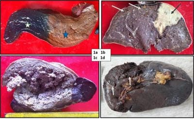
Figure 1a: Spleen shows golden brown hemosiderosis (blue star) confirmed by Prussian blue stain (white star). Figure 1b: Spleen shows multiple grey-brown Gamna Gandy bodies (white arrows) and wedge-shaped pale infarct (black arrow). Figure 1c: Spleen with large ragged cavitatory abscess. Figure 1d: Spleen with the traumatic capsular breach (white arrows).

patho_580_02.JPG
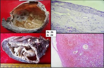
Figure 2a: Primary cyst shows pearly-white inner surface and trabeculations. Figure 2b: Photomicrograph of primary cyst shows cuboidal to the squamous epithelial lining, Hematoxylin Eosin (HE) 100 x. Figure 2c: Pseudocyst shows a ragged cyst wall with hemorrhagic contents. Fig 2d: Photomicrograph of pseudocyst shows hemorrhages and calcification. Epithelial lining absent, HE, 100 x.


patho_580_03.JPG
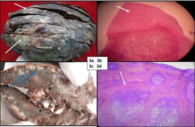
Figure: 3a & 3b: Perisplenitis (white arrows) shows capsule opaque, irregular thickening. Photomicrograph shows capsular fibrinous exudate (white arrow), HE, 100x. Figure 3c, 3d: Non-Hodgkin’s lymphoma shows firm, discrete nodules (blue arrows). Photomicrograph shows enlarged follicles (white arrow) infiltrated by atypical lymphoid cells, HE, 100x.

patho_580_04.JPG
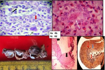
Figure 4a: Photomicrograph shows splenic myelofibrosis with extramedullary hematopoiesis, HE, 400x (myeloid, erythroid cells and megakaryocytes in red, black, green arrows). Figure 4b: Photomicrograph shows microfilaria (white arrow) with eosinophilic infiltrate, HE, 400x. Figure 4c: Splenic artery aneurysmal dilatation. Figure 4d: Photomicrograph of fibromuscular dysplasia shows irregularly thickened arterial wall (black arrows) HE 100 x on left with reduplicated (red arrow) and ruptured (blue arrow) internal elastic laminae on the right, Elastic Von Geison, 100 x.

Discussion

Male dominance and young age (34.4%) were found in other studies, possibly

linked with speed driving, vehicular accidents and splenic laceration. Splenic hilar injury, pulverized parenchyma and hemoperitoneum mandate splenectomy. [2,3].  In hemolytic anemia, splenectomy is advisable if infarction or transfusion exceeds 200 ml/kg/year.[3]. Massive splenomegaly with hypersplenism warrants splenectomy [3]. Splenic cysts need splenectomy, while marsupialization or sclerosant injections have a high recurrence risk. [4]. ITP needs splenectomy if refractory to medical therapy. [3].

Common clinical presentations include anemia, fever, abdominal discomfort, jaundice and bleeding.[5]. Shetty et al. [6] concluded USG is an ideal screening modality. Their spectrum included 60.8% benign lesions (cyst, abscess, infarct). Microscopic findings hemosiderosis, EMH, G.G. bodies, infarcts were similar to our study [2,7]. Spleen, being an in-utero hemopoietic organ, can undergo EMH when marrow shows myelofibrosis or metastasis. Awamleh et al. [8] found 2.1% splenic metastases, while we found none. The phagocytic function makes it a rare metastatic site.

Multipronged investigations like hemogram, serotests for malaria, blood culture, bone marrow, gastroscopy and electrophoresis are essential to investigate splenomegaly.[5]. Our study showed optimum gross-microscopy agreement for capsular breach, cyst, infarct and hilar vessel lesions (Table 2). Spleniculi may cause disease recurrence if not resected. Conversely, retained spleniculi can maintain functions and reduce post-splenectomy complications.[9].

Conclusion

Therapeutic indications are a mainstay for splenectomy. Meticulous gross examination shows excellent concordance with microscopy. Diligent dissection of hilar structures proves fruitful.

Reference

01. Aster, J. C. Disease of white blood cells, lymph nodes, spleen, thymus. Robbins and Cotran Pathologic basis of disease 679 (2005). [Crossref][PubMed][Google Scholar]

02. Gupta D, Gupta A, Suri J, Singh K. Histomorphological spectrum in splenectomy specimens- A 5-year retrospective


study. Indian Journal of Pathology and Oncology 2017; 4:418-421. DOI: 10. 1823/2394-6792.2017.0089 [Crossref][PubMed][Google Scholar]

03. Aliwi AJ, Al-Faddagh ZA. The Pattern of Indications for Splenectomy in Basrah. Basrah Journal of Surgery 2011; 17: 6 p. [Crossref][PubMed][Google Scholar]

04. Maskey P, Rupakheti S, Regmi R, Adhikary S, Agrawal CC. Splenic epidermoid cyst. Kathmandu Univ Med J (KUMJ). 2007 Apr-Jun;5(2):250-2. [Crossref][PubMed][Google Scholar]

05. Deepankar P, Roshan R, Kumar G, Pritam A. Clinical-Etiological Profile of Patients with Splenomegaly in a Tertiary Care Centre. Journal of Medical Science and Clinical Research 2018; 6:268-274. [Crossref][PubMed][Google Scholar]

06. Shetty, Chandrakant M. , et al. Role of ultrasonography and computed tomography in the evaluation of focal splenic lesions. Indian Journal of Radiology and Imaging 15. 02 (2005): 183-190 [Crossref][PubMed][Google Scholar]

07. Khalid, Ahmareen, Anwar Ul Haque, and Lubna Naseem. Spectrum of disease entities in splenectomy specimen. International Journal of Pathology 4. 2 (2006): 88-93. [Crossref][PubMed][Google Scholar]

08. Awamleh, Ahlam A. , et al. Pathological Analysis Of Splenectomy Specimens At King Hussein Medical Center: A 12-Year Study. Tuberculosis 4: 0-7. [Crossref][PubMed][Google Scholar]

09. Vancauwenberghe T, Snoeckx A, Vanbeckevoort D, Dymarkowski S, Vanhoenacker FM. Imaging of the spleen: what the clinician needs to know. Singapore Med J. 2015 Mar;56(3):133-44. doi: 10.11622/smedj.2015040 [Crossref][PubMed][Google Scholar]