E-ISSN:2456-1487
P-ISSN:2456-9887
RNI:MPENG/2017/70771

Research Article

Lesions

Tropical Journal of Pathology and Microbiology

2021 Volume 7 Number 4 July-August
Publisherwww.medresearch.in

Histopathological study of lower gastrointestinal tract lesions

Patel V.1*, Goyal A.2
DOI: https://doi.org/10.17511/jopm.2021.i04.07

1* Vibhaben Kantilal Patel, MD Pathology, Smt. NHL Municipal Medical College, V S. General Hospital, Ahmedabad, Gujarat, India.

2 Anjali Deepak Goyal, Associate Professor, Smt. NHL Municipal Medical College, V S General Hospital, Ahmedabad, Gujarat, India.

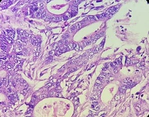
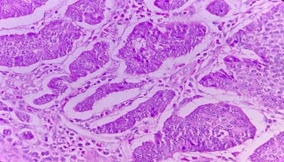
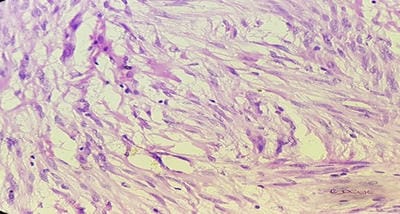
Background: The benign lesions of the lower gastrointestinal tract are responsible for a large number of morbidities. The microscopic examination of and determination of histological types of malignant lesions help to decide treatment options and to predict prognosis. The histopathological study is the Gold standard for the diagnosis of intestinal lesions. Aims and Objectives: To study the prevalence of various lower gastrointestinal tract lesions site-wise, age-wise and gender-wise and to compare the obtained results with other studies. Materials and methodology: A retrospective study of 600 various lower gastrointestinal tract lesions sent for histopathological examination at Pathology department of tertiary care centre, VS General Hospital, Ahmedabad is carried out. Results: Among all the 600 cases, non-neoplastic lesions 572 (95.34%) are far more common than neoplastic lesions 28 (4.66%). Conclusion: Non-neoplastic lesions are common in the small intestine, while the large intestine harbors most malignant lesions.

Keywords: Lower gastrointestinal tract, Malignant, Non-malignant

Corresponding Author How to Cite this Article To Browse
Vibhaben Kantilal Patel, MD Pathology, Smt. NHL Municipal Medical College, V S. General Hospital, Ahmedabad, Gujarat, India.
Email:
Vibhaben Kantilal Patel, Anjali Deepak Goyal, Histopathological study of lower gastrointestinal tract lesions. Trop J Pathol Microbiol. 2021;7(4):194-200.
Available From
https://pathology.medresearch.in/index.php/jopm/article/view/557

Manuscript Received Review Round 1 Review Round 2 Review Round 3 Accepted
2021-07-06 2021-07-24 2021-08-02 2021-08-16 2021-08-23
Conflict of Interest Funding Ethical Approval Plagiarism X-checker Note
No Nil Yes 6%

© 2021 by Vibhaben Kantilal Patel, Anjali Deepak Goyal and Published by Siddharth Health Research and Social Welfare Society. This is an Open Access article licensed under a Creative Commons Attribution 4.0 International License https://creativecommons.org/licenses/by/4.0/ unported [CC BY 4.0].

Introduction

Broadly the whole gastrointestinal tract can be divided into upper and lower segments by taking the insertion of the ligament of Treitz as a landmark. The ligament of Treitz is a suspensory muscle of the duodenum, a thin muscle connecting the junction between the duodenum, jejunum and duodenojejunal flexure to the connective tissue surrounding the superior mesenteric artery and coeliac artery.[1]. The benign lesions of the lower gastrointestinal tract are frequently found and are responsible for a large number of morbidities. Infectious and inflammatory processes often involve the lower gastrointestinal tract. Precise gross and microscopic examination is beneficial in the differential diagnosis of lesions, having a similar clinical presentation and deciding treatment options.

It is crucial to diagnose benign-looking lesions yet having malignant potential. By thorough histopathological evaluation, such pre-malignant lesions can be diagnosed, treated and it can be very helpful to surgeons to decide and plan treatment options. Malignant lesions of the lower gastrointestinal tract show various gross and microscopic presentations. By precise histopathological examination of the margins of the excised specimen, adjacent lymph nodes and the adjacent tissues, exact grading and staging of the malignant lesion can be given. Early diagnosis of malignant lesions can prevent the growth of the tumor to the advanced stage and avoid metastasis.

Special stains are also there to enhance the sensitivity and specificity for detecting pathological abnormalities, especially metaplasia and infections. Frozen section analysis has very few indications in the evaluation of lower gastrointestinal tract lesions. These include the adequacy of tumour resection and determination of a diagnosis in the case where an unusual finding is encountered at the time of surgery.

The histopathological study is the gold standard for the diagnosis of intestinal lesions. The microscopic analysis of and determination of histological types of lesions help to decide treatment options, to predict prognosis and to conduct epidemiological studies and research.

Aims and Objectives

  1. To study the prevalence of various lower gastrointestinal tract lesions - Benign and Malignant.
  1. To give site-wise, age-wise and gender-wise distribution of various neoplastic and non-neoplastic lesions and compare their distribution.
  2. To compare the obtained results of the present study with other studies done.

Materials and Methodology

Setting: A study was carried out at the Histopathology section of the Pathology Department of Tertiary Care Centre, hu General Hospital, Ahmedabad, Gujarat.

Duration and type of study: The study duration is one year, and the study type is a retrospective study.

Sample size: 600 Specimens

Sampling method: Lower gastrointestinal specimens received, processed, examined and diagnosed during the one year were considered. Due importance was paid to brief clinical history with patient's age, inpatient number and presenting signs and symptoms. Sections were stained with H & E stain. Special stains were applied wherever needed. Gross and light microscopic evaluations were done. The diagnosis, typing of and staging of tumors were made following the latest guidelines of WHO.

Inclusion criteria:

Endoscopic biopsies: Endoscopic biopsies are the most commonly performed screening procedure for diagnosing lower gastrointestinal tract diseases and colonic malignancies. Endoscopic biopsies are small fragments of mucosal tissue ranging from 1 to 7-8 mm. When numerous fragments are present, an estimate for the number and dimension in aggregate should be given.

Polypectomy Specimens: Can be sessile or pedunculated. The specimen is sectioned depending on its size and entirely submitted. Sectioning of the pedunculated polyps is ideally done in the vertical plane of the stalk to maximize evaluation of the polypectomy margins.

Bowel resection Specimens: Bowel resections include segmental resections of a part of the small or large intestine, ileocolectomy, low anterior resection, abdominoperineal resection (APR), total colectomy and total mesorectal excision (TME). The resected bowel is oriented, and the length, diameter and wall thickness are measured.


While grossing for the malignant lesion, the external surface (serosa in most cases) of the bowel is inspected for any lesion, tumour involvement, perforation or adhesion. The maximum size of the tumour and distance to proximal and distal resection margins or closest margin is documented. The tumor is then sectioned to assess the depth of invasion and the relationship to adjacent non-neoplastic mucosa. The mesentery and adjacent soft tissue are also dissected. Lymph nodes in the number and sizes are recorded.

Exclusion criteria:

-Improperly preserved specimens.

-Inadequate specimens.

Data collection procedure: Data of the received, processed and diagnosed specimens at the histopathology section was preserved correctly. It was easier to collect data of the reported lesions of the lower gastrointestinal tract. Original images of the microscopic presentation of various lesions were captured.

Results

Table 1: Distribution of lower gastrointestinal tract lesions according to type.

Lesions No. of cases Percentage
Non-neoplastic 572 95.34%
Neoplastic 28 4.66%
Total 600 100%

The table shows that among all the 600 cases, non-neoplastic lesions 572 (95.34%) are far more common than neoplastic lesions 28 (4.66%).

Table 2: Distribution of Lower gastrointestinal tract Lesions according to type and site of lesion

Site Non-neoplastic Neoplastic Total (%)
Small intestine and caecum 148 10 158 26.33%
Colon 82 11 93 15.50%
Rectum 36 3 39 6.50%
Anal canal 59 2 61 10.16%
Appendix 247 2 249 41.50%
Total 572 28 600 100%

The table shows that among 600 cases, 249 (41.50%) cases were of lesions of the appendix, followed by small intestine and caecum, colon and rectum.

Table 3: Distribution of Non-Neoplastic lesion according to histopathology

Type No. of cases Percentage
Non-specific inflammation 138 24.12%
Gangrene 26 4.54%
Gangrene with perforation 1 0.17%
Inflammation with perforation 19 3.32%%
Inflammation with ulceration 48 8.39%
Tuberculous inflammation 19 3.32%
Amoebiasis 4 0.69%
Hirschsprung’s disease 4 0.69%
Meckel's diverticulum 10 1.74%
Polypoidal lesions 7 1.22%
Coeliac disease 3 0.52%
Ulcerative colitis 3 0.52%
Fistula 9 1.57%
Crohn's disease 3 0.52%
Tubulo-villous adenoma 1 0.17%
Atresia 1 0.17%
Hemorrhoids 29 5.06%
Non-neoplastic appendicial lesions 247 43.18%
Total 572 100%

Table Shows that out of 572 non-neoplastic lesions, most non-neoplastic lesions were of appendiceal lesions 247 (43.18%), followed by non-specific inflammation 138(24.12%).

Table 4: Distribution of various lower gastrointestinal tract neoplasms according to their histopathological type

Type No. of cases Percentage
Adenocarcinoma 13 46.42%
Squamous cell carcinoma 2 7.14%
Signet ring adenocarcinoma 3 10.71%
Mucinous adenocarcinoma 5 17.85%
Malignant GIST 4 14.28%
Non-Hodgkin's lymphoma 1 3.57%
Total 28 100%

The above table shows that out of 28 neoplastic lesions, the most common were adenocarcinoma 13 cases (46.42%).

Table 5- Distribution of malignant neoplastic lesions according to age and sex of patients

Years Male Female Percentage
0-10 Years 0 0 0%
11-20 Years 0 0 0%
21-30 Years 2 1 10.71%
31-40 Years 2 3 17.85%
41-50 Years 3 4 25%
51-60 Years 2 3 17.85%
61-70 Years 5 1 21.42%
71-80 Years 0 1 3.57%

81-90 Years 1 0 3.57%
Total 15 13 100%

The table shows that most neoplastic lesions occur between 41 to 70 years with male preponderance (1.15:1).

Discussion

Benign lesions of the lower gastrointestinal tract include Congenital anomalies (Hirschprung's disease, Intestinal atresia, Malrotation with volvulus), Coeliac disease, ulcers, benign inflammatory lesions, benign polyps, lipomas, neurofibromas etc. [2].

patho_557_01.jpg

Figure 1-Aganglionosis of the colon (Hirschsprung's Disease). No ganglion cells in the submucosa of the rectum and hypertrophy of nerve bundles are seen. (H&E Stain, 40X)

patho_557_02.jpg

Figure 2-Giant cells in granuloma in a case of appendiceal tuberculosis. (H&E Stain, 40X)

Pre-malignant lesions include Adenomas, Inflammatory bowel diseases (Crohn's disease and Ulcerative Colitis), Chronic infections and hereditary non-polyposis lesions.

Polyps: need to be briefed as some polyps are benign while some have malignant potential.

Hyperplastic polyps: are benign epithelial proliferation, which needs to be differentiated from

sessile serrated adenomas that are histologically similar but have malignant potential.

Inflammatory polyps: form as a part of solitary rectal ulcer syndrome, an example of pure inflammatory lesion. The clinical triad comprises of rectal bleeding, mucus discharge and an inflammatory lesion of the anterior rectal wall.

Hamartomatous polyps: Occur sporadically or as components of various genetically or acquired syndromes. Many hamartomatous polyp syndromes are caused by germline mutations in tumor suppressor genes or proto-oncogenes. Thus in some cases, they can be considered pre-malignant.

Peutz-Jeghers polyps: are part of Peutz-Jeghers syndrome, an autosomal dominant syndrome, having a heterozygous germline loss of function mutations in the gene STK11. This syndrome is associated with an increased risk of several malignancies, including small and large intestine malignancies.

Neoplastic polyps: The most common are adenomas, which are precursors to the majority of colorectal adenocarcinomas.

The adenoma is a circumscribed benign epithelial tumor with malignant potential. Adenomas show male preponderance, and their incidence progressively increases with age. They occur more frequently in the rectum and distal colon. Grossly they are single or multiple, pedunculated or sessile. Microscopically, Adenomas are classified into three types: Tubular adenoma, Villous adenoma and Tubulovillous adenoma. Adenomas are classified according to the grade of epithelial dysplasia into mild, moderate or severe or low or high quality. The risk of malignant change in an adenoma is related to size, presence of villi and grade of epithelial dysplasia.

(A)patho_557_03.jpg


(B)patho_557_04.jpg

Figure 3 – (A) Villous Adenoma (B) Tubular Adenoma (H&E, 40X)

Serrated adenoma of the large intestine has intermediate morphology between hyperplasic and neoplastic polyp. The diagnostic feature of serrated adenomas includes large size (from 2 to greater than 10 mm), exaggerated crypt infolding, crypt branching, eosinophilic cytoplasm, absence of endocrine cells, focal mucus overproduction, crowding, elongation and mild stratification of nuclei, prominence of nucleoli and occasional superficial mitosis.[3,4]. Gross and microscopic features of Crohn's disease and Ulcerative Colitis should be examined carefully to differentiate them. [5,6].

Features Crohn's disease Ulcerative Colitis
Macroscopic
Bowel region Ileum with caecum Colon
Distribution Skip lesions Diffuse
Stricture More commonly Rare
Wall appearance Thick Thin
Microscopic
Inflammation Transmural Limited to mucosa
Pseudopolyps Moderate Marked
Ulcers Deep, Knife-like Superficial, Broad based
Lymphoid reaction Marked Moderate
Fibrosis Marked Moderate
Serositis Marked Mild to none
Granulomas Yes No
Fistulas/Sinuses Yes No

Lower gastrointestinal tract malignancies include adenocarcinomas mainly and small numbers of squamous cell carcinomas, small cell carcinomas and hematopoietic malignancies.

Adenocarcinoma: is the most commonly found malignant lesion of the lower gastrointestinal tract, so its pathogenesis, in brief, is described.

The combination of molecular events that lead to colonic adenocarcinoma is heterogeneous and includes genetic and epigenetic abnormalities. Two distinct genetic pathways are the APC/β-catenin pathway, which is associated with WNT and the classic adenoma-carcinoma sequence and the microsatellite instability pathway, which is related to defects in DNA mismatch repair. The third group of colon cancers with increased CpG island methylation in the absence of microsatellite instability also exists. [7,8] Grossly, it may be flat, stenosing, ulcerative, infiltrative or polypoid.

Microscopically: Adenocarcinoma can be divided into three grades, based primarily on an overview of the arrangement of cells about the degree of tubular (acinar) formation.

  • Grade-I: These are well-differentiated or low-grade tumors. They are composed mainly of simple tubules in which the nuclei are of uniform size, and the polarity is maintained.
  • Grade-II: These tumors are moderately differentiated. They are composed of tubules that may be simple, complex or slightly irregular, in which the nuclear polarity is barely discernible or is lost.
  • Grade–III: These tumors are poorly differentiated. They are characterized by the absence of epithelial differentiation (solid-like pattern), as well as by loss of polarity. They have a significantly poorer prognosis.

(A)patho_557_05.jpg

Figure 4- Moderately differentiated adenocarcinoma. Irregular tubules with nuclear pleomorphism are seen. (A) H&E Stain, 10 X (B) H&E Stain, 40X


(B)patho_557_06.jpg

patho_557_07.jpg

Figure 5- Signet ring cell adenocarcinoma. Cells have intracellular mucin and displaced nuclei. (H&E Stain, 40X)

patho_557_08.jpg

Figure 6–Neuroendocrine tumour of the ileum composed of uniform cells arranged in nests. Cells have nuclear pleomorphism and hyperchromasia. (H&E stained, 40x)

Figure 7 - Gastrointestinal stromal Tumors, Spindle cell type. The tumor consists of fascicles of bland spindle cells with abundant fibrillary cytoplasm. (H & E Stain, 40 X)

patho_557_09.jpg

Table 6: Comparative study of non-neoplastic and neoplastic lower gastrointestinal tract lesions

Lesion Nanavati M et al. [9] (2014) Present study (2018)
No. of cases (%) No. of cases (%)
Non-neoplastic 157 78.5% 572 95.34%
Neoplastic 43 21.5% 28 4.66%
Total 200 100% 600 100%

In the present study, 572 cases of non-neoplastic lesions were found, comprising 95.34% of the total 600 cases, and 28 cases of neoplastic lesion were seen, comprising 4.66% of actual patients. In the comparative study, there were 157 cases of non-neoplastic lesions containing 78.5% of the total 200 cases, which is lower than the present study.

Table 7: Comparative histological types of lower gastrointestinal neoplasms

Type of neoplasms Abdul Kareem Et Al [10] (2008) Patel Mandakini Et Al [11] (2012) Present Study (2018)
Adenocarcinoma 366 (87.1%) 61 (76.2%) 13 (46.42%)
Mucinous Adenocarcinoma 06 (24%) - 5 (17.85%)
Signet ring cell carcinoma 02 (8%) 03 (3.7%) 3 (10.71%)
Non-Hodgkin’s lymphoma 01 (4%) 02 (2.5%) 1 (3.57%)

In the present study, Adenocarcinomas comprise 46.42% of neoplastic lesions of the lower GI tract compared to 87.1 % and 76.2 % of comparative studies.

Table 8: Comparison of age and sex-wise distribution of neoplastic lesions

  Priyanka Sharma Et Al [12] Present study
Male:Female 1.9:1 1.15:1
Age group 61-70 Years 41-70 Years

Conclusion

  • Non-neoplastic lesions are common in the small intestine, while the large intestine harbours most malignant lesions.
  • Adenocarcinomas are the most common malignant lesion of the large intestine, with a peak incidence between 41 to 70 years.
  • With the aid of special stains and immunohistochemistry, differential diagnoses of lesions are becoming more precise.

Limitations: As the study was conducted for a specified duration, malignant lesions were found in much smaller numbers than non-malignant lesions.

What does the study add to the existing knowledge?

With adequate knowledge and thorough gross and light microscopic examination, a precise diagnosis can be concluded, which can be very useful in deciding treatment options and predicting prognosis.

Author's contribution

Both the authors, Dr Vibha Kantilal Patel and Dr Anjali Deepak Goyal, have contributed to the concept, data acquisition, data analysis and review.

Reference

01. Gray, H. Standring S. Gray's anatomy: the anatomical basis of clinical practice. (2005): 1357Á71. [Crossref][PubMed][Google Scholar]

02. Rosai, Juan. Rosai and Ackerman's surgical pathology e-book. Elsevier Health Sciences. 2011. [Crossref][PubMed][Google Scholar]

03. Perzin KH, Bridge MF. Adenomas of the small intestine: a clinicopathologic review of 51 cases and a study of their relationship to carcinoma. Cancer. 1981 Aug 1;48(3):799-819. doi: 10.1002/1097-0142(19810801)48:3<799::aid-cncr2820480324> ;3.0.co;2-q [Crossref][PubMed][Google Scholar]

04. Hiraoka S, Kato J, Tatsukawa M, Harada K, Fujita H, Morikawa T, et al. Laterally spreading type of colorectal adenoma exhibits a unique methylation phenotype and K-ras mutations. Gastroenterology. 2006 Aug;131(2):379-89. doi: 10.1053/j.gastro.2006.04.027 [Crossref][PubMed][Google Scholar]

05. Lockhart-Mummery He, Morson Bc. Crohn's Disease of The Large Intestine. Gut. 1964 Dec;5(6):493-509. doi: 10.1136/gut.5.6.493 [Crossref][PubMed][Google Scholar]

06. Canavese G, Villanacci V, Zambelli C, Bernardi A, Candelaresi G, Berardengo E, et al. Gastric metaplasia and small bowel ulcerogenesis in a case of ulcerative jejunitis not related to celiac disease. Int J Surg Pathol. 2004 Oct;12(4):415-9. doi: 10.1177/106689690401200418 [Crossref][PubMed][Google Scholar]

07. Hinoi T, Tani M, Lucas PC, Caca K, Dunn RL, Macri E, et al. Loss of CDX2 expression and microsatellite instability are prominent features of large cell minimally differentiated carcinomas of the colon. Am J Pathol. 2001 Dec;159(6):2239-48. doi: 10.1016/S0002-9440(10)63074-X [Crossref][PubMed][Google Scholar]

08. McGregor DK, Wu TT, Rashid A, Luthra R, Hamilton SR. Reduced expression of cytokeratin 20 in colorectal carcinomas with high levels of microsatellite instability. Am J Surg Pathol. 2004 Jun;28(6):712-8. doi: 10.1097/01.pas.0000126757.58474.12 [Crossref][PubMed][Google Scholar]

09. Nanavati M,Parikh J et al. Histopathological study of intestinal lesions. International Journal OF Scientific Research. 3(9)Sep 2014. [Crossref][PubMed][Google Scholar]

10. Abdulkareem FB, Abudu EK, Awolola NA, Elesha SO, Rotimi O, Akinde OR, et al. Colorectal carcinoma in Lagos and Sagamu, Southwest Nigeria: a histopathological review. World J Gastroenterol. 2008 Nov 14;14(42):6531-5. doi: 10.3748/wjg.14.6531 [Crossref][PubMed][Google Scholar]

11. Patel Mandakini M, Gamit B, Patel PR. Analysis of gastrointestinal malignancy: A 5 years study. Natl J, Community Med. 2012; 3(3): 555-7. [Crossref][PubMed][Google Scholar]

12. Sharma, Priyank, and M Dekaa. A study of neoplastic lesions of colorectum in a tertiary care hospital. IJSS. 3;8(2015):88-91. [Crossref][PubMed][Google Scholar]