E-ISSN:2456-1487
P-ISSN:2456-9887
RNI:MPENG/2017/70771

Research Article

Histopathological spectrum

Tropical Journal of Pathology and Microbiology

2021 Volume 7 Number 5 September October
Publisherwww.medresearch.in

Histopathological spectrum of premalignant and malignant endometrial lesions

Abbas F.1, Manzoor F.2, Abas R.3*, Farooq S.4, Ashraf S.5, Latief M.6
DOI: https://doi.org/10.17511/jopm.2021.i05.07

1 Farhat Abbas, Senior Resident, Pathology Division, GMC Srinagar, Srinagar, Jammu and Kashmir, India.

2 Farzana Manzoor, Senior Resident, Pathology Division, GMC Srinagar, Srinagar, Jammu and Kashmir, India.

3* Rahat Abas, Post graduate, Obstetrics and Gynaecology, SKIMS Medical College Srinagar, Srinagar, Jammu and Kashmir, India.

4 Summyia Farooq, Lecturer, Pathology Division, GMC Srinagar, Srinagar, Jammu and Kashmir, India.

5 Sabiha Ashraf, Lecturer, Pathology Division, GMC Srinagar, Srinagar, Jammu and Kashmir, India.

6 Muzamil Latief, Senior Resident, Nephrology, GMC Srinagar, Srinagar, Jammu and Kashmir, India.

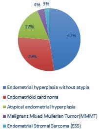
Introduction: The current study was conducted to see the frequency of epithelial malignancies of the endometrium with a focus on the precursor lesions, including non- atypical and atypical endometrial hyperplasia. Methods: It is a prospective descriptive study carried out on 80 specimens of endometrial biopsies and hysterectomy specimen received in GMC Srinagar for two years (2019–2020). Patients were divided into 6 age groups: <30,31-40, 41–50, 51–60, 61–70, and >70 yrs. Tissues were fixed in 10% formalin and processed and stained with haematoxylin-eosin. Stained slides were examined to determine the histological types by WHO classification. Results: Benign endometrial hyperplasia was seen in38(47.5%) atypical hyperplasia 14(17.50%), endometrial adenocarcinoma in 23(28.75%), MMMT in 3(3.75%), endometrial stromal sarcoma in2(2.5%) cases. The most common malignant lesion in the study was endometrial carcinoma, and most of the patients with endometrial carcinomas fall in the age range of 51–60 yrs. Conclusion: The most common age group in endometrial hyperplasia was 41-50 years, thus presenting earlier than malignant pathologies.

Keywords: Endometrium, Endometrial hyperplasia, Carcinoma

Corresponding Author How to Cite this Article To Browse
Rahat Abas, Post graduate, Obstetrics and Gynaecology, SKIMS Medical College Srinagar, Srinagar, Jammu and Kashmir, India.
Email:
Farhat Abbas, Farzana Manzoor, Rahat Abas, Summyia Farooq, Sabiha Ashraf, Muzamil Latief, Histopathological spectrum of premalignant and malignant endometrial lesions. Trop J Pathol Microbiol. 2021;7(5):254-260.
Available From
https://pathology.medresearch.in/index.php/jopm/article/view/554

Manuscript Received Review Round 1 Review Round 2 Review Round 3 Accepted
2021-10-01 2021-10-02 2021-10-09 2021-10-16 2021-10-23
Conflict of Interest Funding Ethical Approval Plagiarism X-checker Note
Nil Nil Yes 18%

© 2021by Farhat Abbas, Farzana Manzoor, Rahat Abas, Summyia Farooq, Sabiha Ashraf, Muzamil Latiefand Published by Siddharth Health Research and Social Welfare Society. This is an Open Access article licensed under a Creative Commons Attribution 4.0 International License https://creativecommons.org/licenses/by/4.0/ unported [CC BY 4.0].

Introduction

The uterus has two major components: the endometrium and the myometrium. The internal cavity is lined by endometrium, composed of glands embedded in a cellular stroma. Endometrial Hyperplasia is defined as the abnormal proliferation of endometrium under the influence of prolonged estrogenic stimulation unopposed by progesterone. There is a wide spectrum of architectural and cytological abnormalities existing in endometrial hyperplasia. These range from disordered proliferative endometrium (anovulatory cycles) to a complex proliferation that resembles well-differentiated adenocarcinoma of the endometrium. [1].

Endometrial cancer is the most common invasive cancer of the female genital tract. It is also known as corpus uterine cancer or corpus cancer. [2]. There are two distinctly different forms of endometrial carcinoma based on light microscopic appearance, clinical behaviour, and epidemiology, risk factors and prognosis. This hypothesis was first proposed in 1983. [3].

Type 1 is oestrogen-related endometrioid carcinoma, and type 2 is non oestrogen-related non-endometrioid carcinoma. Type1 is divided into adenocarcinomas with squamous differentiation, secretory, ciliated cell, and villoglandular variants. Adenocarcinoma with squamous differentiation is further subdivided into adenocarcinoma with squamous metaplasia (adenoacanthoma) and adenosquamous carcinoma. [4].

Uterine papillary serous carcinoma (UPSC), clear cell carcinoma (CC), mucinous and squamous cell carcinoma are included in type 2. [5]. The average age at presentation is about 63 years. [[6]. Macroscopically, endometrial carcinoma can form one or more discrete tan nodules while others are diffuse and exophytic. [7].

Leiomyosarcoma is the most common uterine sarcoma accounting for 1-2% of all uterine malignancies. [8]. Macroscopically, they can be single masses or the largest of all masses when associated with leiomyomas. [7].

Endometrial Stromal Sarcoma (ESS) is divided into low & high grades. Low-grade ESS represents less than 1% of all uterine malignancies but is the second most common uterine malignant

mesenchymal tumour. [8,9]. It occurs over a wide age range with a mean of 52 years. [10]. High-grade ESS is a rare tumour whose true frequency is unknown, as tumours previously considered undifferentiated uterine sarcoma might belong to this category. [11,12,13].

Methods

Setting: This study was done in the department of pathology in government medical college Srinagar, a tertiary care centre.

Type and Duration: It was a retro-prospective study done over two years from January 2019 to December 2020.

Sampling Methods: The specimens were received as endometrial curettage, endometrial biopsy and hysterectomy specimens.

Sample Size: The study included 80 cases of endometrial hyperplasia and endometrial malignancies.

Exclusion Criteria: Cases of disordered proliferative endometrium and endometrial polyps were excluded.

Ethical Consideration and Data Collection:  Detailed clinical history including age, pattern and duration of abnormal bleeding, menstrual history, obstetric history, use of exogenous hormones, physical examination findings including pelvic examination and investigations were recorded. Some of the data was taken from the filing section of the department in case of previously reported cases. The specimen was fixed in 10% formalin. After a detailed gross examination, tissue paraffin blocks were made; sections were cut and stained with haematoxylin and eosin. Histopathological examination of endometrial biopsies and hysterectomy specimens were done. The lesions were classified into benign endometrial hyperplasia, atypical endometrial hyperplasia, endometroid carcinoma, endometrial stromal sarcoma, malignant mixed Mullerian tumour as per WHO classification.

Results

The present study has been conducted on 80 specimens (endometrial curetting/biopsy and hysterectomy specimens) received in the Pathology Department of a tertiary care institution.


Patients' age ranged from 26-75 years, and most of them were seen in the age group of 41-50 years, followed by 51-60 years. The commonest complaint was menorrhagia in33 (41.25%), followed by postmenopausal bleeding in 26(32.5%), polymenorrhea in 21(26.25%) patients. The Majority, 48 (60%) of the patients, were multiparous (para 3-4).17(21.25%) of cases were of low parity (para 1-2), and 15(18.75%) were nulliparous.  Benign endometrial hyperplasia was seen in38(47.5%) atypical hyperplasia 14(17.50%), endometroid adenocarcinoma 23(28.75%), MMMT3(3.75%), endometrial stromal sarcoma in2(2.5%) s.

patho_554_01.JPG

Table 1: Showing benign and malignant endometrial pathologies obtained in our study

Lesions  Premenopausal  Postmenopausal Total  no of cases with %age 
Benign hyperplasia 27 71.05% 11 28.95% 38 47.5%
Complex hyperplasia 10 71.43% 28.57% 14  17.5%
Endometroid carcinoma 6 26.09%  17  73.91% 23  28.75
Endometrial stromal sarcoma     2   2.5%
MMMT     3   3.75%

patho_554_02.JPG

Figure 1: Showing an Endometrial Stromal Sarcoma Showing Variegated Cut Section Obliterating Endometrial Cavity and Invading Myometrium.

patho_554_03.JPG

Figure 2. Microscopic picture of atypical endometrial hyperplasia showing back to back arrangement of glands with minimal intervening stroma and cells showing high grade dysplasia.

Table 2: shows the age distribution of various endometrial pathologies 

Lesions Age groups
  <30 31-40 41-50 51-60 61-70 >70
Simple hyperplasia 3 10 19 6 0 0
Complex hyperplasia   3 7 4    
Endometroid carcinoma     6 13   4
MMMT       3    
Endometrial stromal sarcoma       2    

patho_554_04.JPG

Figure3. Microscopic picture of endometrial stromal sarcoma showing atypical stromal cells with a mild lymphoid response.

Endometrial hyperplasia was primarily seen in the 41-50 years age group in almost (50%) cases, followed by the 51-60 years age group. However, the most common age group in malignant endometrial pathologies was 51-60 years in (64.28%) cases.

Discussion

Endometrial hyperplasia is a precursor of endometrial carcinoma. Abnormal proliferation of endometrium under the influence of prolonged exposure to estrogen unopposed by progesterone is has been called endometrial hyperplasia. [14].

The overall risk of progression of endometrial hyperplasia to carcinoma is considered to be 5-10%. The classification used by the World Health Organization (WHO) designates four different types of hyperplasia. Hyperplasia is classified as simple or complex based on the absence or presence of architectural abnormalities such as glandular complexity and crowding. They are further designated as atypical if they demonstrate nuclear atypia. The new classification classifies endometrial hyperplasia in 2 categories only. These include benign endometrial hyperplasia (BH) and atypical endometrial hyperplasia/EIN (endometrial intraepithelial neoplasia.[15].

In our study, benign hyperplasia was seen in 38/52(73.07%) patients of hyperplasia and was primarily seen in the age group 41-50 years (15 cases). Atypical hyperplasia was seen in 14/52 (26.92%) cases and was primarily seen in

the age group of 41-50(7 patients). Thus, in our study, most of the cases of hyperplasia belonged to the category of simple endometrial hyperplasia. Similar results were found in a survey by lacey JR et al. They found half of all community-based EH diagnoses are SH, and almost a third are CH, leaving approximately 20% as AH. [16]. Similar to the data in other studies, the incidence of hyperplasia peaked in the peri-menopausal age group. In a study done by Mahjabeen et al., Incidence of Endometrial Hyperplasia was found to be more in the 4th decade of life followed by 3rd decade. [17]. Gusberg & Kaplan, in their study (1963) of 191 cases, found that the peak incidence of Endometrial Hyperplasia was noticed in the 4th decade followed by the 5th decade. [18].

Endometrial carcinoma can occur due to excess estrogenic stimulation and developing against a background of endometrial hyperplasia, or it can arise de novo combined with insufficient progesterone levels. It is simple to diagnose different subtypes of endometrial carcinomas based on their characteristic morphology. Our institution observed the same experience, and morphology is the key factor in diagnosing endometrial carcinomas. Well to moderately differentiated adenocarcinoma resembles normal endometrium showing architectural complexity, cribriform pattern and overcrowding of the glands. [19-22].

On the contrary serous carcinomas typically have irregular, branching papillae with budding small papilla. The neoplastic cells show large pleomorphic nuclei. [20-22].

Endometrioid endometrial carcinoma is the most common form of endometrial cancer, and in our study, all the epithelial malignancies belonged to the endometroid variant. In a study done in Pakistan it 80% of specimens was of endometrioid adenocarcinomas, 11% of serous tumours, 4% of clear cell carcinoma, and 4% of squamous cell carcinomas involving both cervix and endometrium. [23].

In our study, the most common age group of malignant lesions was 51-60 years. Similarly, Imrana et al. found that the commonest age group in endometrial carcinomas was 51-60 years (28.8%); however, this percentage is meagre compared to our study. In another study done by Modi et al., 57% of endometrial carcinomas were 41-60 years. [24].


In our study, two cases of endometrial stromal sarcoma were noted in the 51-60 years of age group—both the patients presented with postmenopausal bleeding and mass in the pelvis. In a previously reported study of 14 cases of low-grade ESS, the most common presentation was vaginal bleed (86%), followed by pelvic mass (7%) and pelvic pain (7%). [25]. In our study, endometroid carcinoma was mostly seen in postmenopausal females (17 cases), as reported in the literature. [26-30].

Conclusion

  1. The present study showed that endometrioid adenocarcinomas are the most common malignancies of the endometrium and are more common in postmenopausal women.
  2. Endometrial malignancies were more common in the 5th decade.
  3. Endometrial hyperplasia presented earlier compared to endometrial malignancies.
  4. Endometrial hyperplasia were more common in the 4th decade.
  5. Benign hyperplasia are more common compared to atypical hyperplasia.

Addition to existing knowledge: Our study includes the premalignant and malignant pathologies of the endometrium; however previous studies have not included both things.

Reference

01. Care TAL, Kempson RL, Hendrickson MR: Haines & Taylor obstetrical & Gynecological Pathology. H Fox & M Wells (eds. ) 4th edition, Volume I,CH: 132 Churchill Livingstone, Edinburg – London 1995; P. 421-510. [Crossref][PubMed][Google Scholar]

02. de Haydu, Christopher, et al. "An update on the current pharmacotherapy for endometrial cancer. " Expert opinion on pharmacotherapy 17. 4 (2016): 489-499. [Crossref][PubMed][Google Scholar]

03. Bokhman JV. Two pathogenetic types of endometrial carcinoma. Gynecol Oncol. 1983 Feb;15(1):10-7. doi: 10.1016/0090-8258(83)90111-7 [Crossref][PubMed][Google Scholar]

04. Silverberg SG. Problems in the differential diagnosis of endometrial hyperplasia

and carcinoma. Mod Pathol. 2000 Mar;13(3):309-27. doi: 10.1038/modpathol.3880053 [Crossref][PubMed][Google Scholar]

05. Arafa, Mohammad, et al. "Current concepts in the pathology and epigenetics of endometrial carcinoma. " Pathology 42. 7 (2010): 613-617. [Crossref][PubMed][Google Scholar]

06. Creasman WT, Odicino F, Maisonneuve P, Quinn MA, Beller U, Benedet JL, et al. Carcinoma of the corpus uteri. FIGO 26th Annual Report on the Results of Treatment in Gynecological Cancer. Int J Gynaecol Obstet. 2006 Nov;95 Suppl 1:S105-43. doi: 10.1016/S0020-7292(06)60031-3 [Crossref][PubMed][Google Scholar]

07. Kurman, Robert J. , Maria Luisa Carcangiu, and C. Simon Herrington. World Health Organisation classification of tumours of the female reproductive organs. International Agency for Research on Cancer, 2014 [Crossref][PubMed][Google Scholar]

08. Abeler VM, Røyne O, Thoresen S, Danielsen HE, Nesland JM, Kristensen GB. Uterine sarcomas in Norway. A histopathological and prognostic survey of a total population from 1970 to 2000 including 419 patients. Histopathology. 2009 Feb;54(3):355-64. doi: 10.1111/j.1365-2559.2009.03231.x [Crossref][PubMed][Google Scholar]

09. Harlow BL, Weiss NS, Lofton S. The epidemiology of sarcomas of the uterus. J Natl Cancer Inst. 1986 Mar;76(3):399-402. [Crossref][PubMed][Google Scholar]

10. Chan JK, Kawar NM, Shin JY, Osann K, Chen LM, Powell CB, Kapp DS. Endometrial stromal sarcoma: a population-based analysis. Br J Cancer. 2008 Oct 21;99(8):1210-5. doi: 10.1038/sj.bjc.6604527 [Crossref][PubMed][Google Scholar]

11. Amant F, Tousseyn T, Coenegrachts L, Decloedt J, Moerman P, Debiec-Rychter M. Case report of a poorly differentiated uterine tumour with t(10;17) translocation and neuroectodermal phenotype. Anticancer Res. 2011 Jun;31(6):2367-71. [Crossref][PubMed][Google Scholar]

12. Kurihara S, Oda Y, Ohishi Y, Iwasa A, Takahira T, Kaneki E, et al. Endometrial stromal sarcomas and related high-grade sarcomas: immunohistochemical and molecular genetic study of 31 cases. Am J Surg Pathol. 2008 Aug;32(8):1228-38. doi: 10.1097/PAS.0b013e31816a3b42 [Crossref][PubMed][Google Scholar]


13. Lee CH, Mariño-Enriquez A, Ou W, Zhu M, Ali RH, Chiang S, et al. The clinicopathologic features of YWHAE-FAM22 endometrial stromal sarcomas: a histologically high-grade and clinically aggressive tumor. Am J Surg Pathol. 2012 May;36(5):641-53. doi: 10.1097/PAS.0b013e31824a7b1a [Crossref][PubMed][Google Scholar]

14. Trimble, C. L. "Method M, Leitao M, Lu K, Ioffe O, Hampton M, et al. " Management of endometrial precancers. Obstet Gynecol 120.5 (2012): 1160-75 [Crossref][PubMed][Google Scholar]

15. Lacey JV Jr, Chia VM. Endometrial hyperplasia and the risk of progression to carcinoma. Maturitas. 2009 May 20;63(1):39-44. doi: 10.1016/j.maturitas.2009.02.005 [Crossref][PubMed][Google Scholar]

16. Lacey JV Jr, Ioffe OB, Ronnett BM, Rush BB, Richesson DA, Chatterjee N, et al. Endometrial carcinoma risk among women diagnosed with endometrial hyperplasia: the 34-year experience in a large health plan. Br J Cancer. 2008 Jan 15;98(1):45-53. doi: 10.1038/sj.bjc.6604102 [Crossref][PubMed][Google Scholar]

17. Salma, Mahjabeen, Tazyeen Kouser, and Mohammed Abdul Nasar. "Endometrial Hyperplasia: a 5-Years Retrospective Study. " Indian Journal of Pathology and Oncology 3. 2 (2016): 221-225. [Crossref][PubMed][Google Scholar]

18. Gusberg Sb, Kaplan Al. Precursors Of Corpus Cancer. Iv. Adenomatous Hyperplasia As Stage O Carcinoma Of The Endometrium. Am J Obstet Gynecol. 1963 Nov 1;87:662-78 [Crossref][PubMed][Google Scholar]

19. Azueta, Ainara, Sonia Gatius, and Xavier Matias-Guiu. "Endometrioid carcinoma of the endometrium: pathologic and molecular features. " Seminars in diagnostic pathology. Vol. 27. No. 4. WB Saunders, 2010 [Crossref][PubMed][Google Scholar]

20. Böcker W. WHO-Klassifikation der Tumoren der Mamma und des weiblichen Genitale: Pathologie und Genetik [WHO classification of breast tumors and tumors of the female genital organs: pathology and genetics]. Verh Dtsch Ges Pathol. 2002;86:116-9. German [Crossref][PubMed][Google Scholar]

21. Clement PB, Young RH. Endometrioid carcinoma of the uterine corpus: a review of its pathology with emphasis on recent advances and problematic

aspects. Adv Anat Pathol. 2002 May;9(3):145-84. doi: 10.1097/00125480-200205000-00001 [Crossref][PubMed][Google Scholar]

22. . . Adv Anat Pathol. 2002 May;9(3):145-84. doi: 10.1097/00125480-200205000-00001 [Crossref][PubMed][Google Scholar] [Crossref][PubMed][Google Scholar]

23. Ambros RA, Ballouk F, Malfetano JH, Ross JS. Significance of papillary (villoglandular) differentiation in endometrioid carcinoma of the uterus. Am J Surg Pathol. 1994 Jun;18(6):569-75. [Crossref][PubMed][Google Scholar]

24. . . Am J Surg Pathol. 1994 Jun;18(6):569-75. [Crossref][PubMed][Google Scholar] [Crossref][PubMed][Google Scholar]

25. Tanvir I, Riaz S, Hussain A, Mehboob R, Shams MU, Khan HA. Hospital-based study of epithelial malignancies of endometrial cancer frequency in lahore, pakistan, and common diagnostic pitfalls. Patholog Res Int. 2014;2014:179384. doi: 10.1155/2014/179384 [Crossref][PubMed][Google Scholar]

26. Modi M, Nilkanthe R, Trivedi M. Detailed Histopathological Study of Endometrial Carcinoma, and Importance of Immunohistochemistry. American Journal of Clinical Pathology, Volume 146, Issue suppl_1, September 2016, 74, https://doi. org/10. 1093/ajcp/aqw162.012 [Crossref][PubMed][Google Scholar]

27. Ashraf-Ganjoei T, Behtash N, Shariat M, Mosavi A. Low grade endometrial stromal sarcoma of uterine corpus, a clinico-pathological and survey study in 14 cases. World J Surg Oncol. 2006 Aug 9;4:50. doi: 10.1186/1477-7819-4-50 [Crossref][PubMed][Google Scholar]

28. Sajitha, K. , et al. "Study of histopathological patterns of endometrium in abnormal uterine bleeding. " CHRISMED journal of health and Research 1. 2 (2014): 76 [Crossref][PubMed][Google Scholar]

29. Bolde, Saroj A. , et al. "Histopathological study of endometrium in cases of abnormal uterine bleeding. " (2014). [Crossref][PubMed][Google Scholar]

30. Vaidya S, Lakhey M, Vaidya S, Sharma PK, Hirachand S, Lama S, KC S. Histopathological pattern of abnormal uterine bleeding in endometrial


biopsies. Nepal Med Coll J. 2013 Mar;15(1):74-7. [Crossref][PubMed][Google Scholar]

31. Doraiswami S, Johnson T, Rao S, Rajkumar A, Vijayaraghavan J, Panicker VK. Study of endometrial pathology in abnormal uterine bleeding. J Obstet Gynaecol India. 2011 Aug;61(4):426-30. doi: 10.1007/s13224-011-0047-2 [Crossref][PubMed][Google Scholar]

32. Bhatta, S. , and A. K. Sinha. "Histopathological study of endometrium in abnormal uterine bleeding." Journal of pathology of Nepal 2.4 (2012): 297-300 [Crossref][PubMed][Google Scholar]