E-ISSN:2456-1487
P-ISSN:2456-9887
RNI:MPENG/2017/70771

Research Article

Breast

Tropical Journal of Pathology and Microbiology

2021 Volume 7 Number 3 May-June
Publisherwww.medresearch.in

Aberrant expression of E-cadherin in infiltrating ductal and lobular breast carcinomas and its correlation with clinicopathological parameters – A hospital-based study

Thota S.1, Chavan R.2, Jaikumar Kulkarni N.3*
DOI: https://doi.org/10.17511/jopm.2021.i03.02

1 Shruthi Thota, Assistant Professor, Department of Pathology, Government Medical College, Nizamabad, Kaloji Narayana Rao University of Health Sciences, Warangal, Telengana, India.

2 Ramesh Chavan, Professor, Department of Pathology, Jawaharlal Nehru Medical College, KLE University, Belagavi, Karnataka, India.

3* Naresh Jaikumar Kulkarni, Assistant Professor, Department of Pathology, Mahatma Gandhi Mission Medical College, Mahatma Gandhi Mission Institute of Health Sciences, Navi Mumbai, Maharastra, India.

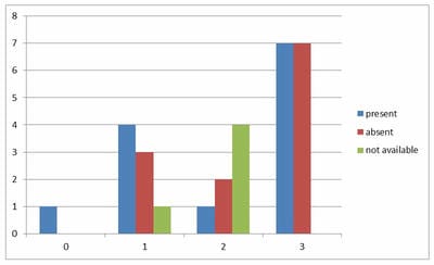
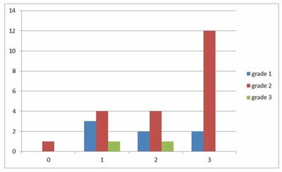
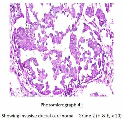
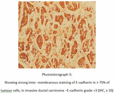
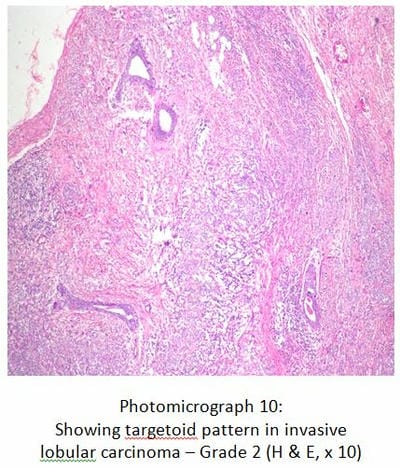
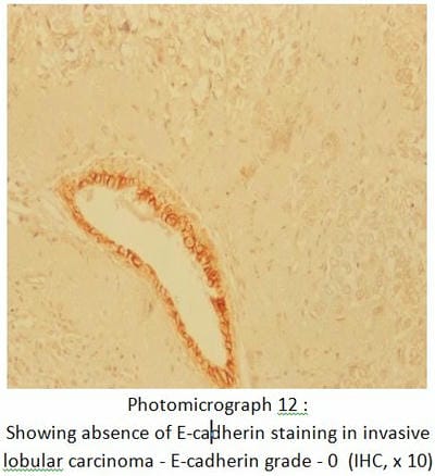
Introduction: Breast carcinoma is one of the commonest malignant tumours in women, leading to premature deaths and morbidity. E-cadherin is a 120kDa calcium-dependent transmembrane glycoprotein encoded by the CDH1 gene located on chromosome 16q21 and is expressed in most epithelial cells. Loss of E Cadherin expression implies cell discohesion and favours metastasis. Materials and Methods: A total of 30 cases of breast carcinomas were studied, over two years. Histological grade and type were assessed by staining the paraffin-embedded sections with H & E. Using IHC technique, E-cadherin antigen was retrieved by Heat-Induced Epitome Retrieval method, and immunostaining was scored semiquantitatively. Cases were grouped as ‘preserved,’ when positivity was strong membranous, and occurred in more than 75% of the neoplastic epithelial cells and ‘aberrant’ in all the remaining cases. Results: E-cadherin was found to be preserved in 46.7% of all the breast carcinomas and aberrant in 51.7% of invasive ductal carcinomas (IDC) alone, while 100% of invasive lobular carcinomas showed aberrant expression. No significant correlation was found with E-cadherin grading and histological type of carcinoma, histopathological grade or involvement of deep surgical margin. Conclusion: Differentiation between invasive ductal and invasive lobular carcinoma based on the loss of E-cadherin has to be done cautiously given its aberrant expression in ductal carcinomas as well.

Keywords: Invasive Ductal Carcinoma (IDC), Invasive Lobular Carcinoma (ILC), E-cadherin, IHC, Grading of E-cadherin, Aberrant E-cadherin expression, Breast carcinoma

Corresponding Author How to Cite this Article To Browse
Naresh Jaikumar Kulkarni, Assistant Professor, Department of Pathology, Mahatma Gandhi Mission Medical College, Mahatma Gandhi Mission Institute of Health Sciences, Navi Mumbai, Maharastra, India.
Email:
Thota S, Chavan R, Kulkarni NJ. Aberrant expression of E-cadherin in infiltrating ductal and lobular breast carcinomas and its correlation with clinicopathological parameters – A hospital-based study. Trop J Pathol Microbiol. 2021;7(3):99-109.
Available From
https://pathology.medresearch.in/index.php/jopm/article/view/540

Manuscript Received Review Round 1 Review Round 2 Review Round 3 Accepted
2021-06-06 2021-06-08 2021-06-12 2021-06-16
Conflict of Interest Funding Ethical Approval Plagiarism X-checker Note
No Nil Yes 9%

© 2021 by Shruthi Thota, Ramesh Chavan, Naresh Jaikumar Kulkarni and Published by Siddharth Health Research and Social Welfare Society. This is an Open Access article licensed under a Creative Commons Attribution 4.0 International License https://creativecommons.org/licenses/by/4.0/ unported [CC BY 4.0].

Introduction

Breast carcinomas are the most common of the malignant tumours and the leading cause of cancer-related death in women with more than one lakh cases occurring worldwide annually [1]. It accounts for 25-32 percent of all cancers in women in India and 12.5 percent worldwide[2]. The important morphologic prognostic factors in invasive carcinoma of the breast include the size of the primary tumour, microscopic grade, axillary lymph node metastasis, blood and lymph vessel emboli, tumour necrosis, skin invasion and nipple invasion[3]. Other possible prognostic parameters like cell proliferation index, estrogen receptor, progesterone receptor, Her2 neu receptor status, p53, bcl-2 are of growing interest [4]. Some studies have shown an independent prognostic significance of histologic grade in breast cancer[5-7] although grading is a subjective evaluation that may have problems with reproducibility[8,9].

The grading of breast cancer was first suggested in 1957 by Bloom and Richardson which was based mainly on architectural features. It was modified by Nottingham and the grade is now obtained by adding up the score for the tubule formation, nuclear pleomorphism, mitotic count.10 The Nottingham modification of the Scarff- Bloom- Richardson (NSBR) grading system provides a platform for uniformity in grading[10,12]. The Nottingham Prognostic Index (NPI), is calculated from three parameters namely tumour size, lymph node stage and histologic grade[13]. It predicts prognosis and helps to distinguish groups of patients suitable for different forms of therapy[13].

The mechanism of progression is not completely recognised. Research suggests that the extracellular part of cadherin, via the ions of calcium, influences catenin with similar molecules on the neighbouring cell. Thus, they create an adhesive complex that maintains cells in compact connection [14]. The cancerous process results in the reduction of the amount of cadherin, which in turn results in disorder in the transition of intracellular signals, and loosened intercellular connections. This leads to increased cells’ invasiveness and ability to migrate[15]. Previous data confirm that diminished E-cadherin expression is seen in patients suffering from breast, lung, prostate and stomach cancer[16]. and this correlated with histological grading/reduced variations/of cancer, presence of distant metastasis, and worse prognosis [17].

The results estimating E-cadherin in patients with breast cancer are contradictory. In most of them, it was proved that the loss of E-cadherin expression was a bad prognostic factor[18] and was connected with a higher level of malignancy, metastasis in axillary lymph nodes, absence of estrogen and progesterone receptors, and presence of recurrence[19]. The objective of this study is to study analyse the E-cadherin expression in both infiltrating ductal and lobular carcinomas of the breast and to determine its usefulness in distinguishing one from the other.

Materials and methods

Setting: Department of Pathology, Jawaharlal Nehru Medical College, Belagavi.

Duration and type of study: A cross- sectional study conducted for a period of two years from January 2014 to December 2015.

Sampling methods: This study was conducted on 30 patients who were histologically diagnosed as either infiltrating ductal carcinoma or infiltrating lobular carcinoma.

Sample size: 30 cases

Inclusion criteria: Histologically proven carcinoma of the breast

Exclusion criteria: Breast cancer specimens received:

  • From patients who have undergone chemotherapy for breast carcinoma
  • From patients with recurrent carcinoma breast
  • Where there are no sufficient viable tumour cells for the accurate evaluation of the immunohistochemical results.

Sample Collection procedure: The specimens were thoroughly examined and clinical details were analysed. The specimen sent in formalin was sliced at 1cm intervals and fixed immediately in 10% NBF (Neutral Buffered Formalin). One dedicated block from the tumour, fixed in formalin for not more than 24 hours, was used for IHC. Four-micron thickness sections were cut and taken on amino silane coated slides and stained for evaluating E- Cadherin expression. Also, the sections were routinely stained with H & E and histological grading of the tumour was done on these sections according to Modified Bloom- Richardson grading.


Table-1: Histological grading using Nottingham modification of Scarff Bloom Richardson system [10].

Criteria Score- 1 Score- 2 Score- 3
Tubule formation >75% of tumour 10-75% of tumour <10% of tumour
Nuclear pleomorphism Minimal variation in size and shape of nuclei Moderate variation in size and shape of nuclei Marked variation in size and shape of nuclei
Mitotic count per 10 hpf (0.44mm) filled diameter 0-5 6-10 >11

Table-2: The scores are of the histological grading[10]

Grades Scores
Grade 1 3 – 5
Grade 2 6, 7
Grade 3 8, 9

Scoring of E-cadherin: The procedure followed for IHC staining is according to standard guidelines. IHC stained sections were observed under light microscopy and the staining was scored. E-cadherin staining interpretation has been adapted from a study done by Esposito N and David J Dabbs, published in Modern Pathology in 200724.

  • The immunoreactivity with E-cadherin was scored semiquantitatively as follows:
  • Strong inter-membranous staining in >75%= 3+; moderate staining in 26- 75%= 2+, weak staining in 1-25%= 1+, absence of membranous staining= 0 Cases were grouped as ‘preserved,’ when positivity was strong membranous, and occurred in more than 75% of the neoplastic epithelial cells and ‘aberrant’ in all the remaining cases, including the ones where staining was absent[26].

Statistical analysis: It was done using “R” software and the relationship between clinicopathological parameters, IHC grading, and histopathological grading was established using Fischer exact test.

Results

In the present study, age ranged from 32 to 70 years and the mean age ± SD was 49.7± 10.29 years. Majority, 10 cases (33.4%) occurred in 41- 50 years age group followed by 9 (30%) in 51-60 years, 7(23.3%) in 31-40 years, and 4(13.3%) in 61-70 years age group (Table – 3).

29 cases (97%) were women and one case (3%) was a 67-year-old man. 24 patients (80%) presented with breast lump alone, followed by breast lump associated with nipple retraction in 3 patients (10%), breast lump with nipple discharge in 2 patients (6.7%) and only one patient (3.3%) had breast lump associated with pain.

One case (3.3%) had a family history of carcinoma. The size of tumor measured between 2.0 - 5.0 cms in 14 cases (46.7%), followed by ≤ 2 cms in 9 cases (30%) and > 5 cms in 7 cases (23.3%).

Table 3: Age distribution of carcinoma breast

Age in years Number of patients %
31-40 7 23.3
41-50 10 33.4
51-60 9 30
61-70 4 13.3
Total 30 100

Mean age ± SD = 49.7± 10.29

Table – 4: Scores of histological grade: (Elston and Ellis modification of Scarff- Bloom- Richardson grading system)

Score Tubule formation Nuclear pleomorphism Mitotic counts
1 1 (3.3%) 0 (0%) 27 (90%)
2 17 (56.7%) 12 (40%) 3 (10%)
3 12 (40%) 18 (60%) 0 (0%)
Total 30 30 30

In the current study, 17 cases (56.7%) had a score of 2, 12 (40%) had a score of 3 and one cases (3.3%) had a score of 1 for tubule formation. Concerning nuclear pleomorphism, eighteen cases (60%) had a score of 3, 12 (40%) has a score of 2 and none had a score of 1.

Twenty-seven cases (90%) had score 1, 3 (10%) had score 2 and none had a score of 3 of mitotic count (Table – 4).

Table-5: Histological grade in various types of carcinoma breast (Elston and Ellis modification of the Scarff- Bloom- Richardson grading system)annexure V

Histological grade No. of patients %
Grade 1 7 23.3
Grade 2 21 70
Grade 3 2 6.7
Total 30 100

In the present study, 21 cases (70%) were of histological grade 2, followed by 7 cases (23.3%) of grade 1 and 2 cases (6.7%) of grade 3 (Table – 5).


The predominant histologic subtype was infiltrating ductal carcinoma (NOS) amounting to 27 cases (90%), followed by 2 cases (6.7%) of intracystic papillary carcinoma and one case (3.3%) of invasive lobular carcinoma.

Table-6: Clinical and Gross features in 27 cases of IDC NOS

  Number ( n=27) %
Gender distribution
Female 27 100
Male 0 0
Gross appearance
Nipple retraction 3 11.1
Peaude orange 0 0
Ulceration 1 3.7
Cut surface
Infiltrating 26 96.3
Circumscribed 1 3.7
Nodular 0 0
Diffuse 0 0
Consistency
Firm 26 96.3
Gritty 1 3.7

Table-7: Microscopic features of 27 cases of IDC-NOS

Histological grade No. of cases %
Grade 1 6 22.2
Grade 2 19 70.4
Grade 3 2 7.4
Other features
DCIS component 5 18.5
Necrosis 9 33.3
Hemorrhage 4 14.8
Calcification 0 0
Osseous metaplasia 1 3.7
Lymphocyte reaction 7 25.9
Lymohovacular invasion 9 33.3
Deep surgical margin involved 4 14.8
Lymphnode status
Positive 12 44.4
Negative 10 37
Not- available 5 19

Of the 27 IDC NOS specimens, 19 were of MRM, 7 were of lumpectomy, and one was of TRM. In the majority of the cases, the cut surface showed a grey-white tumour with infiltrating border and firm to hard in consistency (Table – 6). Histologically, 19 cases (70.4%) are grade 2 tumours, followed by 6 (22.2%) of grade 1, and 2 (7.4%) of grade 3.

Necrosis and haemorrhage were seen in 9 (33.3%) and 4 cases (14.8%) respectively. Calcification was not seen in any of these cases. In one case (3.7%), osseous metaplasia was observed. Insitu component was noticed in 5 (18.5%) of cases, with comedo necrosis as the most common pattern.  Four cases (14.8%) showed involvement of deep surgical margin. Twelve out of 25 cases in which lymph nodes were available showed metastasis and the rest showed reactive changes (Table – 7).

Out of 30 cases, 2 were IDC variant of intracystic papillary carcinoma and one was invasive lobular carcinoma, of which one intracystic papillary carcinoma was seen in a 67-year male. Both the papillary carcinomas had shown microcalcification histologically, and no involvement of the lymph nodes. Whereas, one case had shown an association with in situ component and necrosis while the other had a lymphovascular invasion. The case of invasive lobular carcinoma showed lymphocytic infiltration, Indian file pattern, targetoid pattern, lymphovascular invasion but no necrosis, haemorrhage or any association with in situ component was noted. The lymph nodes showed metastatic deposits.

Male breast carcinoma: 1 case (3.3%)In the present study, one case of intracystic papillary carcinoma was seen in a 67-year-old male patient. The specimen was of MRM which showed a grey-white tumour with infiltrating border. Histologically, the tumour was of grade 1 and showed large round to oval cells arranged predominantly in papillary fashion, also in groups, cords and tubules at places, separated by fibrous tissue. Lymphovascular invasion was present, though the lymph nodes did not show any metastatic deposits.

Table-8: Lymph node status in various types of carcinoma breast

Lymph node status No. of patients (n=30) %
Positive 13 43.3
Negative 12 40
Not available 5 16.7

In the current study, 13 cases (43.3%), showed nodal metastasis, 12 (40%) were negative for tumour deposits while in 5 (16.7%) cases, nodes were not available for examination (Table – 17).

Immunohistochemical profile: In the present study, 14 cases (47%) showed +3, E-cadherin grade, 7 (23%) showed +2, 8 cases(27%) showed +1, and one case (3%) showed grade 0


immunostaining (Table – 17) IHC scoring: The interpretation of E-cadherin immunostaining was adapted from a study done by Esposito and David Dabbs [24].

Table-9: Interpretation of E-cadherin

Grade No. of cases (n=30) %
+3 14 46.7
+2 7 23.3
+1 8 26.7
0 1 3.3
Total 30 100

In the present study, 14 cases (46.7%) showed +3 E-cadherin grade, i.e, strong intermembranous expression in > 75% of tumor cells, 7 cases (23.3%) showed +2, i.e, moderate staining in 26- 75 % of cells, and 8 cases (26.7%) showed +1, i.e, weak membranous staining in < 25 % of tumor cells. One case (3.3%) was invasive lobular carcinoma which showed a complete absence of membrane staining (Table – 9). Grade of +3 (46.7%) was considered as a preserved expression of E-cadherin while +2, +1 and 0 grades, were considered as aberrant expression of E-cadherin, which accounted for 16 cases (53.3%).

Graph – 1: Correlation between the E-cadherin immunostain grading and the lymph node status

patho_540_01.JPG

In this study, 1 (3.3%) case showed 0 grade, 8 (26.7%) cases showed + 1, 7 (23.3%) showed + 2 and 14 (46.7%) showed + 3E-cadherin immunostain. The case which showed 0 grade had a positive lymph node. Of the 8 cases (26.7%) of +1, 4 showed lymph node positivity, of the 7 cases of +2 grade, only 1 showed positivity and of 14 cases (46.7%) of +3, 7 showed positive and 7 showed negative lymph nodes. In 5 cases, lymph nodes were not retrieved. The p-value was at 0.046, hence not correlating with the E-cadherin loss (graph 1).

Graph-2: Correlation between E-cadherin grading and the histopathological grade

patho_540_02.JPG

In this study, one case (3.3%) of grade 0 E-cadherin was of histopathological grade 2. Of the eight cases (26.7%) of +1, 3 were grade 1, four were grade 2 and 1 was grade 3. Of the 7 (23.3%) cases of +2 E-cadherin grade, 2 were grade 1, 4 were grade 2, and one was grade 3. Of the 14 cases (46.7%) of +3 immunostain, 2 were grade 1, 12 were grade 2. There was no correlation with the E-cadherin immunostaining with that of the histopathological grade as the p-value was 0.405 which is not significant. (graph 2).

patho_540_03.JPG

patho_540_04.JPG


patho_540_05.JPG

patho_540_06.JPG

patho_540_07.JPG

patho_540_08.JPG

Discussion

In the present study, mean age was in concordance with the study conducted by Munjal. K et al.27 and was lesser when compared to the other studies done by Amboise et al [28]. and Kowalski et al [26]. The mean age of Indian breast cancer patients is found to be lower when compared to the western countries with an average difference of 5 years. In the study conducted by Raina V et al.[29]. 7% of the carcinoma breast cases had a first degree relative with a history of breast cancer. In the present study, there is only one case of carcinoma breast with a family history, which accounts for 3.3%.In the current study, all the 30 cases (100%) presented with a breast lump, of which, 3 cases (10%) presented with associated nipple retraction, 2 cases (6.7%) with associated nipple discharge and one case (3.3%) presented with lump associated with pain. In the study conducted by Raina V et al.29, 96.5% of cases presented with a breast lump, 15.8% had pain, 9% had nipple retraction and 4.9% had nipple discharge. Pervin et al. 30observed that 100% of patients presented with a breast lump, of which 74% had pain, 20% had nipple discharge and 14% had nipple retraction. The findings of our study are more similar to that of Raina et al.[29].In the study conducted by Ambroise et al.[28]. and Azizum – Nisa et al.[31]. the left breast was more commonly involved accounting for 59.2% and 57 % respectively. In the present study, an equal number of cases (15 cases, 50%) were seen involving the right and left breast.


In the present study, most of the tumours ranged between 2-5cm, which correlated with the studies done by Ayadi L et al.[35]. and Muddawa et al.[22].The second commonest size was found to be < 2cm which correlated with Muddawa et al. [22]. The least number of cases showed tumour size of > 5cm which correlated with Ayadi et al. [35].

27 cases (90%) were Infiltrating ductal carcinoma (NOS) was the commonest histological type with 27 cases (90%) correlating with the other studies like that of Zafrani et al. [21].and Satti MB et al. [36]. In the present study, 21 cases (70%) were of histological grade 2, followed by 7 cases (23.3%) of grade 1 and 2 cases (6.7%) of grade 3 of the modified Scarff Bloom Richardson grading system. Similar observations were made by P Querzoliet al.25.

Kowalski et al.26 had 23% of tumours of grade 3 and 20%of tumours of grade. In the current study, 13 cases (43.3%), showed nodal metastasis, 12 (40%) were negative for tumour deposits while in 5 (16.7%) cases, nodes were not available for examination. Kowalski et al.[26]. found majority of lymph node status as positive (72%) while Onitilo AA et al.[37].

found majority of lymph nodes status as negative (61.2%).

Table-10:Comparison of E-cadherin expression in breast carcinoma with other studies

E Cadherin grade Authors
Kanithilatha Pai et al.[23] (n=28) (%) P Querzoli et al.[25] (n=204) (%) Present study (n=30) (%)
0 0 45.1 3.3
+1 7.1 31.4 26.7
+2 7.1 18.1 23.3
+3 85.8 5.4 46.7

In the current study, 46.7% of cases showed +3 grading, which does not correlate with other studies[23,25] which showed 85.8% and 5.4% of carcinoma breast cases with +3 respectively. A study that was done by Kanithilatha et al.[25] had a sample size of 28, which is closer to our study but all of which were IDC, which probably explains why none of their cases showed grade 0 E-cadherin. But there is a huge difference in the percentage of cases showing +3 between their study and the current study and thus no correlation (Table – 10).


Table–11: Correlation of E-cadherin expression with histological grade of the tumor

Histological Grade Authors
Sekar et al.38 (% of E-cadherin expression) p-value The present study (% of E-cadherin expression) p-value
Preserved Aberrant 0.4 Preserved Aberrant 0.27
1 50 50 28.6 71.4
2 46.7 53.3 57.1 42.9
3 16.7 83.3 0 100

In our study, histological grade 3, showed 100% aberrant expression of E-cadherin which is comparable with Sekar et al. [38]. followed by histological grade 1 with 71.4% and grade 2 with

42.9% of aberrant expression of E-cadherin. Hence E-cadherin expression of histological grade 1 and 2 does not correlate with Sekar et al.[38] (Table – 11).


Table-12: Correlation between histological type and E-cadherinexpression

Histological type Kanithilatha et al.[23] Preserved expression (%) Sauer et al.[39] Preserved expression (%) Kowalski et al. [26] Preserved expression (%) Present study Preserved expression (%)
IDC NOS 85.7 15 55 48.3
ILC 0 0 0 0

Correlation between E-cadherin expression and histological type: In our study, 48.3% of IDC NOS showed preserved expression of E-cadherin which is comparable with the study of Kowalski et al.[26] that showed 55% preserved expression.

While Kanithilatha et al.[23] found that 85.7% of IDC NOS showed preserved expression, Sauer et al.[39] found that only 15% showed preserved expression, neither of which is correlating with our study. However, 0% of ILC in our study shows preserved E-cadherin expression, which is comparable with all other studies (Table – 12).


Hence in our study, we found that there was no significant correlation of E-cadherin expression with the histological type (p = 0.19).In our study, we found that there was no significant correlation of the E- Cadherin expression with the size of the tumour (p = 0.4) as 48% of tumours with tumour size ≥ 2 cm showed aberrant expression while it was preserved in 52%. Horne et al.[40] demonstrated that, 55% of tumours with tumour size ≥ 2cm showed aberrant expression vs. 45% with preserved E-cadherin expression. Sekar et al.[39]. observed, that 70% of tumours with size ≥ 2cm showed aberrant expression while only 30% showed preserved expression, which is not comparable to our study.

Conclusion

Our study was an attempt to understand the E-cadherin expression, its ability to distinguish between infiltrating ductal and lobular breast carcinomas, and its correlation with clinicopathological parameters. No correlation has been found with E-cadherin expression and histological type of carcinoma, or histopathological grade, necrosis, calcification, lymphovascular invasion and lymph node status. In contrary to the previous studies[24] which established that aberrant E-cadherin expression skews the diagnosis towards invasive lobular carcinoma rather than invasive ductal carcinoma of the breast, our study revealed 51.7% of IDC cases with aberrant expression poses a question as to how reliable a marker, E-cadherin is, in differentiating ductal and lobular invasive carcinomas in breast, hence it may not have significance in routine use.

What does this study add to existing knowledge?

Differentiation between infiltrating lobular and ductal carcinomas cannot be made by using E-Cadherin alone. There is a necessity to use newer markers to make this distinction. Recent research has shown the switch of E-cadherin to N-cadherin expression in invasive ductal carcinomas. Further studies on these emerging markers have to be done as there is a potential for newer cancer therapies.

Authors Contribution

Dr. Shruthi Thota: Data collection, data compiling, literature review, manuscript preparation, manuscript editing, final approval.

Dr. Naresh Jaikumar Kulkarni: Data collection, manuscript editing

Dr. Ramesh Chavan: Literature review, data compiling

Reference

01. Rosai J. Breast In- Rosai and Ackerman’s surgical pathology, 9th ed. Philadelphia- Churchill Livingston. 2004;1787. [Crossref][PubMed][Google Scholar]

02. Raj P. Review of cancer statistics in India. International Journal of Advances in Signal and Image Sciences. 2015 Jan 29;1(1)1-4. [Crossref][PubMed][Google Scholar]

03. Ahmad Z, Khurshid A, Qureshi A, Idress R, Asghar N, Kayani N. Breast carcinoma grading, estimation of tumor size, axillary lymph node status, staging, and nottingham prognostic index scoring on mastectomy specimens. Indian J PatholMicrobiol. 2009 Oct-Dec;52(4)477-81. doi: 10.4103/0377-4929.56123 [Crossref][PubMed][Google Scholar]

04. Lari SA, Kuerer HM. Biological Markers in DCIS and Risk of Breast Recurrence- A Systematic Review. J Cancer. 2011 May 1;2;232-61. doi: 10.7150/jca.2.232 [Crossref][PubMed][Google Scholar]

05. Symmers W C. "Assessment of histological grade". Edinburgh, United Kingdom, Churchill Livingstone. 1998. [Crossref][PubMed][Google Scholar]

06. Pereira H, Pinder SE, Sibbering DM, Galea MH, Elston CW, Blamey RW, et al. Pathological prognostic factors in breast cancer, IV- Should you be a typer or a grader?- A comparative study of two histological prognostic features in operable breast carcinoma. Histopathology. 1995 Sep;27(3)219-26. doi: 10.1111/j.1365-2559.1995.tb00213.x [Crossref][PubMed][Google Scholar]

07. Sundquist M, Thorstenson S, Brudin L, Nordenskjöld B. Applying the Nottingham Prognostic Index to a Swedish breast cancer population- South East Swedish Breast Cancer Study Group. Breast Cancer Res Treat. 1999 Jan;53(1)1-8. doi: 10.1023/a:1006052115874 [Crossref][PubMed][Google Scholar]


08. Ugnat AM, Xie L, Morriss J, Semenciw R, Mao Y. Survival of women with breast cancer in Ottawa, Canada- variation with age, stage, histology, grade and treatment. Br J Cancer. 2004 Mar 22;90(6)1138-43. doi: 10.1038/sj.bjc.6601662 [Crossref][PubMed][Google Scholar]

09. Dalton LW, Page DL, Dupont WD. Histologic grading of breast carcinoma- A reproducibility study. Cancer. 1994 Jun 1;73(11)2765-70. doi:10.1002/1097-0142(19940601)73:11<2765::aid-cncr2820731119>3.0.co;2-k [Crossref][PubMed][Google Scholar]

10. Bloom Hj, Richardson Ww. Histological grading and prognosis in breast cancer- a study of 1409 cases of which 359 have been followed for 15 years. Br J Cancer. 1957 Sep;11(3)359-77. doi: 10.1038/bjc.1957.43 [Crossref][PubMed][Google Scholar]

11. Elston CW, Ellis IO. Pathological prognostic factors in breast cancer, I, The value of histological grade in breast cancer: experience from a large study with long-term follow-up. Histopathology. 1991 Nov;19(5)403-10. doi: 10.1111/j.1365-2559.1991.tb00229.x [Crossref][PubMed][Google Scholar]

12. Genestie C, Zafrani B, Asselain B, Fourquet A, Rozan S, Validire P, et al. Comparison of the prognostic value of Scarff-Bloom-Richardson and Nottingham histological grades in a series of 825 cases of breast cancer- major importance of the mitotic count as a component of both grading systems. Anticancer Res. 1998 Jan-Feb;18(1B)571-6. [Crossref][PubMed][Google Scholar]

13. Ellis IO, Pinder SE, Lee AHS. Tumours of the breast, In- Fletcher CDM, editor, Diagnostic histopathology of tumours. 3rd ed, Philadelphia- Churchill Livingston. 2007;903-60. [Crossref][PubMed][Google Scholar]

14. Takeichi M. Cadherin cell adhesion receptors as a morphogenetic regulator. Science. 1991 Mar 22;251(5000)1451-5. doi: 10.1126/science.2006419 [Crossref][PubMed][Google Scholar]

15. Okegawa T, Pong RC, Li Y, Hsieh JT. The role of cell adhesion molecule in cancer progression and its application in cancer therapy. Acta Biochim Pol. 2004;51(2)445-57. [Crossref][PubMed][Google Scholar]

16. Umbas R, Isaacs WB, Bringuier PP, Schaafsma HE, Karthaus HF, Oosterhof GO, Debruyne FM, Schalken JA. Decreased E-cadherin expression is associated with poor prognosis in patients with prostate cancer. Cancer Res. 1994 Jul 15;54(14)3929-33. [Crossref][PubMed][Google Scholar]

17. Batistatou A, Peschos D, Tsanou H, Charalabopoulos A, Nakanishi Y, Hirohashi S, et al. In breast carcinoma dysadherin expression is correlated with invasiveness but not with E-cadherin. Br J Cancer. 2007 May 7;96(9)1404-8. doi: 10.1038/sj.bjc.6603743 [Crossref][PubMed][Google Scholar]

18. Charpin C, Garcia S, Bonnier P, Martini F, Andrac L, Choux R, et al. Reduced E-cadherin immunohistochemical expression in node-negative breast carcinomas correlates with 10-year survival. Am J Clin Pathol. 1998 Apr;109(4)431-8. doi: 10.1093/ajcp/109.4.431 [Crossref][PubMed][Google Scholar]

19. De Leeuw WJ, Berx G, Vos CB, Peterse JL, Van de Vijver MJ, Litvinov S, et al. Cleton-Jansen AM, Simultaneous loss of E-cadherin and catenins in invasive lobular breast cancer and lobular carcinoma in situ. J Pathol. 1997 Dec;183(4)404-11. doi: 10.1002/(SICI)1096-9896(199712)183:4<404::AID-PATH1148>3.0.CO;2-9 [Crossref][PubMed][Google Scholar]

20. Female Reproductive System. In- Young B, Lowe JS, Stevens A, Heath JW, editors, Wheater’s functional histology, A text and colour atlas. 5th ed, Churchill Livingstone, Elseveir. 2009;359-391. [Crossref][PubMed][Google Scholar]

21. Zafrani B, Aubriot MH, Mouret E, De Crémoux P, De Rycke Y, Nicolas A, et al. High sensitivity and specificity of immunohistochemistry for the detection of hormone receptors in breast carcinoma- comparison with biochemical determination in a prospective study of 793 cases. Histopathology. 2000 Dec;37(6)536-45. doi: 10.1046/j.1365-2559.2000.01006.x [Crossref][PubMed][Google Scholar]

22. Siitonen SM, Kononen JT, Helin HJ, Rantala IS, Holli KA, Isola JJ. Reduced E-cadherin expression is associated with invasiveness and unfavorable prognosis in breast cancer. Am J Clin Pathol. 1996 Apr;105(4)394-402. doi: 10.1093/ajcp/105.4.394 [Crossref][PubMed][Google Scholar]


23. Pai K, Baliga P, Shrestha BL. E-cadherin expression- a diagnostic utility for differentiating breast carcinomas with ductal and lobular morphologies. J Clin Diagn Res. 2013 May;7(5)840-4. doi: 10.7860/JCDR/2013/5755.2954 [Crossref][PubMed][Google Scholar]

24. Esposito NN, Chivukula M, Dabbs DJ. The ductal phenotypic expression of the E-cadherin/catenin complex in tubulolobular carcinoma of the breast- an immunohistochemical and clinicopathologic study. Mod Pathol. 2007 Jan;20(1)130-8. doi: 10.1038/modpathol.3800721 [Crossref][PubMed][Google Scholar]

25. Friedl P, Hegerfeldt Y, Tusch M. Collective cell migration in morphogenesis and cancer. Int J Dev Biol. 2004;48(5-6)441-9. doi: 10.1387/ijdb.041821pf [Crossref][PubMed][Google Scholar]

26. Kowalski PJ, Rubin MA, Kleer CG. E-cadherin expression in primary carcinomas of the breast and its distant metastases. Breast Cancer Res. 2003;5(6)R217-22. doi: 10.1186/bcr651 [Crossref][PubMed][Google Scholar]

27. Munjal K, Ambaye A, Evans MF, Mitchell J, Nandedkar S, Cooper K. Immunohistochemical analysis of ER, PR, Her2 and CK5/6 in infiltrative breast carcinomas in Indian patients. Asian Pac J Cancer Prev. 2009;10(5)773-8. [Crossref][PubMed][Google Scholar]

28. Ambroise M, Ghosh M, Mallikarjuna VS, Kurian A. Immunohistochemical profile of breast cancer patients at a tertiary care hospital in South India. Asian Pac J Cancer Prev. 2011;12(3)625-9. [Crossref][PubMed][Google Scholar]

29. Raina V, Bhutani M, Bedi R, Sharma A, Deo SV, Shukla NK, et al. Clinical features and prognostic factors of early breast cancer at a major cancer center in North India. Indian J Cancer. 2005 Jan-Mar;42(1)40-5. doi: 10.4103/0019-509x.15099 [Crossref][PubMed][Google Scholar]

30. Pervin, Mosammat Mira, et al. "Study on Clinical Presentation of Breast Carcinoma of 50 Cases". Chattagram Maa-O-Shishu Hospital Medical College Journal. 2014;13(2)8-11. [Crossref][PubMed][Google Scholar]

31. Azizun-Nisa, Bhurgri Y, Raza F, Kayani N. Comparison of ER, PR and HER-2/neu (C-erb B 2) reactivity pattern with histologic grade, tumor size and lymph node status in breast cancer. Asian Pac J Cancer Prev. 2008 Oct-Dec;9(4)553-6. [Crossref][PubMed][Google Scholar]

32. Meena SP, Hemrajani DK, Joshi N. A comparative and evaluative study of cytological and histological grading system profile in malignant neoplasm of breast--an important prognostic factor. Indian J Pathol Microbiol. 2006 Apr;49(2)199-202. [Crossref][PubMed][Google Scholar]

33. Costa MJ, Tadros T, Hilton G, Birdsong G. Breast fine needle aspiration cytology, Utility as a screening tool for clinically palpable lesions. Acta Cytol. 1993 Jul-Aug;37(4)461-71. [Crossref][PubMed][Google Scholar]

34. Joshi K, Mehtani VG, Mehrotra GC. The pathologic profile of invasive breast cancer I, Factors intrinsic to the tumour. Indian J Cancer. 1983 Jan-Feb;20(1)15-22. [Crossref][PubMed][Google Scholar]

35. Ayadi L, Khabir A, Amouri H, Karray S, Dammak A, Guermazi M, et al. Correlation of HER-2 over-expression with clinico-pathological parameters in Tunisian breast carcinoma. World J Surg Oncol. 2008 Oct 22;6;112. doi: 10.1186/1477-7819-6-112 [Crossref][PubMed][Google Scholar]

36. Satti MB. Oestrogen receptor/progesterone receptor and human epidermal growth factor receptor 2 status in breast cancer- a 9-year study at Princess Noorah Oncology Center, Saudi Arabia. Histopathology. 2011 Sep;59(3)537-42. doi: 10.1111/j.1365-2559.2011.03883.x [Crossref][PubMed][Google Scholar]

37. Onitilo AA, Engel JM, Greenlee RT, Mukesh BN. Breast cancer subtypes based on ER/PR and Her2 expression- comparison of clinicopathologic features and survival. Clin Med Res. 2009 Jun;7(1-2)4-13. doi: 10.3121/cmr.2009.825 [Crossref][PubMed][Google Scholar]

38. Sekar P, Bharti JN, Nigam JS, Sharma A, Soni PB. Evaluation of p53, HoxD10, and E-Cadherin Status in Breast Cancer and Correlation with Histological Grade and Other Prognostic Factors. J Oncol. 2014;702527. doi: 10.1155/2014/702527 [Crossref][PubMed][Google Scholar]


39. Sauer T, Boudjema G, Jebsen PW, Naess O. Immunocytochemical expression of E-cadheri on fine-needle aspirates from breast carcinomas correlate with the cell dissociation pattern seen on smears. Diagn Cytopathol. 2001 Dec;25(6)382-8. doi: 10.1002/dc.10030 [Crossref][PubMed][Google Scholar]

40. Horne HN, Sherman ME, Garcia-Closas M, Pharoah PD, Blows FM, Yang XR, et al. Breast cancer susceptibility risk associations and heterogeneity by E-cadherin tumor tissue expression. Breast Cancer Res Treat. 2014 Jan;143(1)81-7. doi: 10.1007/s10549-013-2771-z [Crossref][PubMed][Google Scholar]