E-ISSN:2456-1487
P-ISSN:2456-9887
RNI:MPENG/2017/70771

Research Article

Parasites

Tropical Journal of Pathology and Microbiology

2021 Volume 7 Number 3 May-June
Publisherwww.medresearch.in

Parasites in histopathology: A tertiary care hospital experience

Inbasekaran P.1*, Chandrasekar M.2, Ramachandran T.3, Lenin Kulandaivel A.4
DOI: https://doi.org/10.17511/jopm.2021.i03.03

1* Poovizhi Inbasekaran, Assistant Professor, Department of Pathology, Vinayaka Missions Kirupananda Variyar Medical College and Hospital, Salem, Tamil Nadu, India.

2 Megala Chandrasekar, Assistant Professor, Department of Pathology, Vinayaka Missions Kirupananda Variyar Medical College and Hospital, Salem, Tamil Nadu, India.

3 Thamilselvi Ramachandran, Professor and Head, Department of Pathology, Vinayaka Missions Kirupananda Variyar Medical College and Hospital, Salem, Tamil Nadu, India.

4 Anbu Lenin Kulandaivel, Associate Professor, Department of Pathology, Vinayaka Missions Kirupananda Variyar Medical College and Hospital, Salem, Tamil Nadu, India.

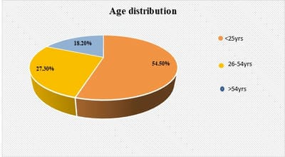
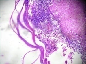
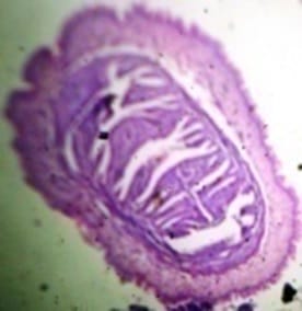
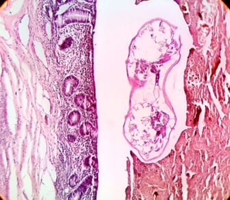
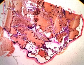
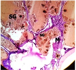
Background: Parasitic infections are a major public health problem worldwide with one-quarter of the world’s population is suffering from it. Intestinal and extraintestinal parasitic infestations are rising in developing countries. There is a raise in the immunocompromised state in which tissue parasitic infestations are increasing which necessitates this type of study. Method: A retrospective descriptive study with data collected from histopathology register from January 2018 to December 2020, all cases diagnosed as parasitic infestation with age, gender, location and histopathological evaluation with tissue response was analysed. Results: In the present study over 3 years 11 parasitic infestations were identified. About 3(27.3%) cases of hydatid cyst, 5(45.5%) cases of Enterobius vermicularis, 2 (18.2%) cases of cysticercosis and 1(9%) case of hard tick was identified. The most common age group affected was <25 years of age (54.5%). The most common parasite found is Enterobius vermicularis in our study. Conclusion: A careful histopathological examination to identify parasitic infestations in tissue sections will help to decrease morbidity and mortality by providing specific treatment to the patient.

Keywords: Hydatid cyst, Enterobius vermicularis, Hard tick

Corresponding Author How to Cite this Article To Browse
Poovizhi Inbasekaran, Assistant Professor, Department of Pathology, Vinayaka Missions Kirupananda Variyar Medical College and Hospital, Salem, Tamil Nadu, India.
Email:
Inbasekaran P, Chandrasekar M, Ramachandran T, Kulandaivel AL. Parasites in histopathology: A tertiary care hospital experience. Trop J Pathol Microbiol. 2021;7(3):110-115.
Available From
https://pathology.medresearch.in/index.php/jopm/article/view/537

Manuscript Received Review Round 1 Review Round 2 Review Round 3 Accepted
2021-05-22 2021-06-02 2021-06-11 2021-06-16
Conflict of Interest Funding Ethical Approval Plagiarism X-checker Note
No Nil Yes 6%

© 2021 by Poovizhi Inbasekaran, Megala Chandrasekar, Thamilselvi Ramachandran, Anbu Lenin Kulandaivel and Published by Siddharth Health Research and Social Welfare Society. This is an Open Access article licensed under a Creative Commons Attribution 4.0 International License https://creativecommons.org/licenses/by/4.0/ unported [CC BY 4.0].

Introduction

The parasitic infestation has worldwide prevalence and it affects almost all age groups and both the sex. The developing countries are more prone to intestinal and extra-intestinal parasitic infestations. The morbidity and mortality due to parasitic infections are higher due to an increase in the prevalence of immunocompromised conditions, nutritional deficiencies, chronic diarrhoea, and impaired development in children. Parasite lives in (endoparasite) or on (ectoparasite) another organism called its host. The host suffers various diseases, infections and discomfort as the organism obtain nourishment and protection [1,2]. The tissue damage caused by the parasite can be either due to physical pressure or toxic secretory products which may lead to hypersensitivity reactions. Tissue response produced by these parasites on histopathology provides a clue in the search of parasites and confirm the diagnosis. Eosinophilic infiltration, abscess formation and granulomatous reaction are the most common tissue responses encountered due to parasites. In the case of dead or calcified parasites there is no tissue response identified [3,4]. The relevant clinical information and histomorphological features in the tissue section would help to identify a particular organism.

Methods

Setting: Department of pathology, Vinayaka Missions Kirupananda Variyar Medical College & Hospital, a tertiary care centre in Salem, Tamil Nadu.

Duration and type of study: A Retrospective descriptive study conducted for a period of 3 years from January 2018 to December 2020.

Sampling methods: All parasitic infestations reported on histomorphology in the department of pathology.

Sample size: 11 cases

Inclusion criteria: All parasitic infestation diagnosed in histopathology on tissue sections.

Exclusion criteria: Parasitic infestations diagnosed in fine needle aspiration cytology. Parasitic infestation diagnosed in brush and exfoliative cytology smears. Parasitic infestations diagnosed in smears from fluid cytology and peripheral blood smears.

Data collection procedure: Data of all parasitic infestations reported in histopathology was retrieved from archives of the department of pathology from January 2018 to December 2020. Details was collected mainly based on age, gender, location, clinical diagnosis and histopathology report with tissue response done on routine Hematoxylin and Eosin stained slides

Statistical Analysis: The data was entered into Microsoft Excel worksheet and analysed with the help of frequency, proportion, mean and standard deviation wherever applicable.

Results

In the present study 11 parasitic lesions were identified on histopathological examination over 3 years. Out of 11 cases, 3 (27%) cases was hydatid cyst and the most common site was liver, 2(18%) cases was cysticercosis with the most common site being skeletal muscle, 5(45.50%) cases was Enterobius vermicularis with the most common site being appendix (GIT) and 1(9%) case was hard tick was seen in lower eyelid (Tab 1). The age group affected by the parasite was 14 -62 years and the mean age was 28.72±17.64 (Fig 1). The most common age group is <25years with 6(54.50%) cases. One case of hard tick which was a very rare and unusual case was found in the lower eyelid in the present study.

Table 1: Clinical presentation of parasitic lesions in the present study.

HP Diagnosis Age Sex Site Clinical diagnosis
Hydatid cyst 60 F Liver Hydatid cyst
Hydatid cyst 62 M Liver Hydatid cyst
Hydatid cyst 55 F Liver Hydatid cyst
Cysticercosis 35 F Intramuscular Mass in epigastric region
Cysticercosis 15 F Intramuscular (hypochondrium) Muscular cysticercosis
Enterobius Vermicularis 16 M Appendix Resolving appendicitis
Enterobius Vermicularis 26 M Appendix Acute appendicitis
Enterobius Vermicularis 14 F Appendix Acute appendicitis
Enterobius Vermicularis 20 M Appendix Acute appendicitis
Enterobius Vermicularis 17 F Appendix Acute appendicitis
Hard Tick 16 M Lower eyelid ? Tricoepithelioma/ Neurilemmoma

patho_537_01.jpg

Figure 1: Age distribution

Table 2: Comparison of incidence of parasitic infestation in various studies [5,6,7,8,9].

Authors Site Occurrence
Vora SH et al[5] Soft tissue cysticercosis 88 %
Rao SS et al [6] Hydatid cyst in Liver 72%
Manoharan A et al [7] Hydatid cyst in the liver, soft tissue cysticercosis. Each 22.2%.
Bhawana Choudhary et al [8] Hydatid cyst liver
Enterobius vermicularis &
Cysticercosis
77.42%
6.44%
3.22%
Shah KP et al [9] Hydatid cyst
Enterobius vermicularis
Cysticercosis
48%
8%
4%
Present study Hydatid cyst liver
Enterobius vermicularis
Cysticercosis
Hard tick
27.30%
45.50%
18.20%
9%

Discussion

Parasitic infestation is a public health problem in endemic countries with a temperate climates. Very few studies have been conducted on the presence of different parasites in the tissue section as biopsies are usually not necessary in most cases. This is done for diagnostic purposes, to identify other entities and in complicated cases resection is done. Many studies have been conducted on stool samples, as stool microscopy being the faster and preferred method but is not relevant in all types of parasites, serological tests can be used for supporting diagnosis [4,10]. Early diagnosis of a parasitic infestation can help for the immediate onset of treatment. Hydatid cyst or Echinococcosis caused by dog tapeworm is a zoonotic helminth that affects mainly the liver and lungs in humans. The feco-oral route is the common mode of spread where after ingestion of eggs by a human they penetrate the intestinal mucosa they enter the portal circulation and carried to the liver, lung and other organs to develop into fluid-filled cyst [11].

The hydatid cyst has an external membrane and internal germinal layer where brood capsules and daughter cysts develop. Tissue response results from multiple host-parasite relationships such as mononuclear cell infiltration, fibrosis, necrosis and areas of calcification. All the 3 cases were clinically diagnosed as hydatid cysts in the liver and all 3 cases showed cysts with surrounding fibrosis, mononuclear cell infiltrate [7,8] and haemorrhage, one case showed necrosis and multinucleated giant cell in the present study (Fig.2).

patho_537_02.jpg

Figure 2: Hydatid cyst with presence of necrosis and mixed inflammatory infiltrate and laminated membranous structure [H -E stain, x10].

Human Cysticercosis is due to food contamination caused by Cysticercus cellulose, the larvae of the pork tapeworm, Taenia solium. The common sites are skeletal muscle, subcutaneous tissue, brain and eye. The most common tissue response being inflammatory infiltrate and xanthogranulomatous reaction [7,12]. Both cases were seen intramuscular, one case was clinically diagnosed as cysticercosis which showed xanthogranulomatous reaction around the parasite [7,9]. other cases diagnosed as mass lesions showed mixed inflammatory cells, multinucleated giant cells and fibroblasts in our study (Fig.3).

The most common helminthic infection of the gastrointestinal tract worldwide is due to Enterobius vermicularis, they are seen in all ages and socioeconomic levels but have a high predilection for children and youngsters similar in our study. E. vermicularis infestation of the appendix can cause acute appendicitis referred to as appendicealcolic independent of histological acute inflammation [13].


patho_537_03.jpg

Figure 3: Intramuscular cysticercosis [H -E stain, x10]

The luminal obstruction can cause an increase in intraluminal pressure which impairs appendix wall blood circulation with mucosal damage, inflammation, sepsis, necrosis and perforation [14]. All 5 cases in our study were clinically diagnosed as appendicitis where the intraluminal parasite was an incidental finding, 3 cases showed eosinophilic with acute inflammatory infiltrate, 2 cases showed only acute inflammatory infiltrate (Fig.4). It is very important to receive antihelminthic treatment even after appendicectomy as the root cause of the disease is not treated [13]. In cases of appendicitis, appendicectomy and unnecessary surgery can be avoided if pre-operative diagnosis of the luminal parasite can be made because clinical management of these infections is different from that for classical appendicitis [13,15].

patho_537_04.jpg

Figure 4: Presence of E. Vermicularis in the lumen of the appendix [H -E stain, x10]

Ticks are blood-sucking ectoparasites that attach to the skin of vertebrates. They transmit serious illnesses such as Lyme disease or severe fever with thrombocytopenia syndrome and tick paralysis caused by their poisonous secretions. In the present study clinical diagnosis was Trichoepithelioma/Neurilemmoma but in other studies tick infestations were diagnosed as a dermoid cyst or epidermoid cyst [16,17]. The presence of capitulum, nonsegmented body, well developed salivary gland and skeletal muscle with midgut was observed (Fig.5). Its external surface was covered with a thick cuticle, including an outermost thin layer with saw-like annulations and the innermost layer with pore canals which was similar to Chang et al and Sung et Al case reports so was diagnosed as a hard tick. However no legs found in our study were Sung et al showed ticks with legs in the external auditory canal. Post histopathological diagnosis patient gave a history of insect hit during travel which could have helped clinicians in early diagnosis.

patho_537_05.jpg patho_537_06.jpg


Figure 5: Hard tick (a) whole section of tick with capitulum (arrow), cuticle structures (arrow head) [H -E stain, x10]. (b) cuticle (arrow), midgut epithelium (arrow head), salivary gland (SG), Musculature (M) [H -E stain, x40].

Conclusion

Parasitic lesions can show different clinical manifestations. Among the 11 cases throughout 3years, only 3 cases of hydatid cyst and one case of cysticercosis were clinically diagnosed. Rest other 7 cases were incidental findings on histopathological examination. With the increase in the immunocompromised state, overuse of antibiotics, malnutrition, change in life habits is a major concern, calling for the cautious eye to look for parasitemia in tissue sections. As there is no vaccine for such infections, early diagnosis and systemic medications will decrease the morbidity and mortality, hospital and monitory burden on the patients too. This study emphasizes the need for careful history taking and histopathological examination of these lesions which helps in diagnosis. It also helps us to understand the interactions between parasites and host tissue and aids in specific treatment for the patient.

What new this study adds to existing knowledge?

Detection of various tissue responses and presence of parasite seen on histopathological examination confirms the primary cause of disease that helps to provide specific treatment for the patients.

Author contribution

Dr. Poovizhi Inbasekaran: data collection, data compiling, literature review, manuscript preparation, manuscript editing. Dr. Thamilselvi R: manuscript editing, final approval. Dr. Megala C: data collection, manuscript editing Dr. Anbu Lenin K: manuscript editing

Reference

01. Papparella S. La istologia nella diagnosi delle parassitosi [Histology in diagnosis of parasitic diseases]. Parassitologia. 2004 Jun;46(1-2)157-8. Italian [Crossref][PubMed][Google Scholar]

02. Manuel A Marcial, Raul A, Marcial-Rojas. Protozoal and helminthic diseases, Damjanov I, Linder J, Anderson’s pathology, tenth edition. Mosby- Missouri. 1996;747-8. [Crossref][PubMed][Google Scholar]

03. Gutierrez, Yezid. Diagnostic pathology of parasitic infections with clinical correlations. Oxford University Press, USA. 2000. [Crossref][PubMed][Google Scholar]

04. Rayan P, Verghese S, McDonnell PA. Geographical location and age affects the incidence of parasitic infestations in school children. Indian J Pathol Microbiol. 2010 Jul-Sep;53(3)498-502. doi: 10.4103/0377-4929.68292 [Crossref][PubMed][Google Scholar]

05. Rao, Siddharth S, Bhupendra Mehra, Ravindra Narang. "The spectrum of hydatid disease in rural central India- An 11-year experience. " Annals of tropical medicine and public health. 2012;5(3)225. [Crossref][PubMed][Google Scholar]

06. Vora SH, Motghare DD, Ferreira AM, Kulkarni MS, Vaz FS. Prevalence of human cysticercosis and taeniasis in rural Goa, India. J Commun Dis. 2008 Jun;40(2)147-50. [Crossref][PubMed][Google Scholar]

07. Manoharan, A. , and S. Sowmya. "Parasitic infections and their tissue response: a histopathological study. " Int J Res Med Sci. 2016;4(6)1938-1942 [Crossref][PubMed][Google Scholar]

08. Bhawana Choudhary, Mohit Gupta, Neelu Gupta, Sunita Kulhari. Parasitic infections and their tissue response- A histopathological study. International Journal of Medical Sciences and Innovative Research. 2019;4(6)102–106. [Crossref][PubMed][Google Scholar]

09. Khushbu P Shah, Mandakini M Patel. Parasites found in surgical pathology- the institutional experience. Int J Res Med Sci. 2019;7(10)3756-3761. Doi: org/10.18203/2320-6012 [Crossref][PubMed][Google Scholar]

10. Behnke J M. "Evasion of host immunity", Parasites- immunity and pathology. The consequences of parasitic infection in mammals. 1990;344-395. [Crossref][PubMed][Google Scholar]


11. Khadidja H, Achour Y, Houcin B, Vasile C. Histological Appearance of Echinococcus Granulosus in the Camel Species in Algeria. Bulletin UASVM Vet Med. 2014;71;79-84. [Crossref][PubMed][Google Scholar]

12. T M K, D M, V R M. Cysticercus of the Breast which Mimicked a Fibroadenoma- A Rare Presentation. J Clin Diagn Res. 2012 Nov;6(9)1555-6. doi: 10.7860/JCDR/2012/4184.2559 [Crossref][PubMed][Google Scholar]

13. Aydin O. Incidental parasitic infestations in surgically removed appendices- a retrospective analysis. Diagn Pathol. 2007 May 24;2;16. doi: 10.1186/1746-1596-2-16 [Crossref][PubMed][Google Scholar]

14. Engin O, Calik S, Calik B, Yildirim M, Coskun G. Parasitic appendicitis from past to present in Turkey. Iran J Parasitol. 2010 Sep;5(3)57-63. [Crossref][PubMed][Google Scholar]

15. da Silva DF, da Silva RJ, da Silva MG, Sartorelli AC, Rodrigues MA. Parasitic infection of the appendix as a cause of acute appendicitis. Parasitol Res. 2007 Dec;102(1)99-102. doi: 10.1007/s00436-007-0735-0 [Crossref][PubMed][Google Scholar]

16. Chang SH, Park JH, Kwak JE, Joo M, Kim H, Chi JG, et al. A case of histologically diagnosed tick infestation on the scalp of a Korean child. Korean J Parasitol. 2006 Jun;44(2)157-61. doi: 10.3347/kjp.2006.44.2.157 [Crossref][PubMed][Google Scholar]

17. Sung WJ, Kim YH. A Case of Engorged Female Hard Tick in the External Auditory Canal of an Infant. Korean J Parasitol. 2017 Oct;55(5)565-568. doi: 10.3347/kjp.2017.55.5.565 [Crossref][PubMed][Google Scholar]