E-ISSN:2456-1487
P-ISSN:2456-9887
RNI:MPENG/2017/70771

Case Report

Disease

Tropical Journal of Pathology and Microbiology

2021 Volume 7 Number 4 July-August
Publisherwww.medresearch.in

Diagnosis of Sinus Histiocytosis with Massive Lymphadenopathy (Rosai-Dorfman Disease) by Fine Needle Aspiration Cytology in a Paediatric patient

Ankur Rana N.1, Vijaykumar Patel D.2*, Samir Shah H.3, Rajiv Daveshwar M.4
DOI: https://doi.org/10.17511/jopm.2021.i04.10

1 Neema Ankur Rana, Assistant Professor, Department of Pathology, Baroda Medical College, Vadodara, Gujarat, India.

2* Durva Vijaykumar Patel, Third-year Resident, Department of Pathology, Baroda Medical College, Vadodara, Gujarat, India.

3 Hiral Samir Shah, Tutor, Department of Pathology, Baroda Medical College, Vadodara, Gujarat, India.

4 Meena Rajiv Daveshwar, Associate Professor, Department of Pathology, Baroda Medical College, Vadodara, Gujarat, India.

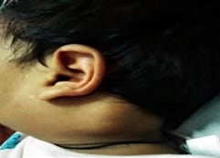
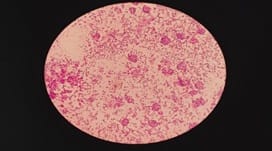
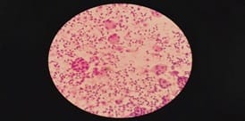
Rosai-Dorfman Disease (RDD) is also known as Sinus Histiocytosis of Massive Lymphadenopathy (SHML). Clinically Rosai-Dorfman Disease is characterized by massive, painless, bilateral cervical lymph node enlargement, often mimics lymphoma. Microscopically, it shows dilatation of lymphatic sinuses occupied by numerous lymphocytes and proliferation of histiocytes with abundant pale eosinophilic cytoplasm containing engulfed lymphocytes or plasma cells- emperipolesis.

Keywords: Rosai-Dorfman Disease, Lymphadenopathy, Emperipolesis

Corresponding Author How to Cite this Article To Browse
Durva Vijaykumar Patel, Third-year Resident, Department of Pathology, Baroda Medical College, Vadodara, Gujarat, India.
Email:
Neema Ankur Rana, Durva Vijaykumar Patel, Hiral Samir Shah, Meena Rajiv Daveshwar, Diagnosis of Sinus Histiocytosis with Massive Lymphadenopathy (Rosai-Dorfman Disease) by Fine Needle Aspiration Cytology in a Paediatric patient. Trop J Pathol Microbiol. 2021;7(4):215-218.
Available From
https://pathology.medresearch.in/index.php/jopm/article/view/532

Manuscript Received Review Round 1 Review Round 2 Review Round 3 Accepted
2021-06-01 2021-06-04 2021-06-30 2021-07-15 2021-08-28
Conflict of Interest Funding Ethical Approval Plagiarism X-checker Note
No Nil Yes 8%

© 2021 by Neema Ankur Rana, Durva Vijaykumar Patel, Hiral Samir Shah, Meena Rajiv Daveshwar and Published by Siddharth Health Research and Social Welfare Society. This is an Open Access article licensed under a Creative Commons Attribution 4.0 International License https://creativecommons.org/licenses/by/4.0/ unported [CC BY 4.0].

Introduction

Rosai Dorfman disease (RDD), also known as sinus histiocytosis with massive Lymphadenopathy (SHML), is a rare benign self-limiting disorder of histiocytic proliferation that commonly involves lymph nodes.[1]. Rosai and Dorfman first described it in 1969. SHML is mainly characterized by massive, painless, bilateral cervical lymph node enlargement, associated with fever, leukocytosis, elevated erythrocyte sedimentation rate, and polyclonal hypergammaglobulinemia.

Other lymph nodes, as well as extranodal sites commonly involved, are eye, ocular adnexa, head and neck region, upper respiratory tract, skin, subcutaneous tissue, skeletal system, central nervous system, gastrointestinal tract, thyroid, genitourinary system, breast, liver, kidney, heart etc. [2]. Most cases occur during the first or second decade of life, but any age group can be affected. RDD has distinct cytological features. Fine needle aspiration cytology plays a significant role in primary diagnosis.

Case History

An 18 months old Indian female child presented with slowly enlarging painless bilateral cervical lymph nodes for one year. There was no history of night sweat, loss of appetite or low-grade fever. On clinical examination, the patient had bilaterally enlarged, soft to firm, mobile, non-tender cervical lymph nodes.

patho_532_01.jpg

Figure:1 shows enlarged lymphadenopathy

The lymph nodes on the left side were two in number, first measuring 1.5x1.5 cm and the other 1.5x1 cm in size, and the lymph node on the right side was 1.5 x 1 cm. The overlying skin was normal.

On haematological examination her haemoglobin was 7.9 gm/dl, total WBC count 15800/mm3, differential count- Neutrophils 48%,lymphocytes 46%,Monocytes 01%,Eosinophils05% and platelet count 4,84,000/ mm3.Peripheral smear examination showed microcytic, hypochromic RBCs. Erythrocyte sedimentation rate was 38 mm/hour. In imaging studies, the x-ray chest showed clear lung fields and enlarged hilar shadow. CT chest showed multiple enlarged non-necrotic pre and paratracheal prevascular lymph nodes and a few sub centimetre sized non-necrotic bilateral axillary lymph nodes. On the CECT abdomen and pelvis, a few enlarged non-necrotic periportal and mesenteric lymph nodes were seen.

patho_532_02.jpg

Figure:2 shows Emperipolesis. (H & E, 100x)

FNAC was performed by 22 G disposable needle of 1-inch length under strict aseptic precaution. Fixation was done by methanol. Wet fixed smears were stained by Haematoxylin and Eosin stain, Pap stain, and Giemsa stained dry fixed smears. AFB was also performed on a heat-fixed smear.

patho_532_03.jpg

Figure: 3 shows emperipolesis with the background showing lymphocytes. (H & E, 400x)

On microscopic examination, the smears were cellular. They showed histiocytes predominantly engulfing lymphocytes-lymphophagocytosis (Emperipolesis). The smears also showed dispersed histiocytes, lymphocytes and few plasma cells.


The histiocytes had fine nuclear chromatin and pale abundant cytoplasm. There was no nuclear atypia or grooving. Caseous necrosis or epithelioid cells were also not seen. AFB stain was negative. Based on this characteristic cytomorphology, a diagnosis of Rosai-Dorfman disease was made.

Discussion

SHML is a rare self-limiting benign histiocytic proliferative disorder of unknown aetiology. The most likely possibilities being infection by a virus or some other microorganism and the manifestation of a subtle undefined immunologic defect. Although Human Herpesvirus 6, Epstein Barr virus and polyomavirus have been suggested, but not been proven. It has been recommended that stimulation of monocytes/macrophages via macrophage colony-stimulating factor(M-CSF) leading to immune suppressive macrophages may be the primary pathogenetic mechanism. Any age group can be affected, including paediatric and elderly patients [1,3]. Involvement of axillary, paraaortic, mediastinal and inguinal lymph nodes have been reported in RDD [3]. In our patient multiple enlarged non-necrotic cervicals, axillary, paratracheal, perivascular, periportal and mesenteric lymph nodes were present. There was no extranodal involvement in our patient.

The other features are fever, leucocytosis, elevated ESR and polyclonal gammaglobulinemia. In the present case, the patient had multiple enlarged lymph nodes with leucocytosis and elevated ESR. Clinically patient was suspected of tuberculosis or lymphoma, and the possibility of RDD was not suspected until FNAC was performed. The specific cytomorphological features are diagnostic for RDD.FNAC shows proliferation of histiocytes with lymphophagocytosis (emperipolesis), which is a characteristic feature in RDD. In lymphophagocytosis, intact lymphocytes, plasma cells and RBCs are engulfed by the histiocytes, which is the hallmark of RDD. With these classical findings on FNAC, a diagnosis of RDD can be made. Hence biopsy may be avoided. [1,4,5,6]. On IHC, these histiocytes show S100 and CD 68 positivity. [2, 6].

The differential diagnosis of RDD includes reactive lymphadenitis with sinus histiocytosis, Tuberculosis, Langerhans cell histiocytosis (LCH), Lymphoma, Hemophagocytic lymphohistiocytosis (HLH) and autoimmune lymphoproliferative syndrome (ALPS) with Massive Lymphadenopathy. [6,7,8,9]

Reactive lymphadenitis with sinus histiocytosis does not show emperipolesis, and S100 protein is harmful. In Langerhans cell histiocytosis, the Langerhans cells are mononuclear or multinucleated with characteristic nuclear features, i.e. nuclei are irregular, elongated with prominent grooves and folded. The cytoplasm of Langerhans cells is abundant and acidophilic. LCH also shows a large number of eosinophils. The Langerhans cell and the cell in LCH are reactive for S100, langerin(CD 207) and CD 1a.[6,7].

Tuberculous lymphadenitis showed caseous necrosis, epithelioid granuloma, and Langhans type of giant cell, absent in our patient's aspirate, and AFB was negative. Smears from Hodgkin's lymphoma show Reed Sternberg cells, lymphocytes, plasma cells, eosinophils and histiocytes. NonHodgkin's lymphoma shows a monomorphic lymphoid cell population. None of the above mention lymphomas shows prominent emperipolesis, which is characteristic findings in RDD.[7,10]. RDD should be differentiated from Hemophagocytic syndrome. In Hemophagocytic lymphohistiocytosis (HLH), there is erythrophagocytosis with cytopenia, hepatosplenomegaly and absence of emperipolesis.[7,10].

Another differential diagnosis of RDD is Autoimmune Lymphoproliferative Syndrome (ALPS).In ALPS, there is a childhood-onset of lymphadenopathy, hepatosplenomegaly, hypergammaglobulinemia and cytopenia and increased risk of lymphoma. There is paracortical hyperplasia, expanded interfollicular areas and polyclonal plasmacytosis.[8,9]. The course of SHML is self-limiting with spontaneous remission in most of the patients. At the same time, some patients have recurrence or persistent disease with asymptomatic lymphadenopathy. Surgery is indicated in symptomatic cases or cutaneous RDD for cosmetic reasons. Other treatment modalities are corticosteroids, chemotherapy and radiotherapy.

Conclusion

RDD is a rare disorder with characteristic clinical, cytological, histological and immunohistochemical findings. RDD is characterized by the expansion of sinuses of lymph nodes. They show proliferation of histiocytes with abundant pale eosinophilic cytoplasm containing engulfed lymphocytes –emperipolesis. The cytological findings should be correlated with appropriate clinical history, and diagnosis should be made.


Recommendation

RDD is a self-limited and specific clinical condition that has a nodal and extranodal presentation. Clinically it looks like lymphoma. The specific cytological features are diagnostic for RDD.FNAC plays a vital role in the diagnosis of RDD and avoiding unnecessary surgery.

Reference

01. Aziz M, Ray PS, Haider N, Rathore SP. Diagnosis of Rosai-dorfman disease in elderly female on fine needle aspiration cytology: a case report. Case Rep Pathol. 2012;806130. doi: 10.1155/2012/806130 [Crossref][PubMed][Google Scholar]

02. Kushwaha R, Ahluwalia C, Sipayya V. Diagnosis of sinus histiocytosis with massive Lymphadenopathy (Rosai-Dorfman Disease) by fine needle aspiration cytology. J Cytol. 2009 Apr;26(2):83-5. doi: 10.4103/0970-9371.55229 [Crossref][PubMed][Google Scholar]

03. Sall A, Touré AO, Ndiaye FS, Sène A, Sall FB, Faye BF, et al. Rosai Dorfman disease diagnosed by fine-needle aspiration cytology in a young man with HIV infection. Clin Case Rep. 2015 Oct;3(10):879-83. Doi: 10.1002/ccr3.391 [Crossref][PubMed][Google Scholar]

04. Das DK, Gulati A, Bhatt NC, Sethi GR. Sinus histiocytosis with massive Lymphadenopathy (Rosai-Dorfman disease): report of two cases with fine-needle aspiration cytology. Diagn Cytopathol. 2001 Jan;24(1):42-5. doi: 10.1002/1097-0339(200101)24:1<42::aid-dc1007>3.0.co;2-n [Crossref][PubMed][Google Scholar]

05. Deshpande AH, Nayak S, Munshi MM. Cytology of sinus histiocytosis with massive Lymphadenopathy (Rosai-Dorfman disease). Diagn Cytopathol. 2000 Mar;22(3):181-5. doi: 10.1002/(sici)1097-0339(20000301)22:3<181::aid-dc10>3.0.co;2-6 [Crossref][PubMed][Google Scholar]

06. Rajyalakshmi R, Akhtar M, Swathi Y, Chakravarthi R, Bhaskara Reddy J, Beulah Priscilla M. Cytological Diagnosis of Rosai-Dorfman Disease: A Study of Twelve Cases with Emphasis on Diagnostic Challenges. J Cytol. 2020 Jan-Mar;37(1):46-52. doi: 10.4103/JOC.JOC_4_19 [Crossref][PubMed][Google Scholar]

07. Garza-Guajardo R, García-Labastida LE, Rodríguez-Sánchez IP, Gómez-Macías GS, Delgado-Enciso I, Chaparro MM, et al. Cytological diagnosis of Rosai-Dorfman disease: A case report and revision of the literature. Biomed Rep. 2017 Jan;6(1):27-31. doi: 10.3892/br.2016.814 [Crossref][PubMed][Google Scholar]

08. Lim MS, Straus SE, Dale JK, Fleisher TA, Stetler-Stevenson M, Strober W, et al. Pathological findings in human autoimmune lymphoproliferative syndrome. Am J Pathol. 1998 Nov;153(5):1541-50. doi: 10.1016/S0002-9440(10)65742-2 [Crossref][PubMed][Google Scholar]

09. Maric I, Pittaluga S, Dale JK, Niemela JE, Delsol G, Diment J, et al. Histologic features of sinus histiocytosis with massive lymphadenopathy in patients with autoimmune lymphoproliferative syndrome. Am J SurgPathol. 2005 Jul;29(7):903-11. doi: 10.1097/01.pas.0000157997.61177.08 [Crossref][PubMed][Google Scholar]

10. Jena, Madhusmita. Diagnosis of Rosai-Dorfman disease by fine needle aspiration cytology in a case with cervical lymphadenopathy and nasal mass. Online Journal of health and Allied Sciences. 10;2(2011). [Crossref][PubMed][Google Scholar]