E-ISSN:2456-1487
P-ISSN:2456-9887
RNI:MPENG/2017/70771

Research Article

Breast

Tropical Journal of Pathology and Microbiology

2021 Volume 7 Number 4 July-August
Publisherwww.medresearch.in

The Spectrum of Malignant Breast lesions by Fine Needle Aspiration Cytology in a Teaching Hospital

Alexander M.1, A. Pattanashetti M.2*
DOI: https://doi.org/10.17511/jopm.2021.i04.05

1 Manika Alexander, MBBS, MD Pathology, Associate Professor, Department of Pathology, Gadag Institute of Medical Sciences, Gadag, Karnataka, India.

2* Mallikarjun A. Pattanashetti, MBBS, MD Pathology, Assistant Professor, Department of Pathology, Gadag Institute of Medical Sciences, Gadag, Karnataka, India.

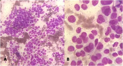
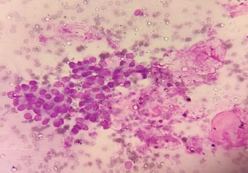
Background: Fine needle aspiration cytology (FNAC) is one of the preliminary tests done to detect malignant breast lesions, which help in early detection and management. Studying the cytology features of various malignant breast diseases was the aim of this study. Methods: This study is a cross-sectional retrospective study conducted in the Department of Pathology from 2015 to 2020. Clinical details and cytology features were collected from the Department records. Results: A total of 75 cases were collected during the study period. All the cases were females. The spectrum of lesions was composed of Ductal carcinoma followed by one point each of Mucinous carcinoma, Malignant Phyllodes tumour and Lobular Carcinoma. Conclusions: FNAC helps in rapid diagnosis and early management of malignant breast lesions.

Keywords: Breast cytology, Malignant breast lesions, FNAC

Corresponding Author How to Cite this Article To Browse
Mallikarjun A. Pattanashetti, MBBS, MD Pathology, Assistant Professor, Department of Pathology, Gadag Institute of Medical Sciences, Gadag, Karnataka, India.
Email:
Alexander M, Pattanashetti MA. The Spectrum of Malignant Breast lesions by Fine Needle Aspiration Cytology in a Teaching Hospital. Trop J Pathol Microbiol. 2021;7(4):181-187.
Available From
https://pathology.medresearch.in/index.php/jopm/article/view/519

Manuscript Received Review Round 1 Review Round 2 Review Round 3 Accepted
2021-01-31 2021-02-10 2021-02-10 2021-02-28 2021-03-08
Conflict of Interest Funding Ethical Approval Plagiarism X-checker Note
No Nil Yes 8%

© 2021 by Manika Alexander, Mallikarjun A. Pattanashetti and Published by Siddharth Health Research and Social Welfare Society. This is an Open Access article licensed under a Creative Commons Attribution 4.0 International License https://creativecommons.org/licenses/by/4.0/ unported [CC BY 4.0].

Introduction

Breast lesions are a heterogeneous group of disorders ranging from inflammatory lesions to invasive cancers. Breast cancer is the most common malignant neoplasm among women worldwide after non-melanoma skin malignancies. Further, it is the most frequent cause of cancer-related death among women worldwide, with an estimated 2.1 million new cases annually. The survival rate varies by continent, with better rates in developed countries[1]. The implementation of breast cancer screening programs is helpful for the early detection of breast lesions that, in turn, translate to a significant decrease in mortality. Lesions suspected of malignancy should be subjected to complementary tests as soon as possible for diagnostic confirmation[2].

The most common modality for the initial evaluation of breast lesions is fine-needle aspiration cytology (FNAC). In addition, some studies recommend that small breast lesions or those suspicious for malignancy should be evaluated through histopathological examination ("core needle biopsy" or excision). However, FNAC is a more simple, low-cost technique with a low risk of complications than that observed with biopsy or excision procedures [4]. FNAC, when performed inadequate conditions, has good accuracy. The aspirated specimen can also be processed as a cell block that can be used for immunohistochemical analysis of related biomarkers (e.g., estrogen receptor, progesterone receptor, and Her2)[5]. The cellblock specimen can also be used for molecular analysis, providing additional information that can be helpful in the diagnosis and treatment by identifying predictive and prognostic markers. Early diagnosis and small lesion size significantly improve the treatment outcomes and prognosis of patients with breast lesions, especially those with malignant lesions

Breast masses are the most common complaint with which females patients present to the hospital. The majority of these lesions are either benign or non-neoplastic [6]. Fine needle aspiration cytology (FNAC) is one of the most common tests for palpable lesions. FNAC is also a part of the triple assessment test for breast masses [7]. It is minimally invasive, cost-effective and diagnostic accuracy is good [8]. Rapid turnaround times help in the same-day diagnosis of breast lesions and early management. Malignant diseases are more common after menopause [4].

Breast cancer is the most common female cancer worldwide. It accounts for nearly a quarter (25%) of all cancers, with an estimated 1.5 lakh new cancer cases diagnosed in 2016. This study was conducted to determine various forms of malignant breast lesions presented to our hospital.

Methods

Setting: Department of Pathology, Gadag Institute of Medical Sciences, Gadag

Duration of Study: 5 years six months from June 2015 to December 2020

Type of Study: Cross-sectional retrospective observational study

Sampling methods: Universal sampling

Sample Size calculation: All the cases as per inclusion criteria were included in the study

Inclusion Criteria: were cases with the diagnosis of neoplastic breast disease in females

Exclusion Criteria: All benign breast diseases in males and females were excluded from the study

Data Collection Procedure: Clinical details and demographic data were obtained from departmental records. FNAC was done by using five cc syringes with 22‑23G needle under all aseptic precautions. Air-dried smears were stained with MGG stain, Leishman stain and wet smears were stained with PAP stain and H and E stain.

Ethical Clearance: Obtained from Institutional Ethical Committee

Statistical Analysis: The data obtained were entered into Microsoft Excel Spreadsheet. Categorical data were expressed in terms of rates, ratios and percentages.

Results

A total of 514 cases were there in which FNAC was done, of which malignancy was detected in 75(14.6%) cases. All the cases were females. The most common presenting complaint was a lump in the breast 72(97%) cases. The mean age of presentation of all malignant breast diseases was 49 years. The mean age of the most common malignant neoplasm (Ductal carcinoma breast) was 49 years. The following were the cytological diagnosis: Ductal carcinoma-72 cases(97%) followed one case each of Mucinous carcinoma


(1%), Malignant Phyllodes tumour (1%) and Lobular Carcinoma(1%) as shown in Table 1.

Table 1: Spectrum of Malignant Breast Lesions on FNAC

Malignant breast neoplasms Number of Cases Percentage (%)
Ductal carcinoma breast 72 97%
Mucinous carcinoma breast 1 1%
Malignant Phyllodes tumour 1 1%
Lobular carcinoma 1 1%

Table 2: Comparison of Malignant Breast lesions among various studies.

Study Malignant breast lesions
Modi et al [25] 16.7%
Georgieva et al[26] 22.5%
Bajwa et al[27] 10.3%
Sunita et al[28] 37.1%
Panwar et al [29] 8.4%
Present Study 14.6%

patho_519_01.jpg

Figure 1: Smears studied shows malignant ductal epithelial cells seen in dyscohesive clusters. These tumour cells have a high N/C ratio with a moderate to scanty amount of cytoplasm. These features are suggestive of Ductal Carcinoma Breast( 1A: Leishman stain, 10 X and 1B: Leishman stain, 40 X)

patho_519_02.jpg

Figure 2: Smear studied shows clusters of tumour cells with mild to moderate nuclear

pleomorphism, floating in extracellular pools of mucin, suggestive of Mucinous carcinoma breast (Leishman stain 40X)

Discussion

Breast lumps are very common, most of the lumps are benign, and the prevalence of malignant lumps increases with age[9]. Breast cancer is one of the commonest cancers amongst women in India, with approximately 75,000 new cases estimated every year[10].

The following are the risk factors for breast cancer: Family history, Genetic predisposition, Estrogen exposure, Radiation exposure, Breast density and Obesity. The following are the Precancerous conditions: Complex fibroadenoma, moderate or florid hyperplasia without atypia, sclerosing adenosis and solitary papilloma without atypical ductal hyperplasia are associated with slightly increased risk (1.5-2times) whereas, atypical ductal hyperplasia and atypical lobular hyperplasia are associated with moderately increased risk (4 -5 times) of breast cancer[11].

The vast majority of breast malignancies are adenocarcinomas arising from the ducts or lobules. Traditionally, these were categorized by morphology. The most common histologic type of breast cancer is invasive ductal carcinoma or carcinoma of no special type (40%-75%) (Figure 1). The remainder of carcinomas is classified as specialized types. These include but are not limited to lobular carcinoma (5%-15%), mucinous carcinoma (2%), tubular carcinoma (2%), medullary carcinoma(< 1%) and metaplastic carcinoma(0.2%-5%) [12].

Lobular carcinoma is the most common type of breast cancer following ductal carcinoma and is composed of dyscohesive monomorphic cells that lack tubule formation [12]. The dyscohesion is due to the inactivation of the cell adhesion molecule E-cadherin. These tumours are invariably ER-positive in 60% to 70% of tumours and generally negative for HER2. The prognosis of this tumor is favorable; however, this type is associated with a worse long-term outcome relative to ductal carcinoma in terms of higher incidences of mortality, recurrence, and distant metastasis [13]. In mucinous carcinoma, well to moderately differentiated neoplastic cells are clustered in small groups within large pools of mucin (see Figure 2). This type of carcinoma is typically ER-positive and HER2-negative.


Mucinous carcinoma has an excellent 5-year survival rate and a low rate of distant and local recurrence.[13]. Tubular carcinoma is composed of well-formed tubules, often shaped like teardrops [12]. This type of carcinoma is uncommon, at only 2% of invasive breast carcinomas, and is associated with an excellent prognosis. Tubular carcinoma is generally ER-positive/HER2-negative[13].

Metaplastic carcinoma is a heterogeneous category of neoplasms that have differentiated into squamous or mesenchymal cells. Greater than 90% of metaplastic carcinomas are ER-negative/HER2-negative. Compared with other TNBCs, metaplastic carcinoma has a worse clinical outcome and lower chemotherapy response rate [13]. Medullary carcinoma comprises solid syncytial sheets of pleomorphic cells with prominent nucleoli, a lymphoplasmacytic infiltrate numerous mitoses, and a pushing border[12]. These tumours are typically triple-negative and often associated with BRCA mutations. Traditionally, these tumors were reported to have a relatively favorable prognosis; however, due to the low level of reproducibility for the diagnosis and the poor prognosis associated with TNBCs, these tumors are now termed "carcinomas with medullary features" and treated with aggressive therapy [13].

Molecular gene expression profiling studies heralded a new era in the classification of breast carcinomas. In addition to morphology, breast carcinomas now are categorized into 1 of 3 subtypes based on the tumour's expression of HER2 and E.R.s. Although testing is almost always performed for P.R. as well as E.R., descriptions of molecular classifications generally use E.R. status as a surrogate marker for P.R. as well. The three subtypes are ER-positive/HER2-negative (50%-65% of tumours). HER2-positive (10%-20% of tumors). ER-negative/HER2-negative (10%-20% of tumors) [14].

ER-positive/HER2-negative breast cancers: These are often associated with losses of chromosome 16q and gains in chromosome 1q, as well as activating mutations in PIK3CA, a growth factor receptor signalling molecule. This type of breast carcinoma often progresses through a sequence that can include flat epithelial atypia, atypical ductal hyperplasia, ductal carcinoma in situ (DCIS), and eventually invasive ductal carcinoma. This subtype is primarily treated with hormone therapy instead of chemotherapy because few of these carcinomas respond to chemotherapeutic drugs.

HER2-Positive Invasive Carcinomas: These tumors are defined by HER-2 positivity, regardless of E.R. status. The HER2-positive carcinomas are characterized by mutations in tumor suppressor TP53, which amplify HER2 on chromosome 17q. The suggested precursor lesion for this type of carcinoma is atypical apocrine adenosis that progresses to DCIS. This subtype is more common in nonwhite and young women and patients with LiFraumeni syndrome (hereditary TP53 mutation). These cancers can metastasize early, often to the brain and viscera, even when small. The drug trastuzumab (Herceptin) is a monoclonal antibody that inhibits HER2 and vastly improves survival. Once metastasized, survival is uncommon[14].

The ER-negative/HER2-negative cancers, also known as triple-negative as P.R., is also negative, are the least understood of the subtypes of invasive ductal carcinoma. This subtype arises through a pathway independent from changes in HER-2 and E.R.s. Most carcinomas in women with BRCA1 mutations are triple-negative. These cancers grow and metastasize quickly to the brain and viscera. Approximately 30% respond to chemotherapy, and they tend to recur within five years. Triple-negative breast carcinomas (TNBCs) tend to be poorly differentiated[14].

In patients presenting with a breast lump, the diagnosis should be made by clinical assessment. Radiological imaging and tissue sample have been taken either for cytology or histology analysis: the so-called Triple Test. There are several competing approaches to a breast biopsy, such as surgical excision biopsy, core needle biopsy and biopsy by aspiration or Fine-needle aspiration (FNA) [15]. FNAC has become an increasingly popular technique for assessing breast lumps. It is a minor invasive technique for obtaining a cytological diagnosis and is highly accurate if done by experienced personnel [16].

The worldwide-accepted protocol for diagnosing breast lumps is the "Triple Assessment", which includes clinical examination, mammography and pathological diagnosis. Fine needle aspiration cytology (FNAC) has become a diagnostic tool to assess the nature of palpable breast lesions [17,18].

It forms an important part of the pathological assessment since it is easy, relatively painless, quick and a cost-effective technique. It also helps in the planning of treatment of breast lump [19].


Moreover, FNAC has good sensitivity, specificity and accuracy in the diagnosis of both malignant and non-malignant breast lumps. Accuracy can be improved by multiple sampling from different angles and by using ultrasound guidance in very small lumps [20]. Malignant breast lesions were found in all age groups. This finding follows a study by Rahman et al.[21], who also concluded that most malignant lesions were found in the middle age group of 31 to 50 years. Similar results from other studies in India like Muddegowda et al.[22].and Khemka et al [23]were reported. However, reports from the western world depict the 5th and 6th decades as the predominant age group for breast cancer [24]. Present study findings of 14.6% cases are similar to a study done by Modi et al. [25], which found 16.7% cases. The other comparison studies are shown in Table 2. The management of breast disease needs a deliberate, synchronized diagnostic and treatment strategy. Fine-needle aspiration cytology (FNAC) is an essential diagnostic tool for the preoperative diagnosis of palpable and non-palpable breast lesions. Our study has the limitation of having a small sample size, and being a retrospective study, there will be deficiencies incomplete data collection. However, it provides baseline data of the patient population presenting to our hospital.

Conclusion

For diagnosing the nature of palpable breast lesions, FNAC is considered a highly accurate procedure with sensitivity and specificity as high as 95%. Its use has been recommended in literature as a preliminary treatment on an outpatient basis. Early diagnosis is of paramount importance to decrease the mortality and morbidity associated with these lesions. FNAC is a relatively simple procedure with good patient acceptance and low morbidity. It is an accurate, safe and repeatable procedure in the diagnosis of various breast lesions –both malignant and non-malignant. Repeated passes should be made for a greater yield of cytological material. FNAC should be used earlier and more frequently to shorten the diagnostic interval and allow more prompt therapy for malignant breast lesions.

What new this study adds to existing knowledge

FNAC helps in rapid diagnosis and early management of malignant breast lesions.

This study gives an insight into the various malignant breast lesions in this country region with a description of few rare lesions.

Authors Contribution

Dr Manika Alxeander: Data collection, Literature review, Manuscript preparation, Manuscript editing, Final approval. Dr Mallikarjun A Pattanashetti: Patients Selection, Collection of Samples, Data Analysis, Statistical analysis and Manuscript preparation.

Reference

01. Globocan Observatory 2019. Globocan Observatory W (2019a) Cancer today - world. Int Agency Res Cancer. 876:2018–2019. Available at: world-fact-sheets.pdf [Article][Crossref][PubMed][Google Scholar]

02. Jessica Aline Tomelin de Cursi, Mariângela Esther Alencar Marques, Cristina Andrea Campos de Assis Cunha Castro, Fernando Carlos Schmitt and Cleverson Teixeira Soares. Fine-Needle Aspiration Cytology (FNAC) is a reliable diagnostic tool for small breast lesions (≤ 1,0 cm): a 20-year retrospective study. Surgical and Experimental Pathology. 2020;3:29:1-8. Doi: 10.1186/s42047-020-00081-0 [Crossref][PubMed][Google Scholar]

03. Manfrin E, Falsirollo F, Remo A, Reghellin D, Mariotto R, Dalfior D, et al. Cancer size, histotype, and cellular grade may limit the success of fine-needle aspiration cytology for screen-detected breast carcinoma. Cancer. 2009 Dec 25;117(6):491-9. doi: 10.1002/cncy.20053 [Crossref][PubMed][Google Scholar]

04. Ali and Parwani, Ali ZS, Parwani AV. Breast cytopathology. Springer, Baltimore. p 189. [Crossref][PubMed][Google Scholar]

05. Bueno Angela SP, Viero RM, Soares CT. Fine needle aspirate cell blocks are reliable for detection of hormone receptors and HER-2 by immunohistochemistry in breast carcinoma. Cytopathology. 2013 Feb;24(1):26-32. doi: 10.1111/j.1365-2303.2011.00934.x [Crossref][PubMed][Google Scholar]

06. Guray M, Sahin AA. Benign breast diseases: classification, diagnosis, and management. Oncologist. 2006 May;11(5):435-49. doi: 10.1634/theoncologist.11-5-435 [Crossref][PubMed][Google Scholar]


07. Ferlay J F. "GLOBOCAN 2000, Cancer incidence, mortality and prevalence worldwide, version 10". IARC cancerbase. (2001). [Crossref][PubMed][Google Scholar]

08. Dixon JM, Mansel RE. ABC of breast diseases, Symptoms assessment and guidelines for referral. BMJ. 1994 Sep 17;309(6956):722-6. doi: 10.1136/bmj.309.6956.722 [Crossref][PubMed][Google Scholar]

09. Smith, Mindy A, and Leslie A Shimp. 20 common problems in women's health care. McGraw-Hill, Health Professions Division. 2000. [Crossref][PubMed][Google Scholar]

10. Notani, Chenoy R. Epidemiology of Breast Carcinoma in Indian Scenario, Guidelines for Breast Pathology reporting. Tata Memorial Hospital. 1998;1-4. [Crossref][PubMed][Google Scholar]

11. Precancerous Breast Disease: Epidemiological, Pathological, and Clinical consideration. In Rosen's Breast pathology, Paul peter Rosen Editor. Chapter 10, 3rd edition, Lippincott Williams and Wilkins. 2009: 264-84. [Crossref][PubMed][Google Scholar]

12. Mehta, Prachi, Srikant Nema, and Sanjeev Narang. "Role of p53 and Ki-67 in prognostication of carcinoma breast". Indian Journal of Pathology and Oncology 6. 2 (2019): 261-265. [Crossref][PubMed][Google Scholar]

13. Lakhani, Sunil R, et al. "WHO Classification of Tumours of the Breast". (2012). [Crossref][PubMed][Google Scholar]

14. Scholl AR, Flanagan MB. Educational Case: Invasive Ductal Carcinoma of the Breast. AcadPathol. 2020 Jan 21;7:2374289519897390. doi: 10.1177/2374289519897390 [Crossref][PubMed][Google Scholar]

15. Koss, Leopold G, and Myron R. Melamed, eds, Koss' diagnostic cytology and its histopathologic bases, Vol, 1. Lippincott Williams & Wilkins. 2006. [Crossref][PubMed][Google Scholar]

16. Saunders C, Baum M. The Breast, In: Russel R, Williams N, Bulstrode C, editors, Bailey and Love's Short Practice of Surgery. 23rd ed, Hachette (UK): Hodder Arnold. 2000. [Crossref][PubMed][Google Scholar]

17. Rahman MZ, Islam S. Fine Needle Aspiration Cytology of Palpable Breast Lump: A Study of 1778 Cases. Surgery. 2013 S12: 001. doi: 10.4172/2161-1076. S12-001 [Crossref][PubMed][Google Scholar]

18. Singh A, A Haritwal, and B M Murali. "Pattern of breast lumps and diagnostic accuracy of fine needle aspiration cytology; a hospital based study from Pondicherry, India". Internet J Pathol. 11;2 (2011):1-6. [Crossref][PubMed][Google Scholar]

19. Khemka A, Chakrabarti N, Shah S, Patel V. Palpable breast lumps: fine-needle aspiration cytology versus histopathology: a correlation of diagnostic accuracy. Internet J Surg. 18;1(2009). [Crossref][PubMed][Google Scholar]

20. Madan M, Sharma M, Mannan R, Manjari M, Kaur J, Garg S. Cytomorphological study of spectrum of breast lesions and determination of efficacy of FNAC in the diagnosis of various breast lesions. J Evol Med Dent Sci. 4;55(2015):9581-7. [Crossref][PubMed][Google Scholar]

21. Rahman MZ, Sikder AM, Nabi SR. Diagnosis of breast lump by fine needle aspiration cytology and mammography. Mymensingh Med J. 2011;20 (4):658-64. [Crossref][PubMed][Google Scholar]

22. Muddegowda PH, Lingegowda JB, Kurpad R, Konapur P, Shivarudrappa A, Subramaniam P. The value of systematic pattern analysis in FNAC of breast lesions: 225 cases with cytohistological correlation. J Cytol. 2011 Jan;28(1):13-9. doi: 10.4103/0970-9371.76942 [Crossref][PubMed][Google Scholar]

23. Khemka A, Chakrabarti N, Shah S, Patel V. Palpable breast lumps: fine-needle aspiration cytology versus histopathology: a correlation of diagnostic accuracy. Internet J Surg. 18;1(2009). [Crossref][PubMed][Google Scholar]

24. Sandhu DS, Sandhu S, Karwasra RK, Marwah S. Profile of breast cancer patients at a tertiary care hospital in north India. Indian J Cancer. 2010 Jan-Mar;47(1):16-22. doi: 10.4103/0019-509X.58853 [Crossref][PubMed][Google Scholar]

25. Modi, Palak, HarenOza, and Jignasa Bhalodia. "FNAC as preoperative diagnostic tool for neoplastic and non-neoplastic breast lesions: A teaching hospital experience". Indian J Med Res. 4(2014) :274-8. [Crossref][PubMed][Google Scholar]

26. Georgieva RD, Obdeijn IM, Jager A, Hooning MJ, Tilanus-Linthorst MM, van Deurzen CH. Breast fine-needle aspiration cytology performance in the high-risk screening population: a study of BRCA1/BRCA2 mutation carriers. Cancer Cytopathol. 2013 Oct;121(10):561-7. doi: 10.1002/cncy.21308 [Crossref][PubMed][Google Scholar]


27. Bajwa, Rakhshindah, and Tariq Zulfiqar. "Association of fine needle aspiration cytology with tumor size in palpable breast lesions". Biomedica. 26(2010):124-9. [Crossref][PubMed][Google Scholar]

28. Sunita H, T Urmila, D C Sharma. "Cytomorphological study breast lesions with sonomammo-graphic correlation". J Evol Med Dent Sci. 4(2015):137-42. [Crossref][PubMed][Google Scholar]

29. Panwar H, Ingle P, Santosh T, Singh V, Bugalia A, Hussain N. FNAC of Breast Lesions with Special Reference to IAC Standardized Reporting and Comparative Study of Cytohistological Grading of Breast Carcinoma. J Cytol. 2020 Jan-Mar;37(1):34-39. doi: 10.4103/JOC.JOC_132_18 [Crossref][PubMed][Google Scholar]