E-ISSN:2456-1487
P-ISSN:2456-9887
RNI:MPENG/2017/70771

Research Article

Breast cytology, Benign breast disease, FNAC

Tropical Journal of Pathology and Microbiology

2021 Volume 7 Number 4 July-August
Publisherwww.medresearch.in

Cytomorphological study of Benign breast lesions in a Tertiary Care Hospital

Alexander M.1, A. Pattanashetti M.2*
DOI: https://doi.org/10.17511/jopm.2021.i04.04

1 Manika Alexander, MBBS, MD Pathology, Associate Professor, Department of Pathology, Gadag Institute of Medical Sciences, Gadag, Karnataka, India.

2* Mallikarjun A. Pattanashetti, MBBS, MD Pathology, Assistant Professor, Department of Pathology, Gadag Institute of Medical Sciences, Gadag, Karnataka, India.

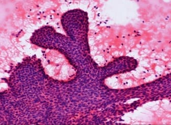
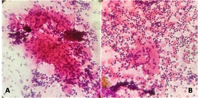
Background: Benign breast disease is one of the most common breast lesions in the reproductive age group. Fine needle aspiration cytology (FNAC) is one of the preliminary tests done to detect breast lesions which help in early detection and management. Studying the cytology features of various benign breast diseases was the aim of this study. Methods: This study is a cross-sectional retrospective study conducted in the Department of Pathology from 2015 to 2020. Clinical details and cytology features were collected from the Department records. Results: A total of 430 cases were collected during the study period. Age groups ranged from 16-40 years. All the cases were females. The spectrum of lesions was composed of fibroadenoma, fibrocystic change, breast abscess, fibroadenosis, granulomatous mastitis, etc. Conclusions: Breast lumps are a common cause of anxiety and apprehension among patients. FNAC helps in rapid diagnosis and early management of lesions. It also helps in preventing unnecessary invasive surgeries in non-neoplastic and benign breast diseases.

Keywords: Alexander M. et al: Cytomorphological study of Benign breast lesions

Corresponding Author How to Cite this Article To Browse
Mallikarjun A. Pattanashetti, MBBS, MD Pathology, Assistant Professor, Department of Pathology, Gadag Institute of Medical Sciences, Gadag, Karnataka, India.
Email:
Alexander M, Pattanashetti MA. Cytomorphological study of Benign breast lesions in a Tertiary Care Hospital. Trop J Pathol Microbiol. 2021;7(4):176-180.
Available From
https://pathology.medresearch.in/index.php/jopm/article/view/518

Manuscript Received Review Round 1 Review Round 2 Review Round 3 Accepted
2021-01-31 2021-02-10 2021-02-20 2021-02-28 2021-03-08
Conflict of Interest Funding Ethical Approval Plagiarism X-checker Note
No Nil Yes 7%

© 2021 by Manika Alexander, Mallikarjun A. Pattanashetti and Published by Siddharth Health Research and Social Welfare Society. This is an Open Access article licensed under a Creative Commons Attribution 4.0 International License https://creativecommons.org/licenses/by/4.0/ unported [CC BY 4.0].

Introduction

Breast lesions are a heterogeneous group of disorders ranging from inflammatory lesions to invasive cancers. Breast masses are the most common complaint with which females patients present to the hospital. The majority of these lesions are either benign or non-neoplastic [1]. Fine needle aspiration cytology (FNAC) is one of the most common tests for palpable lesions. It is one of the preliminary tests done on palpable breast masses. FNAC is also a part of the triple assessment test for breast masses [2].

It is minimally invasive, cost-effective and diagnostic accuracy is good [3]. Rapid turnaround times help in same day diagnosis of breast lesions and early management. The quick identification of benign breast diseases decreases the apprehension of patients [4]. This study was conducted to determine various forms of benign breast lesion presented to our hospital.

Benign breast disease (BBD) has a high prevalence and a noticeable impact on women's quality of life and, for certain histological types, increases breast cancer risk [5]. It has been estimated that one out of every two women may develop some form of fibrocystic breast disease during her lifetime and that one out of every five women may develop fibroadenoma. Although BBD has been extensively studied, the etiology of this disease is still poorly understood.

It appears that endocrine factors are associated with BBD. However, the associations found are weak and inconsistent [5]. Obesity has been identified as one of the only consistent risk factors for BBD.Conflicting results have been found for most risk factors, including exogenous hormone use, smoking, alcohol and caffeine intake. No studies have examined whether radiological tissue density is a risk factor for BBD [6].

Methods

Setting: Department of Pathology, Gadag Institute of Medical Sciences, Gadag

Duration of Study: 5 years 6 months from June 2015 to December 2020

Type of Study: Cross-sectional retrospective observational study

Sampling methods: Universal sampling

Sample Size calculation: All the cases as per inclusion criteria were included in the study

Inclusion Criteria: Cases with a diagnosis of non-neoplastic and benign breast disease.

Exclusion Criteria: All non-benign breast cases in males and females.

Data Collection Procedure: Clinical details and demographic data were obtained from departmental records. FNAC was done by using five cc syringes with 22‑23G needle under all aseptic precautions. Air-dried smears were stained with MGG stain, Leishman stain and Wet smears were stained with PAP stain and H & E stain.

Ethical Clearance: Obtained from Institutional Ethical Committee

Statistical Analysis: The data obtained were entered into Microsoft Excel Spreadsheet. Categorical data were expressed in terms of rates, ratios and percentages.

Results

A total of 430 cases were included in the present study. All the cases were females. The most common presenting complaint was a lump in the breast with 395 (92%) cases, followed by breast pain in 35 (8%) cases. The mean age of presentation of benign breast diseases was 28 years. The youngest patient was 16 years old, and the oldest was 40 years old. The mean age of the most common benign neoplasm fibroadenoma was 25 years. 15 breast lesions were non - diagnostic on FNAC. The following were the cytological diagnosis: Fibroadenoma-244(56.8%), Fibrocystic disease- 48(11.3%), Benign breast disease - 47(10.9%), Fibroadenosis-14(3.2%), Abscess – 6(1.4%), Acute suppurative mastitis- 13(3%), Chronic granulomatous mastitis- 13(3%), Galactocoele-12(2.8%) and various other lesion as shown in Table 1.

Table 1: Cytomorphological Diagnosis of Benign Breast Lesions on FNAC

Diagnosis Number of cases Percentage (%)
Fibroadenoma 244 56.8
Fibrocystic disease 48 11.3
Benign breast disease 47 10.9
Fibroadenosis 14 3.2
Acute suppurative mastitis 13 3.0
Chronic granulomatous mastitis 13 3.0
Galactocoele 12 2.8

Breast disease with atypia 11 2.6
Abscess 6 1.4
Ductal hyperplasia 4 0.9
Accessory breast hyperplasia 2 0.4
Lactational change 1 0.2
Non- diagnostic 15 3.5
Total 430 100

Table 2: Comparison of Cytomorphological findings with other studies

Diagnosis Sangma et al Present Study
Fibroadenoma 48 56.8
Fibrocystic disease 18 11.3
Abscess 12 1.4
Accessory breast hyperplasia 5 0.4
Galactocoele 4 2.8
Breast disease with atypia 3 2.6
Mastitis 5 1.4

Figures

patho_518_01.jpg

Figure 1 : Fibroadenoma: Smear studied show benign ductal epithelial cells arranged in sheet, arranged in staghorn pattern along with myoepithelial cells and bare nuclei in the background. (H& E Stain: 10 X)

patho_518_02.jpg

Figure 2 : Granulomatous Mastitis: Smear studied shows epithelioid granuloma admixed with multinucleated giant cells and lymphocytes. (2A: Granuloma H&E Stain 10 X; 2B: Multinucleate giant cells H&E Stain 10X)

Discussion

Benign breast disease is the most common lesion of the breast in the reproductive age group. Cytology is an essential tool in the preoperative diagnosis of breast lesions. The detection rate of these lesions has increased due to increased awareness among patients and ease of doing FNAC. It has good tolerance among the patients and is relatively inexpensive [7].

Clinical observations in women receiving estrogens and anti-estrogens suggest that hormonal events play a role in the etiology of benign breast lesions. In postmenopausal women receiving estrogens ± progestins for >8 years, the prevalence of benign breast lesions increased by 1.7 fold. In the Women's Health Initiative study (WHI), the use of estrogen plus progestin was associated with a 74% increase in the risk of benign proliferative breast disease [1.35-2.25]. The anti-estrogen, tamoxifen, when used for breast cancer prevention, was associated with a 28 percent reduction in the prevalence of benign breast lesions, including adenosis, cysts, duct ectasia, and hyperplasia [8].

Underlying and acquired genetic changes are also associated with benign breast lesions. Loss of heterozygosity (LOH), a finding caused by deletions of small segments of DNA, is commonly found in benign breast lesions. Women frequently have multi-focal lesions, each of which exhibit loss of heterozygosity (LOH) of different regions of DNA. Women with BRCA1/2 mutations are found to have a high frequency of multiple benign or malignant breast lesions when bilateral mastectomy specimens are meticulously examined. These findings support the current theory of an underlying predisposition to mutations in some patients to cause multiple breast lesions. In the past, this phenomenon was termed a "field effect" and, more recently, a "mutator phenotype"[8].

The clinical presentation of benign breast diseases includes breast pain (cyclical and non -cyclical), non-breast pain (chest wall and non- chest wall),nipple discharge, discrete solitary lump and diffuse lumpiness. A total of 430 cases were included in this study with various types of benign breast disease. All the cases in our study fall in the reproductive age group population. Similar findings have been reported in earlier studies done by Elmadhoun et al., Almobarak et al. and Sangma et al.[1,7,9]. This has been attributed to the influence of hormonal changes on breast tissue.


Fibroadenoma was the most common diagnosis in our study, with 244 (56.8%) cases. Our finding was in agreement with most of the available literature on benign breast lumps, where the frequency of fibroadenoma ranged from 46.6%-55.6% [10-13]. Sangma et al. had reported similar results with 52.74% cases and Albasri et al. 43.3% cases, as shown in Table 2 [9,14]. The slightly higher frequency in our study had no particular contributing factor.

The peak incidence of fi­broadenoma ranged from the 2nd to the 3rddecade of life, which was consistent with the findings of other studies. FNAC was the quickest and the most reliable method which helped in making the diagnosis of breast lumps.

The fibrocystic change was seen in 48(11.3%) cases which were similar tothe study by Sangma et al.[9] with 19.7% of cases, and Albasari et al. [14] had 23.4% cases. The fibrocystic changes were the following common condition in our study, and a majority of the patients belonged to the 3rd and 4thdecades. The incidence varies geographically. Many authors like Adesunkanmi AR and Agbakwuru EA and Ihekwaba FN found that the incidence of the fibrocystic changes ranged from 29.5-42.2% for benign breast lumps [10-11]. We had a slightly smaller figure, with 11.3 %.The mean age at presentation was 31 years. This was almost similar to the observation which was made by Navneet Kaur et al.[15].Our study has the limitation of having a small sample size, and being a retrospective study, there will be deficiencies of incomplete data collection. However, it provides baseline data of the patient population presenting to our hospital.

Conclusion

FNAC is a simple, minimally invasive test that can help in giving rapid diagnosis and triaging patients with non-neoplastic, benign lesions for appropriate management. The purpose of a thorough understanding of benign breast disease is threefold: 1) to alleviate, when possible, symptoms attributable to benign breast disease, 2) to identify patients with an increased risk of breast cancer 3) to distinguish benign from malignant breast disease, and so that increased surveillance or preventive therapy can be initiated. FNAC can achieve this in association with mammography and biopsy. It is advisable for early diagnosis of benign breast diseases to decrease the mortality and morbidity associated with these lesions where treatment can be initiated at the earliest.

What new this study adds to existing knowledge

FNAC helps in rapid diagnosis and early management of benign breast lesions. This study describes the various benign breast diseases along with rare lesions in this region of nation.

Authors Contribution

Dr. Manika Alxeander: Data collection, Literature review, Manuscript preparation, Manuscript editing, Final approval. Dr. Mallikarjun A Pattanashetti: Patients Selection, Collection of Samples, Data Analysis, Statistical analysis and Manuscript preparation and Manuscript Editing.

Reference

01. Elmadhoun WM, Almobarak AO, Ibrahim AM, Bushara S, Noor SK, Husain NE, et al. Cytomorphology of palpable breast lesions: Diagnostic utility of FNAC in a developing country. Diagn Cytopathol. 2015 Oct;43(10):825-9. doi: 10.1002/dc.23320 [Crossref][PubMed][Google Scholar]

02. Garud HT, Sheet D, Mahadevappa M, Chatterjee J, Ray AK, Ghosh A. Breast fine needle aspiration cytology practices and commonly perceived diagnostic significance of cytological features: A pan- India survey. J Cytol. 2012 Jul;29(3):183-9. doi: 10.4103/0970-9371.101168 [Crossref][PubMed][Google Scholar]

03. Liew PL, Liu TJ, Hsieh MC, Lin HP, Lu CF, Yao MS, et al. Rapid staining and immediate interpretation of fine-needle aspiration cytology for palpable breast lesions: diagnostic accuracy, mammographic, ultrasonographic and histopathologic correlations. Acta Cytol. 2011;55(1):30-7. doi: 10.1159/00032 0869 [Crossref][PubMed][Google Scholar]

04. Smith MJ, Heffron CC, Rothwell JR, Loftus BM, Jeffers M, Geraghty JG. Fine needle aspiration cytology in symptomatic breast lesions: still an important diagnostic modality?. Breast J. 2012 Mar-Apr;18(2):103-10. doi: 10.1111/j.1524-4741. 2012.01223.x [Crossref][PubMed][Google Scholar]

05. Goehring C, Morabia A. Epidemiology of benign breast disease, with special attention to histologic types. Epidemiol Rev. 1997;19(2):310-27. doi: 10.1093/oxfordjournals.epirev.a017960 [Crossref][PubMed][Google Scholar]


06. Friedenreich C, Bryant H, Alexander F, Hugh J, Danyluk J, Page D. Risk factors for benign proliferative breast disease. Int J Epidemiol. 2000 Aug;29(4):637-44. doi: 10.1093/ije/29.4.637 [Crossref][PubMed][Google Scholar]

07. Almobarak AO, Elhassan TM, Elhoweris MH, Awadalla HM, Elmadhoun WM, Ahmed MH. Cytomorphologic patterns of breast lesions in Sudanese patients: lessons learned from fine needle aspiration cytology. Asian Pac J Cancer Prev. 2014;15(8):3411-3. doi: 10.7314/apjcp.2014.15.8.3411 [Crossref][PubMed][Google Scholar]

08. Santen R J. "Benign Breast Disease in Women, [Updated 2017 Jan 11]". Endotext [Internet], South Dartmouth (MA): MD Text com, Inc. (2000). [Crossref][PubMed][Google Scholar]

09. Sangma MB, Panda K, Dasiah S. A clinico-pathological study on benign breast diseases. J Clin Diagn Res. 2013 Mar;7(3):503-6. doi: 10.7860/JCDR/2013/5355.2807 [Crossref][PubMed][Google Scholar]

10. Adesunkanmi AR, Agbakwuru EA. Benign breast disease at Wesley Guild Hospital, Ilesha, Nigeria. West Afr J Med. 2001 Apr-Jun;20(2):146-51. [Crossref][PubMed][Google Scholar]

11. Ihekwaba FN. Benign breast disease in Nigerian women: a study of 657 patients. J R Coll Surg Edinb. 1994 Oct;39(5):280-3. [Crossref][PubMed][Google Scholar]

12. Fiorica JV. Fibrocystic changes. Obstet Gynecol Clin North Am. 1994 Sep;21(3):445-52. [Crossref][PubMed][Google Scholar]

13. Greenberg R, Skornick Y, Kaplan O. Management of breast fibroadenomas. J Gen Intern Med. 1998 Sep;13(9):640-5. doi: 10.1046/j.1525-1497.1998.cr188.x [Crossref][PubMed][Google Scholar]

14. Albasri AM. Profile of benign breast diseases in western Saudi Arabia, An 8-year histopathological review of 603 cases. Saudi Med J. 2014 Dec;35(12):1517-20. [Crossref][PubMed][Google Scholar]

15. Kaur N, Agarwal N, Panwar P, Mishra K. Clinicopathologic profile of benign breast conditions in Indian women: prospective study based on aberrations of normal development and involution classification. World J Surg. 2012 Sep;36(9):2252-8. doi: 10.1007/s00268-012-1671-4 [Crossref][PubMed][Google Scholar]