E-ISSN:2456-1487
P-ISSN:2456-9887
RNI:MPENG/2017/70771

Research Article

Fossa

Tropical Journal of Pathology and Microbiology

2021 Volume 7 Number 3 May-June
Publisherwww.medresearch.in

A Rare Case of Rhinosporidiosis in Popliteal Fossa

Gupta R.1, Saishruti.2*
DOI: https://doi.org/10.17511/jopm.2021.i03.04

1 Ranu Gupta, M.D., Department of Pathology, Consultant Pathologist, Centrapath laboratory, Indore, Madhya Pradesh, India.

2* Saishruti, M.D., Department Microbiology, Dr Lalpathlabs, Hyderabad, Telangana, India.

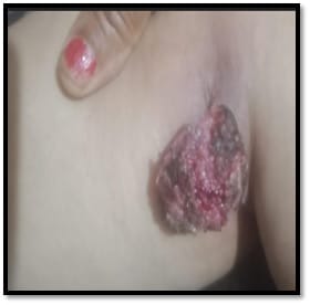
We hereby present a rare case of rhinosporidiosis in the popliteal fossa. Rhinosporidiosis is a chronic granulomatous infection caused by the fungus-like organism Rhinosporidium seebrii most commonly in the nasal cavity, followed by ocular and cutaneous lesions. The following case is of a 45-year-old female who presented to the laboratory with a pedunculated mass which on histopathology revealed a typical sporangium with numerous endospores. The report provides an insight into the clinical suspicion that needs to be kept while handling such cases.

Keywords: Rhinosporidium seebrii, Pedunculated mass, Popliteal fossa

Corresponding Author How to Cite this Article To Browse
Saishruti, M.D., Department Microbiology, Dr Lalpathlabs, Hyderabad, Telangana, India.
Email:
Gupta R, Saishruti. A Rare Case of Rhinosporidiosis in Popliteal Fossa. Trop J Pathol Microbiol. 2021;7(3):116-118.
Available From
https://pathology.medresearch.in/index.php/jopm/article/view/516

Manuscript Received Review Round 1 Review Round 2 Review Round 3 Accepted
2021-02-27 2021-03-09 2021-04-15 2021-04-18 2021-04-30
Conflict of Interest Funding Ethical Approval Plagiarism X-checker Note
No Nil Yes 8%

© 2021 by Ranu Gupta, Saishruti and Published by Siddharth Health Research and Social Welfare Society. This is an Open Access article licensed under a Creative Commons Attribution 4.0 International License https://creativecommons.org/licenses/by/4.0/ unported [CC BY 4.0].

Introduction

Rhinosporidiosis is a chronic granulomatous disease of man characterised commonly by polyposis of the nasal cavity and other mucosal surfaces.[1]. The clinical manifestations of the disease may involve different organs of the body and present in forms such as: Nasal Rhinosporidiosis, Ocular Rhinosporidiosis, Cutaneous Rhinosporidiosis, Disseminated Rhinosporidiosis and Miscellaneous Rhinosporidiosis. The cutaneous manifestations in rhinosporidiosis are rare. There may be wart-like, papillomatous or sessile masses in areas adjoining the nose and face. There could also be subcutaneous scattered nodules, which ulcerate and are seen fungating over the skin.[2]. The mode of infection could either be direct inoculation to the local site or through a hematogenous route as extracutaneous lesions. The dissemination leads to painless, firm to hard, subcutaneous nodules that remain unattached to the skin. The tumour-like masses of bone resemble chondrosarcoma, patients show extensive bony destruction.[3]. Rhinosporidiosis is not a contagious disease, trauma can explain the direct spread of infection from one anatomical site to another. [4]. The popliteal fossa is a very rare site for the disease to occur and thus this paper aims to draw attention towards such rare presentations of the disease.

Case Report

The patient a 45year female who is a known case of Breast carcinoma and on chemotherapy presented to the clinician with a warty swelling in the popliteal fossa, which was painless and pedunculated. On examination, it was about the size of apricot seed. The swelling was ulcerated red to blackish, granular and bled on touch. The clinician suspected it to be a skin metastasis of breast cancer and a biopsy was thus taken for confirmation. After histopathological examination at the laboratory Rhinospirodiosis was confirmed and reported to the clinician and no evidence of metastatic lesion was found which was also informed. Anti-fungal therapy was thus suggested henceforth.

Gross- Pedunculated mass in Right popliteal fossa which was friable and non –bleeding to touch. (Figure1)

Histopathology – we received a biopsy specimen for histopathology, subsequently H & E stain was applied, which showed –giant cells and lymphocytic reaction around the mature sporangium.

The dark coloured chitinous walls of the sporangium were also visible. In stained sections, the epidermis was hyperplastic with edematous connective tissue containing sporangia, having hyaline wall and filled with endospores(sporangiospores). The sporangia which are only known phenotypic structures produced by this pathogen measured 200 micrometres with 300-400 sporangiospores or endospores. Ruptured and degenerating sporangia were also visible. (Figure 2)

patho_516_01.jpg

Figure 1

patho_516_02.jpg

Figure 2

Discussion

Rhinosporidiosis was first described by Seeber, occurs most commonly in the nasal mucosa, and


cutaneous presentation of the disease is very rare, the above case is one such occurrence and draws the attention of the clinician towards such rare presentations which could otherwise be mistaken as squamous cell carcinoma or metastatic carcinoma. Most of the were reported in Srilanka and India mainly from coastal areas. Risk factors include high temperature, warm climate, high humidity provide a good environment for spore formation.[1]. In the present case transmission was not known, however the patient belonged to a rural area of MP where agriculture is the main occupation. Other risk factors include contaminated water, soil and bathing in stagnant water. Diagnosis of rhinosporidiosis is mainly by microscopy.

As culture and serological techniques for confirmation of the disease are minimal other staining methods like PAS or Mayer’s mucicarmine can be used for the same.[5]. Treatment of Rhinosporidiosis is done by the first removal of the mass by electrocautery followed by dapsone 100 mg daily for 6 months. Bigger swellings can be surgically removed and followed by electrocautery to avoid undue bleeding. Antifungal agents usually do not play a, major role in curing the disease but therapy with dapsone arrests maturation of sporangia and promote fibrosis in the stroma when used as an adjunct to surgery thus clearing the disease thoroughly.[6].

Conclusion

Extra nasal Rhinosporidiosis although extremely rare should be a clinical suspicion in similar cases or other suspected skin metastatic lesions as it is very much treatable when diagnosed on time and has good prognostic outcomes unlike malignancies which should be ruled out with a thorough investigation. We aim to provide an insight into such a rare but possible diagnosis through this case study.

Reference

01. Bhargava S, Grover M, Maheshwari V. Rhinosporidiosis- intraoperative cytological diagnosis in an unsuspected lesion. Case Rep Pathol. 2012;101832. doi: 10.1155/2012/101832 [Crossref][PubMed][Google Scholar]

02. Kishan Prasad HL, Rao C, Girisha BS, Shetty V, Permi HS, Jayakumar M, Kiran HS. Subcutaneous rhinosporidiosis masquerading as soft tissue tumor- diagnosed by fine-needle aspiration cytology. Indian J Dermatol. 2015 Mar-Apr;60(2)215. doi: 10.4103/0019-5154.152606 [Crossref][PubMed][Google Scholar]

03. Salim T, Komu F. Varied Presentations of Cutaneous Rhinosporidiosis- A Report of Three Cases. Indian J Dermatol. 2016 Mar-Apr;61(2)209-12. doi: 10.4103/0019-5154.177750 [Crossref][PubMed][Google Scholar]

04. Putthia H, Manjunatha BS, Astekar M, Taufiq S. Palatal rhinosporidiosis- an unusual case report and review of the literature. J Korean Assoc Oral Maxillofac Surg. 2018 Dec;44(6)293-297. doi: 10.5125/jkaoms.2018.44.6.293 [Crossref][PubMed][Google Scholar]

05. Gangneux JP, Lortholary O, Cornely OA, Pagano L. 9th Trends in Medical Mycology Held on 11-14 October 2019, Nice, France, Organized under the Auspices of EORTC-IDG and ECMM. J Fungi (Basel). 2019 Oct 8;5(4)95. doi: 10.3390/jof5040095 [Crossref][PubMed][Google Scholar]

06. Das C, Das SK, Chatterjee P, Bandyopadhyay SN. Series of Atypical Rhinosporidiosis- Our Experience in Western Part of West Bengal. Indian J Otolaryngol Head Neck Surg. 2019 Nov;71(Suppl 3)1863-1870. doi: 10.1007/s12070-018-1270-2 [Crossref][PubMed][Google Scholar]