E-ISSN:2456-1487
P-ISSN:2456-9887
RNI:MPENG/2017/70771

Case Report

Ureters

Tropical Journal of Pathology and Microbiology

2020 Volume 6 Number 4 April
Publisherwww.medresearch.in

Squamous cell carcinoma of the renal pelvis with an associated double ureters-rare case report

Sathiah P.1*, Sarada V.2
DOI: https://doi.org/10.17511/jopm.2020.i04.12

1* Prasath Sathiah, Assistant Professor, PDF Nephropathology, Trichy SRM Medical College Hospital and Research Centre, Trichy, Tamil Nadu, India.

2 Sarada V, Professor and HOD, MD Pathology, Department of Pathology, Trichy SRM Medical College Hospital and Research Centre, Trichy, Tamil Nadu, India.

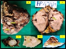
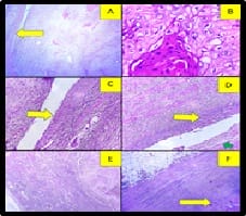
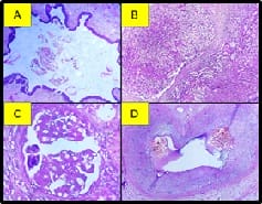
Squamous cell carcinoma is rare in the renal pelvis and kidney than in the lower urinary tract. Mostly it is associated with renal stone. Here a case report of a 72-year-old male who presented with renal mass was reported. Radical nephrectomy was done, shows a tumor involves the middle pole and extends into the renal pelvis. The hilar surface shows the presence of double ureters. Histopathological examination showed a well-differentiated squamous cell carcinoma in the renal pelvis and one of the ureter. Tumor involves more than 50% of the renal parenchyma. There is also squamous metaplasia of lining epithelium of smaller length ureter seen. No evidence of urothelial dysplasia noted in the renal pelvis and both the ureters. Adjacent renal parenchyma, glomeruli show focal segmental glomerulosclerosis with mild Interstitial fibrosis and tubular atrophy(IFTA). Medium size vessels show intimal thickening and mucoid change. The final diagnosis of squamous cell carcinoma of the renal pelvis and smaller ureter infiltrating renal parenchyma, possibly secondary to the risk factor of the double ureter.

Keywords: Urothelial carcinoma, Calculi, Metaplasia

Corresponding Author How to Cite this Article To Browse
Prasath Sathiah, Assistant Professor, PDF Nephropathology, Trichy SRM Medical College Hospital and Research Centre, Trichy, Tamil Nadu, India.
Email:
Sathiah P, Sarada V. Squamous cell carcinoma of the renal pelvis with an associated double ureters-rare case report. Trop J Pathol Microbiol. 2020;6(4):336-340.
Available From
https://pathology.medresearch.in/index.php/jopm/article/view/451

Introduction

Urothelial carcinoma is the most common renal tumor in the renal pelvis representing 90% of renal tumors [1]. Urothelial carcinoma may show a variable amount of squamous cell differentiation. However, Primary squamous cell carcinoma (SCC) of the renal pelvis is a rare malignancy accounting for 0.5 to 15% of

all urothelial carcinoma [2]. It occurs commonly in the older age group [3]. There are many risk factors for this tumor. One of the most common risk factors is a stone disease. Due to stone disease common in older male patients, the development of carcinoma also common in this group. Apart from this, many risk factors are there for squamous cell carcinoma of the renal pelvis.


Manuscript Received Review Round 1 Review Round 2 Review Round 3 Accepted
07-04-2020 17-04-2020 23-04-2020 23-04-2020
Conflict of Interest Funding Ethical Approval Plagiarism X-checker Note
No Nil Yes 14%

© 2020 by Prasath Sathiah, Sarada V and Published by Siddharth Health Research and Social Welfare Society. This is an Open Access article licensed under a Creative Commons Attribution 4.0 International License https://creativecommons.org/licenses/by/4.0/ unported [CC BY 4.0].

Here the current case reportis presenting the case of SCC of renal pelvis and ureter associated with the double ureter.

Case Report

A 72-year-old male with a complaint of lower abdominal pain and distension for 3 months. He also had a history of dysuria and a decreased urine output. Urine examination was done which shows 2+ proteinuria and presence of pus cells(8/10hpf), RBC( 7/10hpf), and granular cast. Peripheral smear examination shows normocytic normochromic anemia (9g/dl) with neutrophilicleucocytosis. CT scan was done which shows a mass lesion in the middle and upper pole of the kidney, also involves renal pelvis. Besides, it shows the presence of double ureters.

Gross examination: Radical nephrectomy was done. On serial section shows a mass predominantly in the middle pole of the kidney and extends into the renal pelvis. This mass is a grey-white firm in consistency with areas of necrosis. The hilar surface shows the presence of 2 ureters measuring 4 cm and 0.5 cm in size respectively. The smaller ureter was dilated and the lumen is filled with grey-white firm mass. Tumor involves the cortex. But overlying renal capsule and perinephric fat are free of tumor. The attached adrenal gland are free of tumor. (Figure 1)

patho_451_01.jpg

Fig-1: A. Radical nephrectomy specimen shows a bulge in the middle lateral aspect of the kidney and the presence of 2 ureters (arrows) in the hilar areas. The lumen of one of the ureters (smaller) is filled with a grey-white lesion (c).B. The cut surface of the kidney shows a tumor predominantly in the middle and also involves the upper pole. Hilarareas( arrow) is involved by tumor. D. Bits from the hilar areas show a dilated pelvicalyceal system filled with a grey-white lesion. E. Bits from the adrenal shows no evidence of tumor infiltration.

Microscopic examination: Section from the renal pelvis and smaller ureter showed tumor occupying the lumen extend into underlying muscularispropria and per pelvic fat. These tumor cells are arranged

in nests having a polygonal shape with vesicular nuclei and prominent nucleoli exhibiting moderate anaplasia. There is also prominent keratin pearls formation seen. This tumor also infiltrates into adjacent renal parenchyma. Adjacent renal parenchyma shows focal segmental glomerulosclerosis with mild chronic interstitial lymphocytic inflammation(10%) and mild Interstitial fibrosis and tubular atrophy(IFTA-15%). Medium size vessels show intimal thickening and mucoid change. Section from the adjacent smaller ureter showed the squamous metaplasia of lining urothelium. However, no urothelial carcinoma in situ or urothelial carcinoma component was identified in the multiple sections studied from the tumor. Section from the other larger ureter shows normal histology (Figure 2, 3).

patho_451_02.jpg

Fig-2: A. Section from the smaller ureter shows a tumor in the lumen of the ureter, arrow pinpoints the wall of the ureter (H and E, 100x) and high power B. shows tumor cells exhibiting classical squamoid morphology (H and E, 400x) and C. show squamous metaplasia in the lining epithelium (yellow arrow (H and E,400x). D. shows a tumor in the pelvicalyceal system(green arrow) with squamous metaplasia of lining epithelium(yellow arrow) (H and E, 400x). E. The section shows tumor-infiltrating whole kidney parenchyma and extends up to the renal capsules(yellow arrow) (H and E, 100x). (F) andperinephric pad of fat is free of tumor (H and E, 100x).

patho_451_03.jpg

Fig-3: Section from the other longer ureter (A) and adrenal (B) are unremarkable, free of tumor (H and E, 100x). C. section from the adjacent kidney parenchyma, glomeruli shows focal segmental glomerulosclerosis(H and E, 400x) and D. blood


vessels show severe intimal thickening and mucoid change(H and E, 400x).

The final diagnosis of squamous cell carcinoma well-differentiated of the renal pelvis and first part of the ureter with extension into adjacent renal parenchyma was given.

Discussion

Squamous cell carcinoma of the renal pelvis is the rare tumor, the most common age group is 50–70 years, similar to the present case and occurs frequently in female patients[3]. Due to the higher prevalence of the stone disease in men now it is seen more commonly occurs in late adulthood and males [4]. The present case is male patient, however not having any stone. Tumor of the renal pelvis and renal parenchyma caused by chronic irritation and inflammation [5]. Although it can occur without any etiological factors [3]. Most of the patients with renal pelvic tumors are associated with the following conditions; chronic infection, long term exposure to pelvicalyceal calculi, endogenous and exogenous chemical, hormonal deficiency, radiotherapy, vitamin deficiency, analgesic abuse and chronic pyelonephritis leading to severe hydronephrosis and pyonephrosis [6-8]. Most of the patients with renal tumors are associated with renal stone and also showed mild hydroureteronephrosis due to dilated ureter which may or may not be a risk factor [9]. Our patient has double ureters which may be the reason for the chronic irritation which leads to carcinoma is long-standing. According to the review done by Chen KSet al, urothelial cancer is the most common tumor associated double ureter [10]. Chronic irritation of urothelium caused by the above-mentioned factors can lead to squamous metaplasia of pelvic urothelium which leading to dedifferentiation, dysplasias, and in the end, into SCC [11,12].The present case also shows squamous metaplasia in the lining urothelium. Patients with SCC renal origin are presented with dull abdominal pain. Fever and leucocytosis occur may be paraneoplastic, secondary to cytokine storm, and severe systemic inflammatory reaction [6]. The present case shows neutrophilic leukocytosis along with normocytic normochromic anemia. If the tumor contains keratinized areas urine examination shows the presence of keratin material [13]. No keratin material was identified in the present caseof urine examination. To call it a renal parenchymal tumor, the renal pelvis should be histologically normal. The grossly renal pelvic tumor tends to be present as

sessile, ulcerated, and infiltrative growth [14]. The present case renal pelvis show infiltrative growth occupy the renal pelvis and ureter and infiltrative into renal parenchyma. Lee et al classified the SCC of the upper urinary tract into central and peripheral types based on tumor location: Central type of tumor is associated with a higher rate of nodal metastasis and lower survival rate and peripheral type of tumor are having parenchymal thickening and adjacent perirenal fat infiltration [15]. In the present case tumor predominantly in the central part. But no lymphovascular invasion and no lymph nodes were included in the specimen sent. Most of the time renal pelvic SCC limited to renal pelvis or shows infiltration into less than half of the kidney due to delay in diagnosis. The involvement of the whole parenchyma of the kidney is unusual but rarely reported [14]. The present case showed involvement of more than half (75%) renal parenchyma which is rare. It is not possible to screen all the patient with renal stone. Screening is necessary for patients with long-standing stone disease associated with poor renal function and hematuria [16]. SCC of renal pelvis appears as an extraluminal and exophytic mass with calcification which leads to hydronephrosis or as the pelvic infiltrative lesion [15]. Radiological differential diagnosis includes other secondary neoplasm and xanthogranulomatouspyelonephritis(XGP). XGP associated with lithiasis which leads to chronic obstruction than to hydronephrosis. besides rarely causes squamous metaplasia which leads to confusion of malignancy [11]. Lmbriaco et al reported the horseshoe kidney with renal stone associated with renal SCC [17]. Because of the non-specific clinical and radiological findings, histopathological examination needed for confirmation for the diagnosis of renal SCC [9]. The most common type of renal pelvic tumor is urothelial carcinoma. if any urothelial carcinoma component or urothelial dysplastic element along with urothelial carcinoma in situ, that tumor is reclassified into urothelial carcinoma with squamous differentiation. Though the rare presence of flattened urothelial keratinized squamous metaplasia adjacent to dysplastic element suggests the diagnosis of primary SCC of the renal pelvis [18].In the present case adjacent ureter showed the presence of squamous metaplasia. Hypertension present in 25- 60 % of renal tumour patients [19,20]. Bijol et al reported the mild to severe vascular changes with parenchymal involvement present in 37% of nephrectomy specimens done for


tumors [21]. Our patient also had intimal thickening, mucoid change, and occlusion of the vascular lumen(30%). Focal segmental glomerulosclerosis has been identified in 9% of tumor nephrectomy patients [21]. Most of the patients are associated with hypertension, arteriosclerosis, and pyelonephritis. Our patient had Focal segmental glomerulosclerosis – NOS with mild IFTA. Surgery is the mainstay of the treatment. Radical nephroureterectomy with excision of the bladder cuff is the treatment of choice for patients with the locoregional disease [22]. Adjuvant treatments like chemotherapy had minimal effect mainly on the treatment of the metastatic site. There is the possibility of anti-EGFR therapy for receptor-positive cases [22].

The prognosis of renal SCC is similar to urothelial cancer in stage-wise. The radiological finding is nonspecific, so the diagnosis of SCC is delayed and they present in an advanced stage with high-grade morphology, locally advanced or metastasis. So it is an aggressive tumor with a poor prognosis [23]. Holmany et al, reported the 5-year survival is under 10% and the average postoperative survival rate is up to 7 months [22].After surgery patient started on chemotherapy with cisplatin and methotrexate. Due to delays in diagnosis, and older age patient condition is not improving.

Conclusion

Although rare in the upper urinary tracts, SCC should be included in the differential diagnosis when evaluating a renal mass. The presence of double ureter may also be one of the risk factors for this tumor. So it is necessary to screen this patient often to prevent delay in diagnosis and start early treatment.

Author’s Contribution

Dr. Prasath Sathiah and Dr. Sarada Vhad an equal contribution in data collection, review of the literature, and preparation of the case report. Both the authors read and signed the final paper.

Reference

  1. Talwar N, Dargan P, Arora MP, Sharma A, Sen AK. Primary squamous cell carcinoma of the renal pelvis masquerading as pyonephrosis- a case report. Indian J Pathol Microbiol. 2006;49(3)418?420.
  1. Mills S, Hornick J, Longacre T, Greenson J and Reuter V. Sternberg's diagnostic surgical pathology. 6th ed, Philadelphia PA- Wolters Kluwer. 2015.
  2. Jain A, Mittal D, Jindal A, Solanki R, Khatri S, Parikh A, et al. Incidentally detected squamous cell carcinoma of renal pelvis in patients with staghorn calculi- case series with sa review of the literature. ISRN Oncol. 2011;620574.
  3. Somani BK, Nabi G, Thorpe P, Hussey J, Cook J. Is percutaneous drainage the new gold standard in the management of emphysematous pyelonephritis?- Evidence from a systematic review. J Urol. 2008;179(5)1844?1849.
  4. Nachiappan M, Litake MM, Paravatraj VG, Sharma N, Narasimhan A. Squamous Cell Carcinoma of the Renal Pelvis, A Rare Site for a Commonly Known Malignancy. J Clin Diagn Res. 2016;10(1)PD04?PD6.
  5. Jiang P, Wang C, Chen S, Li J, Xiang J, Xie L. Primary renal squamous cell carcinoma mimicking the renal cyst- a case report and review of the recent literature. BMC Urol. 2015;15;69.
  6. Nativ O, Reiman HM, Lieber MM, Zincke H. Treatment of primary squamous cell carcinoma of the upper urinary tract. Cancer. 1991;68(12)2575?2578.
  7. Huang WC, Levey AS, Serio AM, Snyder M, Vickers AJ, Raj GV, et al. Chronic kidney disease after nephrectomy in patients with renal cortical tumours- a retrospective cohort study. Lancet Oncol. 2006;7(9)735?740.

  1. Bandyopadhyay R, Biswas S, Nag D, Ghosh AK. Squamous cell carcinoma of the renal pelvis presenting as hydronephrosis. J Cancer Res Ther. 2010;6(4)537?539.
  2. Bhaijee F. Squamous cell carcinoma of the renal pelvis. Ann Diagn Pathol. 2012;16(2)124?127.
  3. Hippargi SB, Nerune SM, Kumar M. Urothelial and Squamous Cell Carcinoma of Renal Pelvis - A Rare Case Report. J Clin Diagn Res. 2016;10(9)ED19?ED20.
  4. Shirasaki Y, Tsushima T, Nasu Y, Kumon H. Long-term consequence of renal function following nephrectomy for renal cell cancer. Int J Urol. 2004;11(9)704?708.
  5. Chen KS, Chuang CK, Wu CH, Liaw CC, Lee N. Upper urinary tract tumor in a duplicated collecting system- report of three cases and review of the literature. Chang Gung Med J. 2003;26(5)377?382.
  6. Ghosh P, Saha K. Primary intraparenchymal squamous cell carcinoma of the kidney- a rare and unique entity. Case Rep Pathol. 2014;256813.
  7. Mardi K, Kaushal V, Sharma V. Rare coexistence of keratinizing squamous cell carcinoma with xanthogranulomatous pyelonephritis in the same kidney- report of two cases. J Cancer Res Ther. 2010;6(3)339?341.
  8. Bernardini S, Adessi GL, Billerey C, Chezy E, Carbillet JP, Bittard H. Immunohistochemical detection of p53 protein overexpression versus gene sequencing in urinary bladder carcinomas. J Urol. 1999;162(4)1496?1501.
  1. Salehipour M, Dastgheib N, Hosseinzadeh M, Makarem A, Rezvani A, Sanati A, et al. Primary renal pelvis and ureter squamous cell carcinoma (SCC)- a rare case report and review of literature. Int Med Case Rep J. 2019;12;189?192.
  2. Lee TY, Ko SF, Wan YL, Cheng YF, Yang BY, Huang DL, et al. Renal squamous cell carcinoma- CT findings and clinical significance. Abdom Imaging. 1998;23(2)203?208.
  3. Raghavendran M, Rastogi A, Dubey D, Chaudhary H, Kumar A, Srivastava A, et al. Stones associated renal pelvic malignancies. Indian J Cancer. 2003;40(3)108?112.
  4. Holmäng S, Lele SM, Johansson SL. Squamous cell carcinoma of the renal pelvis and ureter- incidence, symptoms, treatment and outcome. J Urol. 2007;178(1)51?56.
  5. Imbriaco M, Iodice D, Erra P, Terlizzi A, Carlo RD, Vito CD, et al. Squamous cell carcinoma within a horseshoe kidney with associated renal stones detected by computed tomography and magnetic resonance imaging. Urol. 2011;78(1)54?55.
  6. Tyagi N, Sharma S, Tyagi SP, Maheshwari V, Nath P, Ashraf SM, et al. A histomorphologic and ultrastructural study of the malignant tumours of the renal pelvis. J Postgrad Med. 1993;39(4)197?201.
  7. Bijol V, Mendez GP, Hurwitz S, Rennke HG, Nosé V. Evaluation of the nonneoplastic pathology in tumor nephrectomy specimens: predicting the risk of progressive renal failure. Am J Surg Pathol. 2006;30(5)575?584.