E-ISSN:2456-1487
P-ISSN:2456-9887
RNI:MPENG/2017/70771

Case Report

Paraganglioma

Tropical Journal of Pathology and Microbiology

2020 Volume 6 Number 4 April
Publisherwww.medresearch.in

Paraganglioma of the urinary bladder

Fatima S.1*, Mahdy O.2, Wajih S.S.3
DOI: https://doi.org/10.17511/jopm.2020.i04.11

1* Sohaila Fatima, Assistant Professor, Department of Pathology, King Khalid University, Abha, Saudi Arabia.

2 Omaia Mahdy, Specialist, Department of Laboratory Medicine, Aseer Central Hospital, Abha, Saudi Arabia.

3 Shiza Wajih Siddiqui, Medical Student, Dubai Medical College, United Arab Emirates.

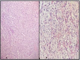
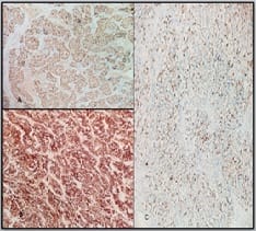
Paragangliomas are extra-adrenal pheochromocytomas. They are catecholamine-secreting neoplasms developing from the chromaffin cells derived from the ectodermic neural system. They are rare neoplasms that account for less than 0.06% of all urinary bladder tumors and 6% of all extra-adrenal pheochromocytomas. They arise from chromaffin tissue of the sympathetic nervous system within the layers of the bladder wall. They can be present in any part of the bladder with a predilection for the detrusor muscle with the most common sites being the dome and trigone.They may be nonfunctional or functional. The most common symptoms are hypertension and hematuria. Functional tumors usually present with symptoms of excessive catecholamine secretion. The patient typically suffers from hypertensive crises that may be accompanied by headache, palpitations, hot flushes, and sweating. Postmicturition hypotension and syncope are another common presentations. They share a genetic basis and can be related to a number of hereditary conditions.They are usually benign but there are no reliable morphologic criteria by which to separate the benign from the malignant forms. Here we present a 22 years old female with hematuria which on histopathology proved to be paraganglioma of the urinary bladder.

Keywords: Paragangliomas, Extra-adrenal pheochromocytomas, Urinary bladder

Corresponding Author How to Cite this Article To Browse
Sohaila Fatima, Assistant Professor, Department of Pathology, King Khalid University, Abha, Saudi Arabia.
Email:
Fatima S, Mahdy O, Siddiqui SW. Paraganglioma of the urinary bladder. Trop J Pathol Microbiol. 2020;6(4):333-335.
Available From
https://pathology.medresearch.in/index.php/jopm/article/view/449

Introduction

Paraganglioma is the generic term applied to nonepithelial tumors of paraganglion cells regardless of location[1]. They are extra-adrenal pheochromocytomas, catecholamine-secreting neoplasms developing from the chromaffin cells derived from the ectodermic neural system. It is a rare neoplasm as such and accounts for less than 0.06% of all uri-

nary bladder tumors. It arises from chromaffin tissue of the sympathetic nervous system within the layers of the bladder wall[2]. The most common presentations are hematuria and hypertension. Hypertension could only manifest at the time of the procedure. These could be mistaken clinically for urothelial carcinoma because of the rarity with histopathology being the diagnostic modality. We present a 22 years old female


Manuscript Received Review Round 1 Review Round 2 Review Round 3 Accepted
07-04-2020 17-04-2020 23-04-2020 28-04-2020
Conflict of Interest Funding Ethical Approval Plagiarism X-checker Note
No Nil Yes 17%

© 2020 by Sohaila Fatima, Omaia Mahdy, Shiza Wajih Siddiqui and Published by Siddharth Health Research and Social Welfare Society. This is an Open Access article licensed under a Creative Commons Attribution 4.0 International License https://creativecommons.org/licenses/by/4.0/ unported [CC BY 4.0].

with hematuria which on histopathology proved to be paraganglioma of the urinary bladder.

Case Report

A 22-year-old female presented 3 days post-delivery with hematuria of one-year duration. Physical examination was unremarkable and magnetic resonance imaging (MRI) of the abdomen which revealed fungating bladder mass implicating the right aspect of posterior uterine wall measuring 3.7x 3.7 cms with extravesical extension. It is encroaching upon the lower ureteric end with subsequent hydronephrotic hydroureteric changes. There is an obliteration of fat planes in between mass and cervix and anterior superior vaginal wall local spread with questionable invasion.There are bilateral enlarged dysmorphic iliac lymph nodes largest measuring 36x22mm. She underwent cystoscopy with fluctuating intraoperative blood pressure 200/108 mmHg and 90/60 mmHg. The hematoma evacuation alongwith transurethral resection of bladder tumor was performed whose histopathology revealed polygonal tumor cells arranged in trabecular, alveolar, and nesting(zellballen) arrangement with inconspicuous sustentacular cells and capillary network at the periphery. They invade the muscularis propria but without a desmoplastic reaction. Areas of hemorrhage present but no necrosis and mitosis are seen. Synaptophysin, chromogranin positive in chief cells, and S100 positivity were observed in sustentacular cells.A diagnosis of paraganglioma of urinary bladder was made.

patho_449_01.jpg

Fig-1A, B: Section showing polygonal tumor cells arranged in nesting pattern with sustentacular cells and capillary network at the periphery (Hematoxylin and Eosin:A- 10X, B-40X).

patho_449_02.jpg

Fig-2: Immunohistochemical study showing A. Chromogranin positivity in chief cells (chromogranin x 20X). B. Synaptophysin positivity in chief cells (synaptophysin x 20X) C. S100 positivity in sustentacular cells (S100 x 20X).

Discussion

Paragangliomas are extra-adrenal pheochromocytomas constituting only about 10% of cases. These may arise in the parasympathetic paraganglia (mostly of the head and neck) or the sympathetic paraganglia (found along the sympathetic nerve chain adjacent to the vertebral bodies in the abdomen, pelvis, and thorax and along sympathetic nerve fibers)[1]. Paraganglioma develops from chromaffin tissue of the sympathetic nervous system. 10% are localized in the bladder wall, accounting for 0.05% of all bladder tumors. They are usually solitary masses presenting in any part of the bladder and at any level of the bladder wall, with a predilection for the detrusor muscle with the most common sites being the dome and trigone[3]. They may be nonfunctional or functional. The most common symptoms are hypertension and hematuria. Functional tumors usually present with symptoms of excessive catecholamine secretion. The patient typically suffers from hypertensive crises that may be accompanied by headache, palpitations, hot flushes, and sweating. Postmicturition hypotension and syncope are another common presentations. Our patient presented with hematuria so the clinical diagnosis was bladder carcinoma. The median age for paraganglioma of UB was found to be much lower - 43 to 45 compared with 60 to 70 years for the patients with urothelial carcinoma with female predominance [4,5]. Paraganglioma shares a genetic basis and can be related to a number of hereditary conditions, includ­ing von Hippel-Lindau, neurofibromatosis type 1, Carney triad, multiple endocrine neoplasia types 2a and 2b, and SDHB(succinate dehydrogenase subunit B), SDHC, and SDHD mutations[6]. They are usually benign, but 15–20% of tumors may show malignant behavior. The only absolute criterion for malignancy is the presence of metastases to sites where chromaffin tissue is not usually found[7]. There are no reliable morphologic criteria by which to separate the benign from the malignant forms; therefore, risk assessment models have been proposed. The grading system for adrenal pheochromocytoma and paraganglioma (GaPP) may be applied to paragan-


gliomas for risk stratification. Increasing tumor size (>5 cm), SDHB mutations, MAX(myc-associated factor X) mutations, and increasing the Ki-67 proliferation rate are also risk factors for metastatic behavior[1]. Thompson proposed the pheochromocytoma of the adrenal scaled score (PASS) system which scores multiple microscopic findings. A PASS of <4 accurately identified all histologically and clinically benign tumors, and a PASS of ≥ 4 correctly identified all tumors that were histologically malignant [8]. Common sites of metastasis include bone, lymph nodes, lung, and liver[1].The patient had no family history and it was considered benign. The most useful imaging tech­niques to localize paragangliomas of the urinary bladder are computerized tomographic scan (CT) or magnetic resonance imaging (MRI). CT is the first choice of imaging howev­er, the sensitivity of CT for paraganglioma may be infe­rior to MRI [9,10]. Histologically differential diagnosis include urothelial carcinoma (nested variant), malignant melanoma, metastatic renal cell carcinoma, prostate carcinoma, carcinoid tumors, and granular cell tumor with confirmation of diagnosis by immunohistochemistry. The treatment of choice is surgical resection either transurethral resection of bladder tumor after control of concomitant hypertension or a partial cystectomy[5].

Conclusion

In conclusion, paraganglioma although rare could be a differential diagnosis of bladder tumors in patients presenting with hematuria. Functional tumors are easy to diagnose but those with silent presentation can create havoc during procedures by leading to marked fluctuations in blood pressure as seen in our case.

Reference

  1. McKenney JK. Bladder In- Goldblum, JR; Lamps, LW; McKenney, JK; Myers, JL, editors, Rosai and Ackermans surgical pathology. 11th ed, Mosby St Louis, Missouri. 2017;p-1066-1096.
  2. Thompson LD. Pheochromocytoma of the Adrenal gland Scaled Score (PASS) to separate benign from malignant neoplasms- a clinicopathologic and immunophenotypic study of 100 cases. Am J Surg Pathol. 2002;26(5)551-566.
  1. Leestma JE, Price EB Jr. Paraganglioma of the urinary bladder. Cancer. 1971;28(4)1063�1073.
  2. MochH , Humphrey PA, Ulbright TM , Reuter V. WHO Classification of Tumours of the Urinary System and Male Genital Organs. International Agency for Research on Cancer, Lyon, France. 2016.
  3. Elgazzar AH, Gelfand MJ, Washburn LC, Clark J, Nagaraj N, Cummings D, et al. I-123 MIBG scintigraphy in adults- A report of clinical experience. Clin Nucl Med. 1995;20(2)147-152.
  4. Hamrahian AH, Ioachimescu AG, Remer EM, Motta-Ramirez G, Bogabathina H, Levin HS, et al. Clinical utility of noncontrast computed tomography attenuation value (hounsfield units) to differentiate adrenal adenomas/hyperplasias from nonadenomas- Cleveland Clinic experience. J Clin Endocrinol Metab. 2005;90(2)871-877.
  5. Lenders JWM, Duh QY, Eisenhofer G,Gimenez-Roqueplo AP, Grebe SKG, Murad MH, et al. Pheochromocytoma and Paraganglioma- An Endocrine Society Clinical Practice Guideline. J Clin Endocrinol Metabol. 2014;99(6)1915-1942.
  6. Cheng L, Leibovich BC, Cheville JC, Ramnani DM, Sebo T J, Neumann RM, et al. Paraganglioma of the urinary bladder Can biologic potential be predicted?. Cancer. 2010;88(4)844-852.
  7. Menon S, Goyal P, Suryawanshi P,Tongaonkar H, Joshi A, Bakshi G, et al. Paraganglioma of the urinary bladder- A clinicopathologic spectrum of a series of 14 cases emphasizing diagnostic dilemmas. Indian J Pathol Microbiol. 2014;57(1)19-23.