E-ISSN:2456-1487
P-ISSN:2456-9887
RNI:MPENG/2017/70771

Research Article

Cell Block

Tropical Journal of Pathology and Microbiology

2020 Volume 6 Number 2 February
Publisherwww.medresearch.in

A comparative study of conventional cytology and cell block method with immunohistochemistry in the diagnosis of serous effusions

Datta P.1, Saha R.2*, Chakraborty J.3
DOI: https://doi.org/10.17511/jopm.2020.i02.06

1 Pratyush Datta, Postgraduate trainee, Department of Pathology, ESI-PGIMSR, Manicktala, Kolkata, West Bengal, India.

2* Rama Saha, Senior Resident, Department of Pathology, ESI-PGIMSR, Manicktala, Kolkata, West Bengal, India.

3 Jayati Chakraborty, Professor, Professor and Head, ESI-PGIMSR, Manicktala, Kolkata, West Bengal, India.

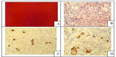
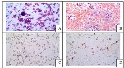
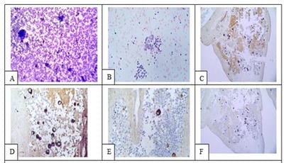
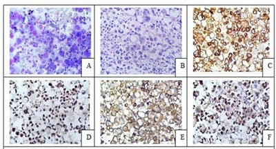
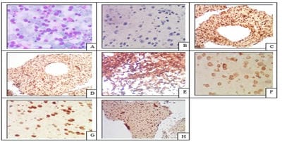
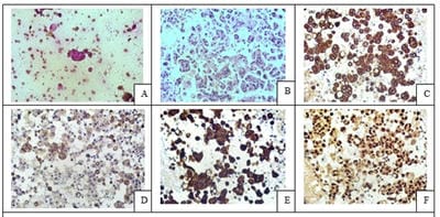
Introduction: Conventional smear (CS) examination of serous effusions is of paramount importance for diagnosis, staging, prognostication, and management of malignancy. The method has some disadvantages which can be overcome by cell block (CB) preparation. CB technique increases the diagnostic accuracy due to increased cellularity, preservation of tissue architecture and feasibility of performing immunohistochemistry (IHC). Aims and Objectives: To assess and compare the diagnostic yields of CS and CB techniques for detection of malignancy in pleural and peritoneal effusions and to study the utility of CB preparation with special emphasis on the feasibility of performing IHC in identifying the primary site of malignancy in the cases of carcinoma of unknown primary (CUP). Materials and Methods: In ESI-PGIMSR, Manicktala, each of 104 fluid samples were divided into two equal parts: one part was subjected to CS technique, the smears were stained with Leishman-Giemsa and Papanicolaou stains while the other part was subjected to Plasma thromboplastin CB technique and the sections stained by Hematoxylin and Eosin. IHC was performed whenever required. Provisional diagnoses made on CSs were compared with the diagnoses revised after examining CB slides. Results: Out of 104 fluid samples, on CS, 19 (18.27%) cases were positive for malignancy, whereas on CB 39(37.5%) cases were diagnosed as malignancy. The additional yield of malignancy was 19.23% more by the CB method. IHC done on CBs could suggest the possible primary site in 31 cases. Conclusions: The current study reports the diagnostic efficiency of the CB method to be superior to that of conventional smear alone as it has various advantages. Hence CB preparation should be routinely incorporated along with the use of IHC, if required, in the evaluation of serous effusions for a more accurate diagnosis.

Keywords: Cellblock, Conventional smear, Immunohistochemistry, Plasma Thromboplastin, Pleural and Peritoneal fluids

Corresponding Author How to Cite this Article To Browse
Rama Saha, Senior Resident, Department of Pathology, ESI-PGIMSR, Manicktala, Kolkata, West Bengal, India.
Email:
Datta P, Saha R, Chakraborty J. A comparative study of conventional cytology and cell block method with immunohistochemistry in the diagnosis of serous effusions. Trop J Pathol Microbiol. 2020;6(2):146-154.
Available From
https://pathology.medresearch.in/index.php/jopm/article/view/435

Manuscript Received Review Round 1 Review Round 2 Review Round 3 Accepted
06-01-2020 16-01-2020 24-01-2020 27-01-2020
Conflict of Interest Funding Ethical Approval Plagiarism X-checker Note
No Nil Yes 12%

© 2020 by Pratyush Datta, Rama Saha, Jayati Chakraborty and Published by Siddharth Health Research and Social Welfare Society. This is an Open Access article licensed under a Creative Commons Attribution 4.0 International License https://creativecommons.org/licenses/by/4.0/ unported [CC BY 4.0].

Introduction

The cytological examination of body fluids is of paramount importance not only for diagnosis but also for the staging, prognosis and further management of the patient [1]. Although the preparation of conventional smears (CSs) of effusions is a much simpler procedure than that of paraffin sections, it has a low sensitivity for detecting malignancy. This is attributed to lack of tissue architecture, overcrowding and overlapping of cells, cell loss, artifacts due to suboptimal processing and delaying, plenty of reactive mesothelial cells, paucity of representative cells, an abundance of inflammatory cells obscuring the morphology of atypical cells, subtle cytomorphological features of some malignant neoplasms and leaving behind useful material during processing [2,3,4,5]. In CS the accurate identification of cells as either malignant or reactive mesothelial cells with markedly atypical morphologic features remains a commonly encountered diagnostic challenge even to the experienced observer. The storage of CS slides is also a practical problem [6,7].

In contrast, cell block (CB), though it takes time, offers the following advantages. After completion of cytological preparations, the residual material often contains valuable diagnostic evidence including tissue fragments. It can be recovered by CB method thus providing additional information essential to resolve the diagnostic dilemmas. The additional benefits of CB technique are cell enrichment, lesser cellular dispersal, preservation of specific tissue architecture, better morphological details, the familiarity of the Haematoxylin and eosin (H and E) stain and feasibility of performing ancillary studies i.e, special histochemical stains and immunohistochemistry (IHC) [2,5,7]. A panel of specific IHC biomarkers can be carried out for establishing the lineage of the malignant cells, especially in a scenario of CUP [1,7,8,9,10,11]. Multiple sections of the same material can be taken for the same purpose which permits evaluation of a large number of antigens, or molecular studies [2, 5, 12, 13, 14]. IHC staining, when applied to the CB preparations, provides the same accuracy as do the histological specimens. [1,7,8]. It is particularly helpful to differentiate reactive mesothelial cells mimicking malignancy from malignant cells [15-22] CB technique is simple, safe, cost-effective and reproducible even in resource-limited settings [2, 23, 24]. Cases that are suspicious or equivocal on

the smears can be diagnosed definitively with the aid of a CB preparation [25]. The storage of slides and blocks of CBs for retrospective studies is easier compared to the smears.

This study was conducted to assess and compare the diagnostic yields of CS and CB techniques for detection of malignancy in pleural and peritoneal effusions and to study the utility of CB preparation with special emphasis on the feasibility of performing IHC in identifying the primary site of malignancy in the cases of carcinoma of unknown primary (CUP).

Materials and Methods

Setting: Department of Pathology, ESI-PGIMSR, Manicktala, Kolkata.

Duration: A period of 2 years from June 2017 to June 2019.

Type of study: Institution‑based prospective observational and analytical cross-sectional study.

Sampling methods: Consecutive 104 fluid samples matching the inclusion criteria were taken.

Sample size: A total of 104 body fluid samples were subjected to evaluation by both CS and CB methods.

Inclusion criteria: All the104 consecutive pleural and ascitic fluid samples received in the department during this period for the diagnostic purpose were included in this study.

Exclusion criteria: All other fluids except pleural and ascitic, clotted fluid specimens, specimens less than 10 ml and sub-optimally preserved fluids were excluded from the study.

Data collection procedure: Relevant information regarding age, sex, presenting clinical features, radiological and laboratory findings were recorded. Type of fluid (pleural or ascitic), smear findings, CB H and E and IHC findings and final diagnosis were assessed.

After routine cell count, cell type and biochemical examination, the fluid samples were homogenized and immediately processed for both CS and CB preparation. Provisional diagnoses made on CSs were compared with the diagnoses revised after examining H and E stained slides and IHC did on the CB preparation. Each of the10 ml of fresh fluid sample was divided into 2 equal parts. One part was subjected to the CS technique and the other part for


the CB technique. About 5 ml was centrifuged at 2500 rpm for 15 minutes. The supernatant was discarded and 2 routine smears were prepared from the sediment. One smear was air-dried and stained with Leishman Giemsa stain. The other smear was immediately fixed in 95% alcohol and stained with Papanicolaou stain. CBs were prepared by plasma–thromboplastin technique. The remaining sample was centrifuged at 2500 rpm for 15 minutes. After pouring off the supernatant, a cell pellet was obtained which was mixed with 250 microlitres of pooled plasma and 250 microlitres of uniplastin and allowed to stand for 2 minutes. The cell button thus obtained was scraped out and wrapped in a filter paper, placed in a cassette, fixed in 10% neutral buffered formalin and processed along with other routine histopathological specimens in automatic tissue processor and a CB was obtained. The CBs were embedded in paraffin, microtomy was done, sectioned at 4 µm thickness, stained by hematoxylin and eosin method, mounted with DPX mountant and examined after drying under the microscope. The suspicious specimens were subjected to IHC by using the standard Horseradish Peroxidase (HRP)- anti peroxidase technique on Poly-L Lysine coated slides. A comprehensive panel of immunomarkers were utilized in doubtful cases to distinguish atypical mesothelial cells from metastatic malignancies and then to categorize the type and the primary site of malignancy using immunomarkers that included panCK, EMA, CK7, CK20, Calretinin, WT-1, TTF-1, Napsin A, CDX2, CK19, ER, PR, PSA, CD45, CD20, CD3, Vimentin, and Synaptophysin, etc. wherever relevant.

Data analysis: The samples were categorized as benign, suspicious for malignancy or malignant. A comparative evaluation of the results of CS versus the CB techniques was conducted.

Statistical analysis was done by using SPSS software.

Ethical consideration and permission: This study was approved by the Institutional Ethics Committee.

Results

In the present study, a total of 104 serous fluid samples were processed both for CS and CB preparation. It had a predominance of pleural effusions (73.08%), followed by peritoneal effusions (26.92%). The age of the patients ranged from 25 to 82 years with a mean of 51.96 years. The

maximum number of samples was from the 61to 70 yrs age group while the least number of patients were in the age group of 11-20 years. Male patients outnumbered female patients (male: female ratio being 1.21:1). The malignant effusions were slightly more common in females than in males (the female-to-male ratio was 1.05:1). The most common malignancy was metastatic adenocarcinoma of the lung. In the case of malignant effusions, the primary origin of the tumor could not be arrived at just based on cytology. However, the architectural pattern of malignant cells in the CB preparation along with the panel of antibodies of IHC helped decide the probable primary origin of tumors in most of these cases. IHC was instrumental in diagnoses of the primary malignancy in cases of CUPs. Out of the total of 104 cases on cytological smears, 19(18.27%) cases were positive and 17(16.35%) cases were suspicious for malignancy, whereas on CB 39(37.5%) cases were diagnosed as malignancy. Out of the 17 samples reported as suspicious for malignancy by the CS method, 16(94.12%) were diagnosed as positive for malignancy and the other 01 was still suspicious even on the CB method due to scanty cellularity. By the CB method, 20 additional cases were detected as malignant, i.e, 19.23% more diagnostic yield for malignancy (Table 1).

Table-1: Analysis of CS and CB methods in a total of 104 fluid samples

Diagnostic category CS Method CB Method
Benign 68 (65.38%) 63 (60.58%)
Suspicious for malignancy 17 (16.35%) 02 (1.92%)
Malignant 19 (18.27%) 39 (37.5%)
Total 104 (100%) 104 (100%)

In 31 cases a possible primary site of origin could be indicated. Figure 1 represented only blood in PAP stained smear, but cell block prepared from it along with immunohistochemistry proves it to be a case of malignancy.

patho_435_01.jpg

Fig.1. A. Only blood in PAP stained smear, B. CB- H; E (40X), C. CK Positive, D. CK7 Positive.


Figure 2 represented a case of pleural effusion which showed scattered single atypical cells having abundant cytoplasm morphologically resembling mesothelial cells. But on CB followed by IHC, it was diagnosed as carcinoma lung.

patho_435_02.jpg

Fig.2. A case of malignant pleural effusion due to lung carcinoma A. PAP (40X), B. CB- H; E (40X), C. Pan CK Positive, D. CK7 Positive, E. Napsin A Positive, F. TTF-1 Positive.

Other primary sites of the tumor were breast in 3 cases (Figure 3), pancreaticobiliary in 1 case, ovary in 1 case, a gastrointestinal tract in 1 case.

In 1 case a few possible primaries could be suggested. One was a poorly differentiated malignant neoplasm, possibly sarcomatoid carcinoma and 5 cases had unknown primary. Carcinoma ovary accounts for most of the cases of malignant ascites (45.45%) followed by one case each of carcinoma of the GIT, lung, biliary and DLBCL. (Figure 4,5,6,7)

patho_435_03.jpg

Fig.3. Malignant pleural effusion in a known case of breast carcinoma A. PAP 10X, B. CB -H; E (40X), C. ER Positive, D. PR Positive.

patho_435_04.jpg

Fig. 4: A case of metastatic pancreatobiliary carcinoma A: LG 10X, B: CB- H; E (40X), C: CK19

Positive, D: CK7 Positive, E: CK20 Positive, F: CK Positive.

patho_435_05.jpg

Fig. 5. A case of metastatic ovarian serous carcinoma A. LG 40X, B. CB- H; E (40X), C. CK7

positive, D. PAX8 positive (diffuse strong), E. EMA positive, F. WT1 positive.

patho_435_06.jpg

Fig. 6. A case of malignant ascites caused by DLBCL. A. LG 40X, B. CB-H; E (40X), C. LCA

positive, D. CD 20 +, E. CD 79a +, F. Bcl2 +, G. MUM 1 positive, H.Ki67- 80%.

patho_435_07.jpg

Fig.7. Metastatic adenocarcinoma of GI origin A. LG 40X, B. CB-H and E (40X), C. Pan-CK Positive, D. CK7 focal scattered positive, E. CK20 Positive, F. CDX2 Positive.

Discussion

The current study studied 104 specimens among which pleural fluid was the commonest (76/104; 73.08%) followed by ascitic fluid (28/104; 26.92%). This is in concordance with Bhanvadia et al [26] who also reported pleural fluid (79/150; 53%) as the commonest of all effusions but in contrast with the result of the study conducted by Thapar et al [2] were the most common fluid was peritoneal (92/190) followed by pleural (88/190). In the present study, the most common age group was 61-70 years, accounting for 25% (26/104) of the cases which can be attributed to the increased incidence of malignancies in the elderly. But this finding contrasted with those found in the studies by Bansode et al [27] and Padmavathi et al,[28] who reported the most number of cases in the age group 41-60 years (54% and 69.3% respectively). In the


present study, the mean age of cases of malignant effusion was found to be 57.9 years which was similar to most of the previous relevant studies. In this study, there was a male preponderance (54.8% of 104 cases), the male: female ratio being 1.21:1. Bansode et al [27] and Padmavathi et al [28] reported male: female ratio of 2.1:1 and1.4:1 in their respective studies. However, in the study by Khan et al, none of the patients were males [29]. Studies by Shivakumarswamy [12] and Fagere et al [30] also reported female preponderance. 45 (59.21%) male and 16 (57.14%) female cases were highest in number in pleural and ascitic fluid effusion category respectively. In the present study, there were 11 cases of malignant peritoneal effusion, among which 8 were from women and 3 were from men, i.e., the male: female ratio is 1:2.67. There were 28 cases of malignant pleural effusion, among which 16 were from men and 12 were from female patients, the male: female ratio, in this case, is 1.33:1. Similarly, others [27,28] have also reported a higher number of peritoneal aspirates from females and more pleural aspirates from males which can be attributed to the different incidences of different malignancies in both sexes. The present study identified additional 19.23% (20 cases) malignancies by CB method when compared to the CS method. The diagnostic yield for malignancy was 18.27% (19/104) on CS examination which was increased to 37.5% (39/104) by CB technique. (Table 1) This is in agreement with findings obtained by the studies done by Dekker et al (38%), [1] Thapar M et al (13%), [2] Shivakumarswamy et al (15%), [12] Bansode et al (6.33%), [27] Padmavathi et al (1.47%), [28] Khan et al (20%), [29] Richardson et al (12%), [31] Liu et al (12%), [32] Shukla et al. (15%), [33] Shivakumarswamy et al (13.63%), [34] Bodele et al (7%), [35] Poorana (5%), [36] Sujathan et al(2.35%), [37] and Nathani et al (5%) [38]. This is higher than the study by Bhanvadia et al where 18/34 (53%) of the malignant cases could be identified by CS alone [26]. Out of 150 cases studied by Archana et al [5], 39 (26%) were positive for malignancy by CB method, while by routine method only 29 samples were reported as positive for malignant cells. According to various studies, an additional diagnostic yield for malignancy was noted if the CS technique was supplemented by the CB method [8]. Compared to Thapar’s study our figures are similar for CBs, but much lower for smears. Among 17 specimens that were suspicious for malignancy on CS, 16 were

diagnosed as malignant and 1 case remained suspicious even on CBs due to scanty cellularity. By the CB method, the number of cases that were suspicious for malignancy was 02/104 (1.92%) cases and by CS analysis, 17/104 (16.35%) cases. But according to the study by Bhanvadia et al, it was 0% by CB method and 11% by CS analysis [26]. Supplementing CB was the use of a case-based panel of antibodies for IHC. This added expression of markers by IHC was instrumental in objectively defining the cell of origin of the malignant cell, which enabled us to clearly ascertain the primary malignancy in 31 cases of CUP. In 25/39 (64.1%) cases, it was possible to narrow down to a single primary malignancy. In 6/39 (15.38%) cases, IHC could suggest 2 or 3 possible primary malignancies. There were 8 cases (20.51%) where a definite primary malignancy could not be identified (Fig. 1) mostly because of low cellularity where the representative cells could not be identified in deeper sections for further IHC. Three cases were negative on CSs but diagnosed as positive for malignancy on CBs as it demonstrated preserved architectural patterns. In two of them, malignant cells were misinterpreted as reactive mesothelial cells on CS, which were correctly diagnosed on CB with the help of IHC. The best results for identifying the primary malignancy were for metastatic Serous Carcinoma Ovary and Adenocarcinoma Lung. Pomjanski N et al in his study also reported similar results in identifying primary malignancies [39]. In the case of pleural fluids, 28 samples (36.84%) were malignant and 48 samples (63.16%) were reactive effusions. The most common cause of malignant pleural effusion was adenocarcinoma of the lung in males as well as in females (53.57%) (15/28 cases). Shivakumarswamy et al [34] noted pericellular lacunae in more than 60% of the cases of adenocarcinoma, characterized by large cell clusters that were absent in this case. Similar to the current study, the most common primary neoplasm causing pleural effusion was carcinoma of the lung in the studies by Nair and Manjula [40] and Gaur DS et al [41] followed by carcinoma of the breast in the former. Khan et al showed carcinoma lung was the most common site of malignant effusion followed by carcinoma ovary and GIT. [29] Sears and Hajdu [42] have reported the most common primary neoplasm causing pleural effusions as carcinoma of the breast (24%), followed by carcinoma of the lung (19%), and malignancies of the lymphoreticular system (16%). Murphy and Ng described the most


common primary lesions were in breast followed by lung and ovary [43]. In 20.51% of cases, though adenocarcinoma was confirmed, the site of primary could not be identified. These results are consistent with Khan et al [29] who determined the primary site in 81.3 % of the serous fluids of unknown origin. Shivakumarswamy et al [12] studied 60 pleural fluid samples where 10 of these fluids were malignant and primary was not known in three of the cases. In a study by Kushwaha et al [9], out of 28 samples with malignancy, the primary site could be confirmed on cytology in 16 (57.14%) of cases while in the remaining 12 cases, the primary was not known. The predominant lesion detected in the various fluids was negative for malignancy 65 (62.5%); malignancy was detected in 39 (37.5%) cases. Out of the 65 cases of reactive effusions, 73.85% of cases were of pleural effusion followed by peritoneal (26.15%). Regarding ascitic fluids, 39.3% were malignant and 60.7% were reactive. Out of 28 ascitic effusions, 5 cases were of ovarian cancers. In the study of Monte SA et al most of the malignant neoplasm in ascitic fluids were also derived from adenocarcinoma of ovarian tumors [43]. In the study by Nair and Manjula,[40] most common primary malignancy in cases of malignant ascitic effusions were adenocarcinoma of the gastrointestinal tract (GIT) followed by carcinoma of the ovary. Common primary malignancies in cases of malignant ascitic effusions were carcinoma of the ovary (32%), carcinoma of the breast (15%), and lymphoreticular malignancies (7%) in the study by Sears and Hajdu [42]. In the present study, most of the malignant effusions were metastatic adenocarcinoma. Our results correlated with the studies done by Khan K et al,[29] Sears and Hajdu, [42] Foot et al [45, 46] and van de Molengraft et al [47]. The most common cause for malignant pleural effusion was adenocarcinoma lung and that of malignant ascites, serous ovarian carcinoma. This is in concordance with the studies by Ghosh et al [24], Bhanvadia et al [26] and Bonito et al [48]. The most common primary malignancy in the fluid samples was Lung Carcinoma (41%) in the present study; similar to the studies by Karki S et al [49], Bjorn et al [50]. Other malignancies included GI Carcinoma, Pancreatobiliary Carcinoma, Mucinous Carcinoma Ovary, Carcinoma Breast, Serous Carcinoma of ovary and DLBCL. Spieler and Gloor [51] stated that common primary lesions identified in their study were in breast, ovary, lung, and GIT. Shivakumarswamy et al [12] reported that common primary lesions in their study were in the lung and

then in GIT. Limitations of the present study are that only pleural and ascitic fluids were included, not any other type of effusion fluids and the limited spectrum of malignancies seen. Also, observation could have been more representative and more statistically significant if the number of cases was more.

Conclusion

The present study concludes that the cell block technique, when used as an adjunct to conventional smear examination, has superior diagnostic yield due to better preservation of the architectural pattern and better morphological features of the cell clusters, which is particularly important in those cases fraught with the diagnostic dilemma between reactive changes and malignancy. There is a limited area of cell dispersal leading to the concentration of the diagnostic material i.e., high cellularity and increased cell density. A number of sections and perform special stains and IHC can also be studied, if required, to identify the primary site of origin in the carcinoma of unknown primary. The blocks can be preserved for molecular pathology too.

What does the study add to the existing knowledge?

The current study reports the diagnostic efficiency of the cell block method to be superior to that of conventional smear alone. Hence cell block preparation should be routinely incorporated along with the use of IHC, if required, in the evaluation of serous effusions for a more accurate diagnosis.

Author’s contribution

All the authors, Dr. Pratyush Datta, Dr. Rama Saha, and Dr. Jayati Chakraborty contributed equally to the conduct of the study and in the preparation of the manuscript.

Acknowledgment

The authors hereby acknowledge the contribution of the clinicians and the technicians of our department.

Reference

  1. Price B, Ehya H, Lee J. Significance of pericellular lacunae in cell blocks of effusions. Acta Cytologica. 1992;36(3)333-337.

  1. Kung I, Yuen R, Chan J. Optimal formalin fixation and processing schedule of cell blocks from fine needle aspirates. Pathology. 1989;21(2)143-145.
  2. Mishra R, Sharma A, Goyal V, Goyal V, Thapar M. Critical analysis of cell block versus smear examination in effusions. J Cytol. 2009;26(2)60-64.
  3. Mezger J, Stotzer O, Schilli G, Bauer S, Wilmanns W. Identification of carcinoma cells in ascitic and pleural fluids Comparison of four panepithelial antigens with the carcinoembryonic antigen. Acta Cytologica. 1992;36(1)75-81.
  4. Grandhi B, Shanthi V, Rao NM, Reddy VC, Mohan K. The diagnostic utility of cell block as an adjunct to cytological smears. Int J Med Res Health Sci. 2014;3(2)278-284.
  5. Dekker A, Bupp P. Cytology of Serous Effusions- An Investigation into the Usefulness of Cell Blocks versus Smears. Am J Clin Pathol. 1978;70(6)855-860.
  6. Leung S, Bedard Y. Simple miniblock technique for cytology. Modern Patholology. 1993;6(5)630-632.
  7. Zito F, Gadaleta C, Salvatore C, Filático R, Labriola A, Marzullo A et al. A modified cell block technique for fine needle aspiration cytology. Acta Cytologica. 1995;39(1)93-99.
  8. Kushwaha R, Shashikala P, Hiremath S, Basavaraj H. Cells in pleural fluid and their value in differential diagnosis. J Cytol. 2008;25(4)138.
  9. Bancroft J, Gamble M. Theory and practice of histological techniques. Philadelphia- Churchill Livingstone. Elsevier. 2011.
  1. Krogerus L, Andersson L. A simple method for the preparation of paraffin-embedded cell blocks from fine needle aspirates, effusions and brushings. Acta Cytologica. 1998;32(4)585-587.
  2. Bahrenburg L. On the diagnostic results of the microscopical examination of the ascitic fluid in two cases of carcinoma involving the peritoneum. Cleveland Medical Gazette. 1896; 11;274-278.
  3. Shivakumarswamy U, Karigowdar M, Arakeri S, Yelikar B. Diagnostic utility of the cell block method versus the conventional smear study in pleural fluid cytology. J Cytol. 2012;29(1)11.
  4. Mason MR, Bedrossian CW, and Fahey CA. Value of immunocytochemistry in the study of malignant effusions. Diagn Cytopathol. 1987;3(3)215-221.
  5. Yang G, Wan L, Papellas J, Waisman J. Compact Cell Blocks. Acta Cytologica. 1998;42(3)703-706.
  6. Esteban J, Yokota S, Husain S, Battifora H. Immunocytochemical Profile of Benign and Carcinomatous Effusions- A Practical Approach to Difficult Diagnosis. Am J Clinic Pathol. 1990;94(6)698-705.
  7. Cibas E, Corson J, Pinkus G. The distinction of adenocarcinoma from malignant mesothelioma in cell blocks of effusions- The role of routine mucin histochemistry and immunohistochemical assessment of carcinoembryonic antigen, keratin proteins, epithelial membrane antigen, and milk fat globule-derived antigen. Human Pathology. 1987;18(1)67-74.
  8. Nance K, Silverman J. Immunocytochemical Panel for the Identification of Malignant Cells in Serous Effusions. Am J Clin Pathol. 1991;95(6)867-874.

  1. Gray W, Kocjan G. Diagnostic cytopathology[Edinburgh]- Churchill. Livingstone/Elsevier. 2010.
  2. Santwani P, Vachhani J. Analysis of Diagnostic Value of Cytological Smear Method Versus Cell Blocks Method in Body Fluid Cytology- Study of 150 Cases. Ethiop J Health Sci. 2014;24(2)125-131.
  3. Doglioni C, Dei A, Laurino L, Iuzzolino P, Chiarelli C, Celio M et al. Calretinin- A Novel Immunocytochemical Marker for Mesothelioma. Am J Surg Pathol. 1996;20(9)1037-1046.
  4. Ko E, Jhala N, Shultz J, Chhieng D. Use of a Panel of Markers in the Differential Diagnosis of Adenocarcinoma and Reactive Mesothelial Cells in Fluid Cytology. Am J Clin Pathol. 2001;116(5)709-715.
  5. Khan N, Sherwani R, Afroz N, Kapoor S. Cytodiagnosis of malignant effusion and determination of primary site. J Cytol. 2005;22(3)107‑108.
  6. Padmavathi A, B. V S, B A. A comparative study of fluid cytology with smear and cell block preparation. J Evid Based Med Healthc. 2016;3(65)3532-3535.
  7. Kulkarni M, Desai S, Ajit D, Chinoy R. Utility of the thromboplastin-plasma cell-block technique for fine-needle aspiration and serous effusions. Diagnos Cytopathol. 2009;37(2)86-90.
  8. Keyhani-Rofagha S, Vesey-Shecket M. Diagnostic value, feasibility, and validity of preparing cell blocks from fluid-based gynecologic cytology specimens. Cancer. 2002;96(4)1204-209.
  1. Barberis M, Faleri M, Veronese S, Casadio C, Viale G. Calretinin. Acta Cytologica. 1997;41(6)1757-1761.
  2. Bansode S, Kumbalkar D, Nayak S. Evaluation of Cell Block Technique in the Cytodiagnosis of Body Fluids. Int J Sci Res. 2015;4(7)87-94.
  3. Richardson H, Koss L, Simon T. An evaluation of the concomitant use of cytological and histocytological techniques in the recognition of cancer in exfoliated material from various sources. Cancer. 1955;8(5)948-950.
  4. Ghosh I, Dey S, Das A, Bhattacharjee D, Gangopadhyay S. Cell block cytology in pleural effusion. J Indian Med Assoc. 2012; 110(6)390-392.
  5. Fagere M. Diagnostic Utility of AgNORs Staining of Serous Effusion among Sudanese Patients. Int J Sci Tech. 2016;5(1)36-42.
  6. Liu K, Dodge R, Glasgow B, Layfield L. Fine-needle aspiration- Comparison of smear, cytospin, and cell block preparations in diagnostic and cost effectiveness. Diagnostic Cytopathol. 1998;19(1)70-74.
  7. Karki S, Jha A, Sayami G. The Role of Argyrophilic Nucleolar Organizer Region (AgNOR) Study in Cytological Evaluation of Fluids, Especially for Detection of Malignancy. Kathmandu Univ Med J. 2012;10(1)34-39.
  8. Sujathan K, Kannan S, Mathew A, Pillai K, Chandralekha B, Nair M. Cyto-diagnosis of serous effusions- A combined approach to morphological features in Papanicolaou and May-Grunwald Giemsa stained smears and a modified cell block technique. J Cytol. 2000;17(2)89-95.

  1. Sears D, Hajdu S. The cytologic diagnosis of malignant neoplasms in pleural and peritoneal effusions. Acta Cytologica. 1987;31(2)85-97.
  2. Monte S, Ehya H, Lang W. Positive effusion cytology as the initial presentation of malignancy. Acta Cytologica. 1987;31(4)448-452.
  3. Foot N. Identification of types and primary sites of metastatic tumors from exfoliated cells in serous fluids. Am J Pathol. 1954;30(4)661-677.
  4. Foot N. The identification of neoplastic cells in serous effusions; critical analysis of smears from 2,029 persons. Am J Pathol. 1956;32(5)961-977.
  5. Fred J, Molengraft V, Peter V. The interval between the diagnosis of malignancy and the development of effusions, with reference to the role of cytologic diagnosis. Acta Cytologica. 1988;32(2)183-187.
  6. Murphy W, Ng A. Determination of Primary Site by Examination of Cancer Cells in Body Fluids. Am J Clin Pathol. 1972;58(5)479-488.
  7. Risberg B. Flow cytometric immunophenotyping of serous effusions and peritoneal washings- comparison with immunocytochemistry and morphological findings. J Clin Pathol. 2000;53(7)513-517.
  8. Spieler P, Gloor F. Identification of types and primary sites of malignant tumors by examination of exfoliated tumor cells in serous fluids Comparison with the diagnostic accuracy on small histologic biopsies. Acta Cytologica. 1985;29(5)753-767.
  9. Bonito L, Falconieri G, Colautti I, Bonifacio D, Dudine S. The positive pleural effusion- A retrospective study of the cytopathologic diagnosis along with the autopsy conformation. Acta Cytologica. 1992;36(3)329-332.
  1. Shivakumarswamy U, Arakeril S, Karigowdar M, Yelikar B. The role of the cell block method in the diagnosis of malignant ascitic fluid effusions. J Clin Diagnos Res. 2012;6;1280–1283.
  2. Bodele A, Parate S, Wadadekar A, Bobhate S, Munshi M. Diagnostic utility of cell block preparation in reporting of fluid cytology. J Cytol. 2003;20(3)133-135.
  3. Poorana P. Cytological analysis of body fluids in conventional smear and cell block technique– Study of 120 cases. Int J Pharma Bio Sci. 2015;6(4)609-615.
  4. Shukla P, Kaur S, Gulwani H. Diagnostic utility of plasma thromboplastin cell block preparation in cytological evaluation of serous effusions. Int J Biomed Res. 2015;6(11)890-896.
  5. Nathani R, Hazari R, Patle Y, Gupta S. Comparative analysis of cavity effusions by cell blocks and smear examination. Int J Rec Trend Sci Technol. 2014;12(1)69-72.
  6. Pomjanski N, Juergen GH, Doganay P, Schmiemann V, Buckstegge B, Böcking, A. Immunocytochemical identification of carcinomas of unknown primary in serous effusions. Diagnos Cytopathol. 2005;33(5)309-315.
  7. Nair G, Manjula A. Comparative study of cell blocks and routine cytological smears of pleural and peritoneal fluids in suspected cases of malignancy. Indian J Pathol Oncol. 2015;2(2)61-68.
  8. Gaur D, Chauhan N, Kusum A, Harsh M, Talekar M, Kishore S et al. Pleural fluid analysis- role in diagnosing pleural malignancy. J Cytol. 2007;24(4)183.