E-ISSN:2456-1487
P-ISSN:2456-9887
RNI:MPENG/2017/70771

Research Article

Granulomatous

Tropical Journal of Pathology and Microbiology

2020 Volume 6 Number 4 April
Publisherwww.medresearch.in

Granulomatous lesions !! Expected guests in unexpected places

Recharla M.1, Ramu R.2, Bindu J. B.3, Murthy C. N.4
DOI: https://doi.org/10.17511/jopm.2020.i04.02

1 Madhuri Recharla, Resident, Department of Pathology, Basaveshwara Medical College and Hospital, Rajiv Gandhi University of Health Sciences, Chitradurga, Karnataka, India.

2* Ramu R., Associate Professor, Department of Pathology, Basaveshwara Medical College and Hospital, Rajiv Gandhi University of Health Sciences, Chitradurga, Karnataka, India.

3 Bindu B. J., Associate Professor, Department of Pathology, Basaveshwara Medical College and Hospital, Rajiv Gandhi University of Health Sciences, Chitradurga, Karnataka, India.

4 Narayana Murthy C., Professor, Department of Pathology, Basaveshwara Medical College and Hospital, Rajiv Gandhi University of Health Sciences, Chitradurga, Karnataka, India.

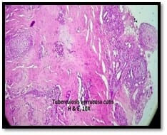
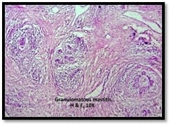
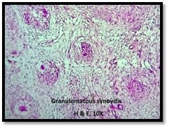
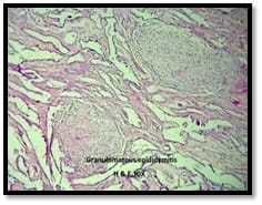
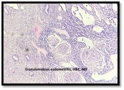
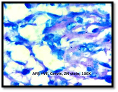
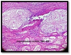
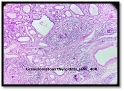
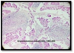
Introduction: Granulomatous lesions in various sites have different modes of presentation, different etiologic factors with identical histological patterns. The aim was to study the occurrence of granulomatous lesions at uncommon sites. Materials and methods: A retrospective study of histopathological specimens received over a period of one year from June 2017 to May 2018 was done and cases of granulomatous lesions of various sites reported on histopathological examination were reviewed along with Ziehl-Neelsen (ZN) and Fite-Faraco (FF) staining. Results: Out of total 3623 histopathology specimens, 61 (1.7%) were granulomatous lesions of which 28 (45.9%) were AFB (Acid-fast bacilli) positive. 20 cases (32.7%) were granulomatous lymphadenitis out of which 12 (19.6%) were positive for AFB. 4 (6.5%) out of 13 (21.3%) granulomatous skin lesions showed AFB positivity. 6 (9.8%) cases of granulomatous mastitis (1 was AFB +ve), 4(6.5%) cases of granulomatous synovitis (2 were AFB +ve), 3 (4.9%) cases of granulomatous endometritis (2 were AFB+ve) were seen. 2 (3.3%) cases each of granulomatous epididymitis and appendicitis were noted. Conclusion: In the present study tuberculosis was the most common cause of granulomatous lesions of various sites. Histopathology plays an important role in the diagnosis and management of a variety of granulomatous lesions. Special stains play a vital role in the diagnosis of infectious granulomatous lesions.

Keywords: Granulomatous lesions, Tuberculosis, Ziehl-Neelsen, Fite-Faraco

Corresponding Author How to Cite this Article To Browse
Ramu R., Associate Professor, Department of Pathology, Basaveshwara Medical College and Hospital, Rajiv Gandhi University of Health Sciences, Chitradurga, Karnataka, India.
Email:
Recharla M, Ramu R, Bindu BJ, Murthy CN. Granulomatous lesions !! Expected guests in unexpected places. Trop J Pathol Microbiol. 2020;6(4):308-317.
Available From
https://pathology.medresearch.in/index.php/jopm/article/view/430

Manuscript Received Review Round 1 Review Round 2 Review Round 3 Accepted
07-04-2020 17-04-2020 22-04-2020 28-04-2020
Conflict of Interest Funding Ethical Approval Plagiarism X-checker Note
No Nil Yes 15%

© 2020 by Madhuri Recharla, Ramu R., Bindu B. J., Narayana Murthy C. and Published by Siddharth Health Research and Social Welfare Society. This is an Open Access article licensed under a Creative Commons Attribution 4.0 International License https://creativecommons.org/licenses/by/4.0/ unported [CC BY 4.0].

Introduction

A granuloma can be defined as a focal compact collection of microscopic aggregation of macrophages transformed into epithelium like cells surrounded by lymphocytes and sometimes associated with central necrosis [1]. Granulomas usually form as a result of the persistence of a non-degradable product or as the result of hypersensitivity responses. It involves a complex interplay between invading organism or antigen, chemical, drug or other irritants, prolonged antigenemia, macrophage activity, a T helper (Th1) cell response, B cell overactivity, circulating immune complexes and a vast array of biological mediators [2].

It is formed as a protective mechanism when an acute inflammatory process cannot destroy invading agents [3]. Macrophages are attracted by areas of inflammation or immunological reactivity. These activated macrophages lead to increased expression of major histocompatibility complex (MHC) class II, CD4+, and Th 1 lymphocytes. Antigen-presenting cells (APC) bearing MHC class II molecules present the protein peptides to T helper cells. T cells activation also requires the B7:CD28/CTLA: 4 costimulatory pathway. With CD28 mediated costimulator, there is active T cell proliferation, without which there are anergy and apoptosis. Excess of Th1 relative to Th2 cells leads to cell-mediated hyperactivity, tissue destruction, and granuloma formation [2].

Several cytokines are produced by T cell subsets, macrophages, and other immune cells. Sub-classification of granulomas is done based on the amounts and types of these cytokines. They are broadly classified into two functional types, Thl and Th2. Thl- type cells produce interleukin 2 (IL- 2), interferon γ (IFN- γ), and tumor necrosis factor (TNF- β) upon stimulation with antigen and they participate in type IV hypersensitivity responses. Th2- type cells produce IL- 4, IL- 5, IL- 6, IL- 9, IL- I0, and IL- 13 cytokines that are important for B cell development and eosinophilia [3]. Macrophages are transformed into epithelioid cells and they fuse to form giant cells. Epithelioid granuloma is formed as an end result which progresses under the influence of transforming and platelet-derived growth factors (TGF and PDGF) to fibrosis [4,5,6].

Granulomatous inflammations are divided into various patterns like a foreign body, necrotizing, non- necrotizing granulomas, suppurative

granulomas, histiocytic response with no granulomas. The necrotizing and non-necrotizing granulomas are called immune granulomas. Necrotizing granulomas distinctly have central necrosis with a lymphohistiocytic reaction and a collar of chronic inflammation. The most common etiology of necrotizing granulomas is Mycobacteria species. Non-necrotizing granulomas are characterized by the collection of epithelioid histiocytes and giant cells with minimal peripheral chronic inflammation, the prototypical example being sarcoidosis [7].

The granulomatous lesions are classified based on infections, vasculitis, immunological aberrations, leucocyte oxidase defects, hypersensitivity pneumonitis, chemicals, neoplasia, and other miscellaneous infections. Infections are the most common cause of disseminated granulomatous disease. Granulomatous fungal infections mimic sarcoidosis, hence fungal infections need to be excluded and distinguished from sarcoidosis [2]. Other non – infectious causes that commonly induce granulomatous inflammations are sarcoidosis, foreign bodies, Wegener’s granulomatosis, and Crohn's disease [8].

In countries that have a high incidence of tuberculosis (TB), it is considered primarily in the differential diagnosis of granulomatous diseases [8]. Mycobacterium tuberculosis granulomas are classically characterized by the presence of caseous necrosis in the center that is referred to as a tubercle. Typically, there is a central area of amorphous caseating granular debris along with loss of cellular detail. This area is encircled by epithelioid cells, lymphocytes, histiocytes, fibroblasts, and occasionally Langhans' giant cells. Ziehl- Neelsen stain demonstrates acid-fast bacilli. These histologic features of the granuloma are characteristic and they allow the exact diagnosis of tuberculosis [3].

Inhalation of Mycobacterium bacilli initiates a histologic granulomatous response mediated by similar cytokine-mediated mechanisms within the lung as described. Chronic granulomatous response limits the extent of inflammation, activates histiocytes, and forms a collar of histiocytes and peripheral lymphocytes around a necrotic center called Ghon’s focus. Mycobacterium tuberculosis can affect any tissue by the direct inoculation or by the hematogenous spread such as miliary tuberculosis or as the localized cavitary form [7].

India has the world’s largest TB cases which are


around 26% of the world's tuberculosis cases [9]. The other mycobacterium (M) which can cause granulomatous reactions are M leprae, M. kansasii, M. marinum, M. gordonae, M. scrofulaceum, Mycobacterium avium-intracellulare complex (MAC), M. ulcerans, M. fortuitum, M. chelonae, and M. Abscessus [2].

Granulomatous lesions in various sites have different modes of presentation, different etiologic factors with identical histological patterns [10]. Tuberculosis and non-tuberculous mycobacterial infections have distinct clinical and histologic presentations but their clinical and radiographic appearance may have overlapping features with other diseases or even malignancy [7]. The commonly affected organ systems by granulomatous lesions are lungs, skin, kidney, liver, and lymph node [8]. The aim was to study the occurrence of granulomatous lesions at uncommon sites.

Materials and Methods

The study was conducted at the central research lab in a tertiary care center. It was a retrospective study done over a period of one year.

Universal sampling was done (All the histopathological specimens received in the department of pathology over a period of one year from June 2017 to May 2018 were included in the study).

Inclusion criteria: Cases of granulomatous lesions among all the histopathological specimens of various sites.

Exclusion criteria: Granulomatous lesions associated with any neoplasms were excluded.

Cases of granulomatous lesions of various sites reported on histopathological examination were reviewed along with Ziehl- Neelsen (ZN) and Fite-Faraco (FF) staining. Ziehl- Neelsen staining procedure is as follows:

  • Sections were kept in xylene for 2 minutes, dehydrated in absolute alcohol for 2 minutes.
  • Slides were stained with concentrated Carbol fuchsin for 10 minutes, heated intermittently, and washed with water.
  • Decolorised with 20% Sulphuric acid until the color was completely drained off.
  • Washed with water and counterstained with Loffler’s methylene blue for 30 seconds.
  • Water washed, air-dried, dehydrated, and mounted.

For Fite- Faraco (FF) staining 5% sulphuric acid was used for decolorizing.

Statistical analysis: Data was entered in the excel spreadsheet. Results were expressed as frequencies and percentages for comparison.

Ethical committee clearance was obtained from the institutional ethical committee.

Results

Out of total 3623 histopathology specimens, 61 (1.7%) were granulomatous lesions of which 28 (45.9%) were AFB (Acid-fast bacilli) positive (+ ve). The most common granulomatous lesion in the present study was granulomatous lymphadenitis accounting for 20 cases (32.7%), out of which 12 (19.6%) were positive for AFB. (Table 1 and Table 2)

Out of 13 granulomatous skin lesions, 2 (3.3%) each of tuberculosis verruca cutis (TVC), and granuloma annulare were noted, 3 (4.9%) were tuberculoid leprosy, 6 (9.8%) were borderline tuberculoid leprosy.

4 (6.5%) out of these 13 cases showed AFB positivity. Among them 3 were Fite- Faraco stain positive, 1 (1.6%) out of 2 (3.3%) tuberculosis verrucosa cutis was Ziehl- Neelsen stain positive.

6 (9.8%) cases of granulomatous mastitis (1 was AFB + ve), 4 (6.5%) cases of granulomatous synovitis (2 were AFB + ve), 3 (4.9%) cases of granulomatous endometritis (2 were AFB + ve) were seen. 2 (3.3%) cases each of granulomatous epididymitis and appendicitis were noted. AFB positivity was seen in 1 (1.6%) case of granulomatous epididymitis and 2 cases of appendicitis (3.3%).

1 (1.6%) case of HIV positive hysterectomy specimen showed granulomas in endometrium and cervix and was concurrently positive for AFB.

1 (1.6%) case each of granulomatous lesions was found at other uncommon sites like a salivary gland, thyroid, testis, parietal pleura, nasal septum, suture granuloma of the fallopian tube (FT), lipoma with foreign body granuloma (LFBG), sinus tract, intestine, and omentum. Acid-fast bacilli positivity was seen in each case of granulomatous lesions of a sinus tract, parietal pleura, and intestine.


Table-1: Relative frequencies of granulomatous lesions seen at different sites.

Granulomatous lesions Total no (n=61) n %
Lymph node 20 32.7
Skin 13 21.3
Breast 6 9.8
Synovium 4 6.5
Endometrium 3 4.9
Epididymis 2 3.3
Appendix 2 3.3
Salivary gland 1 1.6
Thyroid 1 1.6
Testis 1 1.6
Endometrium and Cervix 1 1.6
Parietal pleura 1 1.6
Sinus tract 1 1.6
Fallopian tube suture granulomas 1 1.6
Lipoma with foreign body granulomas 1 1.6
Intestine 1 1.6
Omentum 1 1.6
Nasal septum 1 1.6

Table 2: AFB positive cases.

  Total cases (61) Total AFB +ve cases (28) (45.9%)
Granulomatous lymphadenitis 20 12 (19.6%)
Skin lesions 13 3 (FF+) (4.9%) 1 (AFB+) (1.6%)
Granulomatous mastitis 6 1 (1.6%)
Granulomatous synovitis 4 2 (3.3%)
Granulomatous appendicitis 2 2 (3.3%)
Granulomatous endometritis 3 2 (3.3%)
Granulomatous endometritis and cervicitis 1 1 (1.6%)
Granulomatous lesion of sinus tract 1 1 (1.6%)
Granulomatous lesion- parietal pleura 1 1 (1.6%)
Granulomatous epididymitis 2 1 (1.6%)
Granulomatous lesion- Intestine 1 1 (1.6%)
Granulomatous sialadenitis 1 0
Granulomatous thyroiditis 1 0
Granulomatous orchitis 1 0
Suture granuloma of the fallopian tube 1 0
Lipoma with foreign body granulomas 1 0
Granulomatous lesion of omentum 1 0
Granulomatous lesion of nasal septum 1 0

patho_430_01.jpg

Fig-1: Tuberculosis verrucosa cutis, H and E, 10X.

patho_430_02.jpg

Fig-2: Borderline tuberculoid leprosy, H and E, 10X.

patho_430_03.jpg

Fig-3: Granulomatous mastitis, H and E, 10X.


patho_430_04.jpg

Fig-4: Granulomatous synovitis, H and E, 10X.

patho_430_05.jpg

Fig-5: Granulomatous epididymitis, H and E, 10X.

patho_430_06.jpg

Fig-6 a: Granulomatous endometritis, H and E, 10X.

patho_430_07.png

Fig-6 b: Granulomatous cervicitis, AFB +VE, ZN stain, 100X.

patho_430_08.jpg

Fig-7: Granulomatous orchitis, H and E, 40X.

patho_430_09.jpg

Fig-8: Granulomatous thyroiditis, AFB +VE, ZN stain, 100X.


patho_430_10.jpg

Fig-9: Granulomatous sialadenitis, H and E, 40X.

Discussion

Lymph node tuberculosis (LNTB) is one of the most common extrapulmonary manifestations of tuberculosis. It commonly involves the cervical lymph nodes followed by the mediastinal, axillary, mesenteric, hepatic portal, perihepatic, and inguinal lymph nodes. Tuberculous lymphadenitis most commonly develops from the reactivation of healed focus and from the progressive primary tuberculosis [11]. Out of 20 granulomatous lymphadenites, 12 were positive for acid-fast bacilli in the present study.

Out of 13 granulomatous skin lesions, 2 each of tuberculosis verruca cutis (TVC), and granuloma annulare were noted, 3 were tuberculoid leprosy (23.1%), 6 were borderline tuberculoid leprosy (46.1%). (Figure 1 and Figure 2)

Table-3: Comparison of various granulomatous skin lesions

  Skin lesions
Gautam et al (8) Present study
Tuberculoid/Boderline tuberculoid leprosy 47.6 % 69.2%
Tuberculosis verruca cutis 0.9% 15.4%
Granuloma annulare 3.7% 15.4%
Others 47.8% -

A study conducted by Gautam et al leprosy constituted the majority of granulomatous cases of skin which is in well concordance with the present study. Infections form an important cause of granulomatous skin lesions [10] (Table 3). Histologically, the tuberculoid and lepromatous forms have distinct appearances. Lepromatous leprosy is rich with dermal macrophages filled with bacilli, whereas the tuberculoid form is similar to the

granulomatous inflammation seen elsewhere with the presence of epithelioid histiocytes, multinucleated giant cells, and a lymphohistiocytic collar. Granulomas can persist for 18 months after treatment [7]. Granuloma annulare (GA) is a dermatosis most commonly presenting on the extremities of female patients as an isolated erythematous papule or arcuate plaque. GA may be seen in association with many autoimmune, neoplastic, and drug-induced states including sarcoidosis, Alagille’s syndrome, thyroid disease, hepatitis B and C, Epstein Barr virus (EBV), mycosis fungoides, Hodgkin and non-Hodgkin lymphomas (NHL), gastrointestinal stromal tumors (GIST), adenocarcinomas, allopurinol, amlodipine, and TNF-alpha inhibitors [7].

Breast tuberculosis is a rare disease that accounts for 3% of all surgically treated lesions in developing countries and is less common than carcinoma of the breast. It presents with non-specific clinical, radiological, and histological findings and closely looks like carcinoma of the breast and pyogenic inflammatory disease [12]. In the present study 6 (9.8%) cases of granulomatous mastitis were diagnosed and one case showed positivity for acid-fast bacilli. Other cases of granulomatous mastitis were found to have no significant clinical, radiological history towards any infectious etiologies and thus were considered to be idiopathic in nature. Granulomatous lesions in the breast include other infections like sarcoidosis, granulomatous reaction to the tumor, and foreign body reaction [12] (Figure 3).

According to a study done by Vijay PM, tuberculous synovitis was one of the commonest causes of inflammatory synovial lesions constituting 18.07% [13] and in the present study, 2 (3.3%) out of 4 granulomatous synovitis were positive for AFB (Figure 4). 30% of skeletal TB involves the joints and knee being the third most commonly affected joint after the spine and the hip. The involvement of the musculoskeletal system by tuberculosis is by hematogenous spread often from a primary focus in the lungs. As the bone and joint tuberculosis are paucibacillary, the Ziehl-Nielsen test may yield a negative result. In such cases Lowenstein culture would be an essential tool for the diagnosis of tubercular etiology [14].

The incidence of granulomatous inflammation in the appendix is around 1.3 to 2.3% in developing countries. Diagnostic criteria for granulomatous appendicitis are similar to those of the intestinal


tract which include granulomatous inflammation, lymphoid accumulation in all the layers of the intestine, and fissure type ulcers. Several infectious agents such as Yersinia, Mycobacterium, Blastomycosis, Schistosoma, Actinomyces, Campylobacter, and Histoplasma are responsible for causing granulomatous inflammation of the appendix. Granulomatous appendicitis is also caused by Crohn's disease and other conditions such as systemic sarcoidosis [15]. In the present study 2 cases of granulomatous appendicitis were seen and were positive for AFB.

Granulomatous reaction, particularly tuberculosis, is one of the frequent causes of chronic infectious epididymitis and has a subacute onset. It presents with epididymal swelling which may or may not be painful. Additional features include systemic symptoms, scrotal thickening, and fistula [16]. One (1.6%) out of two cases of granulomatous epididymitis showed acid-fast bacilli positivity in the present study (Figure 5). Other causes of granulomatous epididymitis include intravesical instillations of Bacillus Calmette– Guerin (BCG), Sarcoidosis, and brucellosis [16].

The present study, encountered 4 cases of granulomatous endometritis and one case was found to have coexisting human immunodeficiency virus (HIV) infection that showed caseating granulomas comprising of epithelioid cells, Langan's giant cells and lymphocytes in the endometrium and cervix, ZN staining demonstrated acid-fast bacilli (Figure 6a and Figure 6b). The other 3 granulomatous endometritis showed no acid-fast bacilli. The incidence of tuberculosis in countries with high HIV prevalence has been increasing. HIV targets CD4 T cells resulting in their depletion and dysfunction. Abnormal macrophage function may be seen because of direct infection and lack of macrophage activation factors produced by CD4 T cells promotes rapid progression to TB [17]. Hence testing for TB and HIV should be undertaken even at the slightest suspicion. Granulomatous orchitis is a rare condition, the precise etiology of which is not known. Clinically seminoma and granulomatous orchitis are difficult to distinguish [18]. An orchiectomy specimen showing the grey-white homogenous area which was suspected for malignancy showed chronic inflammatory cells, epithelioid cells, Langan's giant cells, and granulomas with no evidence of malignancy and was reported as granulomatous orchitis. ZN staining was negative for acid-fast bacilli.

The probable causes could be trauma, an autoimmune reaction to sperms, and urinary tract infection. The differential diagnosis of non-caseating granulomas mainly includes sarcoidosis. The granulomas in sarcoidosis are seen in the interstitium whereas in granulomatous orchitis they are confined to seminiferous tubules. Tuberculous infections primarily involve the epididymis [18]. The clinical and ultrasound findings of the disease often simulate malignancy and histopathology plays an important role in diagnosing granulomatous lesion [19] (Figure 7).

Abdominal TB is associated with significant morbidity and mortality because of its late diagnosis due to non-specific clinical symptoms. Approximately 15% - 25% of cases with abdominal TB have associated pulmonary TB. In the areas with high prevalence, a high index of suspicion is an important factor in early diagnosis. The most common site of gastrointestinal TB is the ileocecal region (ileocecal TB) followed by jejunum and colon. Oesophagus, stomach, and duodenum are rarely involved [10]. In the present study a case of granulomatous lesion of intestine was noted which was positive for AFB. Abdominal TB also involves other sites like peritoneum, lymph nodes, and solid viscera [10]. Crohn’s disease is the other commonest cause of granulomatous disease of the gastrointestinal tract [2].

One case (1.6%) of granulomatous thyroiditis was noted in the present study. The incidence of granulomatous thyroiditis is 0.16% of all primary thyroid diseases. Areas of thyroid tissue show intrafollicular cellular infiltration with partial or complete loss of colloid from infiltrated follicles and partial or complete distraction of their lining epithelium. This leads to granuloma formation along with fibrosis and inflammatory cell infiltration in the interstitium [20] (Figure 8).

One (1.6%) case of granulomatous lesion of the nasal septum was noted in the present study which showed negativity for ZN staining. Granulomatous diseases are rare causes of sinonasal inflammatory disease. They are categorized under inflammatory, infective, and neoplastic lesions [21]. In immunocompromised patients fungi may cause granulomatous lesion of the nasal septum. Inflammatory diseases that may affect the nasal septum include sarcoidosis, reparative granuloma, and granulomatosis with polyangiitis (Wegener granulomatosis) [22].


The present study had one (1.6%) case of granulomatous sialadenitis which was negative for AFB. The probable causes of granulomatous sialadenitis other than tuberculosis include mycosis, sarcoidosis, duct obstruction from calculi, or malignant tumors [23] (Figure 9). The present study was limited by additional investigations of diagnosis like nucleic acid amplification tests, a mycobacterial culture that would have prompted in diagnosing tubercular lesions in particular. Since the specimens were formalin-fixed mycobacterial culture will have less sensitivity.

Conclusion

In the present study tuberculosis was the most common cause of granulomatous lesions of various sites. Leprosy was the most common cause confined to the skin. Histopathology plays an important role in the diagnosis and management of a variety of granulomatous lesions. Histologic patterns (foreign body, necrotizing, non-necrotizing, suppurative, and a diffuse histiocytic reaction) can narrow the clinical differential diagnosis. Special stains play a vital role in the diagnosis of infectious granulomatous lesions.

What does the study add to the existing knowledge?

Infections are one of the important causes of granulomatous lesions and it was interesting to find the occurrence of granulomatous lesions at uncommon places as described. Granulomatous inflammation would indicate an effective macrophage and Th1 cell response to the antigenic stimulus involving cytokines and other mediators of inflammation.

Author’s contribution

All the authors; Dr. Madhuri Recharla, Dr. Ramu R., Dr. Bindu B. J., Dr. Narayana Murthy C. have made a substantial contribution to the work reported in the manuscript in terms of the concept, data analysis, manuscript preparation, and manuscript editing.

Reference

  1. Gautam K, Pai RR, Bhat S. Granulomatous lesions of the skin. J Pathol Nepal. 2011;1(2)81-86.
  1. James DG. What makes granulomas tick?. Thorax. 1991;46;734-736.
  2. Kumar V, Abbas AK, Aster JC. Robbins and Cortran pathologic basis of disease. 9th ed; New Delhi, Elsevier. 2016;p 97-99.
  3. Zumla A, James DG. Granulomatous infections- etiology and classification. Clin Infect Dis. 1996;23(1)146-156.
  4. James DG. Granuloma formation signifies a Th1 cell profile. Sarcoidosis. 1995;12(2)95-97.
  5. Roman J, Jeon YJ, Gal A, Perez RL. Distribution of extracellular matrices, matrix receptors, sand transforming growth factor-1 in human and experimental living granulomatous inflammation. Am J Med Sci. 1995;309(3)124-133.
  6. Shah KK, Pritt BS, Alexander MP. Histopathologic review of granulomatous inflammation. J Clin Tuberc Other Mycobact Dis. 2017;7;1-12.
  7. Guler M, Simsek A, Ofluglu R, Erguden HC, Capan N. Are all granulomatous lesions tuberculosis?. Resp Med Case Reports. 2012;5;42-44.
  8. Debi U, Ravisankar V, Prasad KK, Sinha SK, Sharma AK. Abdominal tuberculosis of the gastrointestinal tract- Revisited. World J Gastroenterol. 2014;20(40)14831-14840.
  9. James DG. A clinicopathological classification of granulomatous disorders. Postgrad Med J. 2000;76(898)457-465.

  1. Gandhare A, Mahashur A. Tuberculosis of the lymph nodes- many facets, many hues. Astrocyte. 2017;4(2)80-86.
  2. Sousa RD, Patil R. Breast tuberculosis or granulomatous mastitis- A diagnostic dilemma. Ann Trop Med PH. 2011;4(2)122-125.
  3. Vijay PM, Doddikoppad MM. Clinicopathological study of inflammatory synovial lesions. Int J Biol Med Res. 2011;2(4)882-888.
  4. Williams G, Sindwani R, Chandra R. Granulomatous diseases of sinonasal tract. Rhinology and Facial Plastic Surgery. 2009;321-333.
  5. Valencia MP, Castillo M. Congenital and acquired lesions of the nasal septum- A practical guide for differential diagnosis. Radiographics. 2008;28(1)205-223.
  6. Duggal S, Duggal N, Hans C, Mahajan RK. Female genital tuberculosis and HIV co-infection- case report. India J Med Micro. 2009;27(4)361-363.
  1. Goldblum JR, Lamps LW, Mckenney JK, Myers JL. Rosai and Ackerman’s surgical pathology. 11th ed, Philadelphia, Elsevier. 2018;p-236-237.
  2. Mah EL, Bux S. Tuberculous synovitis of the knee with unusually thick synovial granulation tissue- A Case Report. Internet J Ortho Surg. 2006;6(2)1-5.
  3. Dincel O, Goksu M, Turk BA, Pehlivanoglu B, Isler S. Incidental findings in routine histopathological examination of appendectomy specimens; retrospective analysis of 1970 patients. Indian J Surg. 2016;88(6)1-7.
  4. Cek M, Sturdza L, Pilatz A. Acute and chronic epididymitis. Europe Urol Suppl. 2017;16(4)124-131.
  5. Brunson J, Press HC, Hong IS. Granulomatous orchitis. J Nat Med Assoc. 2000;92(9)458-459.
  6. Gadgil NM, Ranadive NU, Sachdeva M. Granulomatous orchitis - a case report. Indian J Urol. 2001;18(1)92-94.
  7. Meachim G, Young MH. De Quervain's subacute granulomatous thyroiditis: histological identification and incidence. J Clin Path. 1963;16(3)189-199.