E-ISSN:2456-1487
P-ISSN:2456-9887
RNI:MPENG/2017/70771

Research Article

Biofilm

Tropical Journal of Pathology and Microbiology

2020 Volume 6 Number 2 February
Publisherwww.medresearch.in

Association of biofilm production in ESBL and MBL producing clinical isolates of Pseudomonas aeruginosa

Kulkarni D.M.1, Nilekar S.L.2, Vidhya T.3*
DOI: https://doi.org/10.17511/jopm.2020.i02.10

1 Kulkarni D.M., Associate Professor, Department of Microbiology, S.R.T.R Government Medical College, Ambajogai, Maharashtra, India.

2 Nilekar S.L., Professor and HOD, Department of Microbiology, S.R.T.R Government Medical College, Ambajogai, Maharashtra, India.

3* Vidhya T., Resident, Department of Microbiology, S.R.T.R Government Medical College, Ambajogai, Maharashtra, India.

Introduction: Pseudomonas aeruginosa is one of the most prevalent nosocomial pathogens that cause a life-threatening infection. One of the important characteristics of P. aeruginosa is biofilm formation and the most studied bacterium related to biofilm formation so far. The biofilm formation and beta-lactamases production synergistically contribute to the extensive dissemination of multi-drug resistant strains. Aim: The present study was conducted to identify, biofilm-producing isolates of P. aeruginosa along with their antibiotic resistance pattern and ESBL and MBL production and to analyze their antibiogram. Materials and methods: Various clinical specimens were collected and totally 82 clinical isolates of P. aeruginosa were included in this study. Biofilm producing isolates were identified by the tube adherence method. Results: Among the total, 22 [26.83%] isolates were biofilm producers and the maximum number was obtained from blood [100%], followed by ETT [75%], and Drain [66.67%]. Biofilm producing isolates were showing more resistance in comparison to non-biofilm producers. Conclusion: High-level resistance to antimicrobial agents is a characteristic feature of infection caused by biofilm and lead to chronic infections. Knowledge about these biofilm-producing isolates is important in the clinical setting to eradicate these chronic and life-threating infections.

Keywords: Antibiogram, Biofilm, ESBL, MBL, Pseudomonas aeruginosa

Corresponding Author How to Cite this Article To Browse
Vidhya T., Resident, Department of Microbiology, S.R.T.R Government Medical College, Ambajogai, Maharashtra, India.
Email:
Kulkarni DM, Nilekar SL, Vidhya T. Association of biofilm production in ESBL and MBL producing clinical isolates of Pseudomonas aeruginosa. Trop J Pathol Microbiol. 2020;6(2):174-180.
Available From
https://pathology.medresearch.in/index.php/jopm/article/view/423

Introduction

Pseudomonas aeruginosa is remarkably considered one of the most adaptive nosocomial pathogens [1]. Infections resulting from P. aeruginosa are frequently life-threa

tening and hard to treat causing levated stay in a medical institution or even accelerated morbidity and mortality as it exhibits intrinsically excessive resistance to many antimicrobials and the development of multi-drug resistance in health care settings [2].


Manuscript Received Review Round 1 Review Round 2 Review Round 3 Accepted
28-01-2020 09-02-2020 14-02-2020 19-02-2020
Conflict of Interest Funding Ethical Approval Plagiarism X-checker Note
No Nil Yes 14%

© 2020 by Kulkarni D.M., Nilekar S.L., Vidhya T. and Published by Siddharth Health Research and Social Welfare Society. This is an Open Access article licensed under a Creative Commons Attribution 4.0 International License https://creativecommons.org/licenses/by/4.0/ unported [CC BY 4.0].

Pseudomonas aeruginosa is a gram-negative, non-fermenting, obligately aerobic, the saprophytic bacterium which is widely distributed [3]. It is known for its intrinsic resistance to several antimicrobials, disinfectants, and tolerance to a wide range of physical conditions [4]. One of the important characteristics of P. aeruginosa is biofilm formation [1] and the most studied bacterium related to biofilm formation so far [5].

At present, biofilm is a serious worldwide concern due to its extracellular polymeric substances (EPS) which plays a vital role in antimicrobial resistance [6,7]. The biofilm-producing bacteria may show higher Minimal Bactericidal Concentration (MBC) and Minimal Inhibitory Concentration (MIC) of antibiotics up to 100–1000 fold than the planktonic form of bacteria [8].

Biofilm forming bacteria cause chronic persistent infection [8] and life-threatening device-associated infection. The various medical devices are shown to be colonized by biofilm [9] which leads to device-associated nosocomial infection.

This biofilm infection affects millions of people each year and many deaths occur as a consequence [10]. NIH publication showed more than 60% of all kinds of infections are related to the formation of biofilm [11].

In the present day, antibiotic resistance is an emerging problem and especially higher antibiotics like carbapenem are under the threat due to the widespread presence of carbapenemase (mainly Metallo Beta-lactamase). The biofilm formation and beta-lactamases (like ESBL and MBL) production synergistically contribute to the wide distribution of multi-drug resistant strains [12].

So the present study was conducted to identify, biofilm-producing isolates of P. aeruginosa along with their antibiotic resistance pattern and ESBL and MBL production. Also to find out the association between biofilm formation and drug resistance among P. aeruginosa in the hospital set up.

Materials and methods

Place of study: The study was carried in the Department of Microbiology at S.R.T.R Govt Medical College and Tertiary care center, Ambajogai. Maharashtra

Duration: From December 2018 to November 2019.

Type of Study: Observational study

Sampling methods- Specimens like Urine, Pus, Sputum, Drain, Blood, ETT (Endotracheal tube) were collected from patients admitted in various wards and ICUs. All specimens were cultured and identified by the standard conventional method [13]. Antimicrobial susceptibility testing (AST) was performed on Mueller-Hinton agar by Kirby Bauer's disc diffusion technique according to CLSI 2018 guidelines [14].

For ESBL and MBL detection- A Ceftazidime (CAZ 30µg) and Ceftazidime/clavulanic acid (CAZ/CA 30µg/10µg) disk were used to determine ESBL production. If there’s an increase of ≥ 5-mm in zone diameter for Ceftazidime/clavulanic acid compared to the diameter of the Ceftazidime, it is considered as ESBL producing isolates [14].

An imipenem (10 µg) and imipenem /EDTA (10 µg /750 µg) disk were used to determine MBL production. If there’s an increase of ≥ 7mm in zone diameter for imipenem – EDTA disc compared to zone diameter of imipenem disc, it was considered as MBL producing isolates [15].

Detection of Biofilm formation by Tube Adherence method- The isolated colony of P. aeruginosa was inoculated into a test tube contains trypticase soy broth (TSB) and incubated for 24 h at 35 °C. Next day content of the tube was discarded, and phosphate buffer saline was used to wash the tube and it was dried at room temperature. Then the tube was treated using 0.1% crystal violet for staining and then washed with water and dried. The presence of visible biofilm lining sidewall and the bottom of the tube was considered as biofilm producer [16].

Sample size: A total of 82 P. aeruginosa isolates identified were included in this study.

Data analysis: These study results were analyzed in SPSS version 16 software. Chai Square test was applied and p<0.05 was considered as significant.

Ethical consideration and permission: This study was reviewed and approved by the institutional ethical committee.

Results

A total of 82 isolates of Pseudomonas aeruginosa were recovered from various clinical specimens (Table 1).


Table-1: Distribution of Pseudomonas aeruginosa from various clinical specimens

Specimens No. (%)
Urine 29 (35.37%)
Pus 23 (28.04%)
Sputum 21 (25.61%)
ETT 4 (4.88%)
Drain 3 (3.66%)
Blood 2 (2.44%)
Total 82 (100%)

The rate of isolation of Pseudomonas aeruginosa from various clinical specimen has been shown in table 1. A maximum number of isolates were obtained from Urine 29 (35.37%), followed by Pus 23 (28.04%) and sputum 21 (25.61%). All 82 isolates were tested for biofilm production by tube method as mentioned above. Among the total 22 (26.83%) isolates were biofilm producers and 60 (73.17%) were biofilm non-producers. Most of the biofilm-producing isolates were identified from ICU with 14 (63.64%), as compared to ward 8 (36.36%).

Among the total of 22 biofilm-producing, P. aeruginosa isolates, the maximum number was recovered from blood specimen (100%), followed by ETT (75%), and Drain (66.67%) all from invasive sites (Table-2).

Table-2: Distribution of biofilm producer and non-biofilm producers in various clinical specimens

Specimens Biofilm producerN (%) Non-Biofilm producerN (%) Total
Urine 3 (10.34%) 26 (89.66%) 29 (100%)
Pus 7 (30.43%) 16 (69.57%) 23 (100%)
Sputum 5 (23.81%) 16 (76.19%) 21 (100%)
ETT 3 (75%) 1 (25%) 4 (100%)
Drain 2 (66.67%) 1 (33.33%) 3 (100%)
Blood 2 (100%) 0 2 (100%)
TOTAL 22 (26.83%) 60 (73.17%) 82 (100%)

Resistance to Ceftazidime (77% vs. 35%), Cefepime (77% vs.38%), Piperacillin-tazobactam (73% vs. 28%), Ciprofloxacin (68% vs. 22%), Gentamicin (59% vs. 27%), and Amikacin (32% vs. 8.3%) were higher among biofilm producing P. aeruginosa compared to non-biofilm producers (Statistically significant < 0.05) (Table 3).

Table-3: Antibiotic Resistance pattern of Pseudomonas aeruginosa in relation to biofilm production

Antibiotic Biofilm P-Value
ProducerN (%) (n=22) Non-ProducerN(%) (n=60)
Ceftazidime 17 (77.27%) 21 (35%) 0.000
Cefepime 17 (77.27%) 23 (38.33%) 0.002
Piperacillin-tazobactam 16 (72.72%) 17 (28.33%) 0.000
Gentamicin 13 (59.09%) 16 (26.6%) 0.009
Ciprofloxacin 15 (68.18%) 13 (21.6%) 0.000
Meropenem 3 (13.63%) 3 (5%) 0.23
Amikacin 7 (31.8%) 5 (8.3%) 0.015
Colistin 0 0 *
Polymyxin B 0 0 *

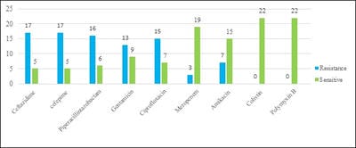
In the present study, biofilm-producing Pseudomonas aeruginosa showed high-level resistance i.e. 77%, 73%, 68%, 59% to an antipseudomonal cephalosporin (Ceftazidime and Cefepime), Piperacillin-tazobactam, Ciprofloxacin, Gentamicin, respectively. Somewhat lower resistance was observed to Amikacin (32%) and Meropenem (14%). All isolates of P. aeruginosa were sensitive to polymyxin B and colistin (Figure 1).

patho_423_01.jpg

Fig-1: Antibiotic susceptibility pattern of biofilm producer.

Table-4: Rate of ESBL and MBL production in relation to biofilm formation

  Biofilm producers (n=22) Non-Biofilm producers (n=60) p-Value
ESBL producer (n=32) 15(68.18%) 17(28.33%) 0.002
MBL producer (n=3) 2(9.09%) 1(1.69%) 0.19

Among 82 Pseudomonas aeruginosa isolates, 32(39.02%) were ESBL producers. Among non-biofilm producers 28.33% and among biofilm producers 68.18% were ESBL producers. THE maximum ESBL producer was biofilm positive with the statistically significant association (p-value


=0.002). Totally in the present study 3(3.65%), isolates were MBL producers among all. Among non-biofilm producers 1.69% and among biofilm producers 9.09% were MBL producers. Although MBL production was comparatively higher in biofilm producers it was not statistically significant (P = 0.19).

Discussion

P. aeruginosa causes the leading and life-threating nosocomial infections, ranking only second among the gram-negative pathogens [4]. As per the CDC statement, the P. aeruginosa infection rate was near about 0.4% in the US hospitals and 4th common nosocomial pathogen accounts for 10.1% of all hospital-acquired infections [17]. The main problem in treating P. aeruginosa infection is its high-level resistance to various antibiotics.

Studies show that infection by drug-resistant P. aeruginosa leads to increased length of hospital stay, morbidity, and mortality, and chronic infection [18]. The biofilm formation along with beta-lactamase production further complicates the scenario [12]. Production of an extracellular matrix is the hallmarks of a mature biofilm that acts as a barrier for any antibiotics and increases resistance to these antibiotics [19].

In the present study, among a total of 82 isolates of P. aeruginosa, 22(26.3%) were biofilm-producer and this finding is comparable with other studies which show (27.05%) [20], (32.3%) [21] and (33%) [22], but in contrast with others who showed higher rate of biofilm production (73.68%) [12] and (83.33%) [23]. This variation in the rate of isolation also may be due to sample size, type of specimen studied because medical devices were frequently colonized by biofilm-forming organisms and the various methods used for biofilm identification like Congo red agar method or Tissue culture plate which were showing a higher rate of detection.

The present study show, maximum biofilm-producing isolates recovered from specimens received from ICU (63.64%) compared to the ward (36.36%) and similar findings was shown in another study (83.3%)[2]. This could be possibly due to the ICU setup uses multiple medical devices for treatment and intervention of patient care although indwelling devices used widely in hospitals [24] and biofilm is known for colonizing these medical devices.

In the present study, the maximum rate of biofilm positive isolates was identified from the blood (100%) and this finding was similar to another study which showed that 100% sterile fluids isolates were biofilm producers [22]. A catheter might have inserted for several purposes and this can be colonized. After blood samples, the ETT showed biofilm formation in 75% isolates and this could be explained by the fact that more specimens were obtained from patients admitted in ICU who were either intubated or needing ventilator support [24]. It was found that 66.67%, 30.43%, and 23.81% biofilm-producing isolates were from the drain, pus, and sputum respectively. This finding supports the fact that biofilm development is aided by tissue lesions, chronic respiratory disease, implanted medical devices, surgical wounds, etc. [9,25].

The antibiotic susceptibility of biofilm-producing bacteria is reduced because of a restricted antibiotic penetration, adaptive response and the occurrence of persisting cells [2]. In the present study, high resistance was noted among biofilm producers to an antipseudomonal cephalosporin (Ceftazidime and Cefepime), Piperacillin-tazobactam, Ciprofloxacin, and Gentamicin with 77%, 73%, 68%, and 59% respectively. These findings were nearly matching with other studies [21,22]. This may be due to the widespread use of these easily available antibiotics without knowing the infection status. All isolates were susceptible to polymyxin-B and colistin like other studies [1,2,21].

In the present study, resistance to Ceftazidime, Cefepime, Piperacillin-tazobactam, Ciprofloxacin, Gentamicin, and Amikacin was comparatively higher in biofilm producer than a non-biofilm producer. The difference was statistically significant (p < 0.05). It is similar to findings from other studies for most of the antibiotics tested [12,21,22]. So Meropenem, colistin, and polymyxin-B remain the treatment of choice for biofilm-producing isolates. However, due to their high toxicity, polymyxin is used for the treatment of only serious infections.

In the present study, it was found that there is an association between ESBL production and biofilm formation (Statistically significant, p= 0.002). It was similar to another study [26] but the contrast study done by Dumaru et al [12]. No statistically significant association could be established between MBL production and biofilm production (Statistically insignificant, p=0.19) which was in agreement with another study [20].


The resistance to antimicrobials in biofilm-producer may be explained by the fact like, there are an increased plasmid transfer and gene transfer among biofilm bacteria which further intensifies the problem of drug resistance [27] and also by the fact that in the process of biofilm development, drug resistance varies bacterium to bacterium [28].

Conclusion

The present study showed, 26.3% isolates of P. aeruginosa were biofilm producers and also showed that there is an association between biofilm formation and drug resistance. The present study emphasizes the relationship between ESBL production and biofilm formation in P. aeruginosa. Identification of biofilm-producing isolates is important because it leads to treatment failure due to the high drug resistance to multiple antibiotics. Biofilm may be controlled by replacing the device that was colonized and also by taking care of the device or wound that was already existing to prevent biofilm formation.

Limitations- The lack of confirmation of biofilm, ESBL, and MBL production by using molecular technologies are the drawbacks of this study.

What does this study add to the existing knowledge?

The present study highlight’s the importance of performing the test for biofilm production in P. aeruginosa isolates. By knowing the resistance pattern of these isolates’ clinicians can able to choose the right empirical antibiotic in life-threatening conditions.

Author’s contribution

Dr. Kulkarni D.M: Data analysis, Manuscript writing

Dr. Nilekar S.L.: Study concept, data analysis

Dr. Vidhya T: Data collection, Manuscript writing

Reference

  1. Haji SH. Detection of Biofilm Formation in Pseudomonas aeruginosa Isolates from Clinical Specimens. Zanco J Pure Appl Sci. 2018; 30[4]83-89.
  1. Bose S, Khodke M. Detection Of Biofilm Producing Staphylococci- Need Of The Hour. J Clin Diagnostic Res. 2009;[3]1915-1920.
  2. Saha S, Devi KM, Damrolien S, Devi KS, K, Sharma KT. Biofilm production and its correlation with antibiotic resistance pattern among clinical isolates of Pseudomonas aeruginosa in a tertiary care hospital in north-east India. Int J Adv Med. 2018;5[4]964.
  3. Vallés J, Mariscal D, Cortés P, Coll P, Villagrá A, Díaz E, et al. Patterns of colonization by Pseudomonas aeruginosa in intubated patients- A 3-year prospective study of 1,607 isolates using pulsed-field gel electrophoresis with implications for prevention of ventilator-associated pneumonia. Intensive Care Med. 2004;30[9]1768-1775.
  4. Chong Y. Imipenem-EDTA disk method for differentiation of metallo-beta-lactamase-producing clinical isolates of Pseudomonas spp and Acinetobacter spp. J Clin Microbiol. 2002 Oct;40[10]3798–801.
  5. Gales AC, Jones RN, Turnidge J, Rennie R, Ramphal R. Characterization of Pseudomonas aeruginosa Isolates- Occurrence Rates, Antimicrobial Susceptibility Patterns, and Molecular Typing in the Global SENTRY Antimicrobial Surveillance Program, 1997–1999. Clin Infect Dis. 2001, 32[s2]S146–S155.
  6. Costerton JW. Cystic fibrosis pathogenesis and the role of biofilms in persistent infection. Trends in Microbiology. 2001 Vo,l 9 p 50-52. [PMID:11173226]
  7. Wayne P. CLSI, Performance Standards for Antimicrobial Susceptibility Testing, In- CLSI Supplement M100. 28th ed. 2018.

  1. Sharma D, Misba L, Khan AU. Antibiotics versus biofilm- An emerging battleground in microbial communities. Antimicrob Resist Infect Control. 2019;8[1]1–10.
  2. Gebreyohannes G, Nyerere A, Bii C, Sbhatu DB. Challenges of intervention, treatment, and antibiotic resistance of biofilm-forming microorganisms. Heliyon. 2019;5[8]:e02192.
  3. Høiby N, Ciofu O, Johansen HK, Song ZJ, Moser C, Jensen PØ, et al. The clinical impact of bacterial biofilms. Int J Oral Sci. 2011;3[2]55–65.
  4. Costerton JW, Stewart PS, Greenberg EP. Bacterial biofilms- A common cause of persistent infections. Science. Vol 284,1999; p-1318–1322.
  5. Ali, Syed Sajeed and Wakte PS. Isolation and identification of biofilm forming pseudomonas aeruginosa pseudomonas aeruginosa from wounds infection. Int J Curr Res. 2016;8[09]38974–7.
  6. Dumaru R, Baral R, Shrestha LB. Study of biofilm formation and antibiotic resistance pattern of gram-negative Bacilli among the clinical isolates at BPKIHS, Dharan. BMC Res Notes. 2019;12[1]1–6.
  7. Patricia M. Tille Traditional cultivation and identification, In- Baily and Scott’s Diagnostic microbiology. Canada- Elsevier, 14th ed. 2017; p- 86–112.
  8. Obritsch MD, Fish DN, MacLaren R, Jung R. Nosocomial infections due to multidrug-resistant Pseudomonas aeruginosa- Epidemiology and treatment options. Pharmacotherapy. 2005, Vol 25, p;1353–64.
  1. Yong D, Lee K, Yum JH, Shin HB, Rossolini GM, Christensen GD, Simpson WA, Younger JJ, Baddour LM, Barrett FF, Melton DM, et al. Adherence of coagulase-negative staphylococci to plastic tissue culture plates- A quantitative model for the adherence of staphylococci to medical devices. J Clin Microbiol. 1985;22[6]996–1006. PMID: 3905855
  2. Aloush V, Navon-Venezia S, Seigman-Igra Y, Cabili S, Carmeli Y. Multidrug-resistant Pseudomonas aeruginosa- Risk factors and clinical impact. Antimicrob Agents Chemother. 2006 Jan;50[1]43–8.
  3. Heydari S and Eftekhar F. Biofilm Formation and β-Lactamase Production in Burn Isolates of Pseudomonas aeruginosa. Jundishapur J Microbiol. 2015;8[[3]]:e15514.
  4. Revdiwala S, Rajdev BM, Mulla S. Characterization of bacterial etiologic agents of biofilm formation in medical devices in critical care setup. Crit Care Res Pract. 2012;1–6.
  5. Baniya B, Pant ND, Neupane S, Khatiwada S, Yadav UN, Bhandari N, et al. Biofilm and metallo beta-lactamase production among the strains of Pseudomonas aeruginosa and Acinetobacter spp at a tertiary care hospital in Kathmandu, Nepal. Ann Clin Microbiol Antimicrob. 2017;16[1]6–9.
  6. Shrestha R, Nayak N, Bhatta DR, Hamal D, Subramanya SH, Gokhale S. Drug Resistance and Biofilm Production among Pseudomonas aeruginosa Clinical Isolates in a Tertiary Care Hospital of Nepal. Nepal Med Coll J. 2019;21[2]110-116.
  7. Nepal et al. Is there correlation of biofilm formation with multidrug resistance and ESBL production in pseudomonas aeruginosa ?. Eur J Biomed Pharm Sci. 2017;4[01]366–72.

  1. Gurung J, Khyriem AB, Banik A, Lyngdoh WV, Choudhury B, Bhattacharyya P. Association of biofi lm production with multidrug resistance among clinical isolates of Acinetobacter baumannii and Pseudomonas aeruginosa from intensive care unit. Indian J Crit Care Med. 2013;17[4]214-218.
  2. Angel Díaz M, Ramón Hernández J, Martínez-Martínez L, Rodríguez-Baño J, Pascual A. Extended-spectrum beta-lactamase-producing Escherichia coli and Klebsiella pneumoniae in Spanish hospitals- 2nd multicenter study [GEIH-BLEE project, 2006]. Enferm Infecc Microbiol Clin. 2003;21[2]503-510.
  1. Rewatkar AR. Staphylococcus aureus and Pseudomonas aeruginosa- Biofilm formation Methods. IOSR J Pharm Biol Sci. 2013;8[5]36–40.
  2. Basak S, Rajurkar MN, Attal RO, Mallick SK. Biofilms- A Challenge to Medical Fraternity in Infection Control. Infection Control. 29th 2013.
  3. Singh S, Singh SK, Chowdhury I, Singh R. Understanding the Mechanism of Bacterial Biofilms Resistance to Antimicrobial Agents. Open Microbiol J. 2017;11[1]53–62.