E-ISSN:2456-1487
P-ISSN:2456-9887
RNI:MPENG/2017/70771

Research Article

Cervical

Tropical Journal of Pathology and Microbiology

2020 Volume 6 Number 3 March
Publisherwww.medresearch.in

A study of liquid-based cytology in cervical smears

Phani Meghana B.1, Venkata Renuka I.2, Ramya C.3*, Durga Prasad B.4, Sireesha K.5
DOI: https://doi.org/10.17511/jopm.2020.i03.05

1 Bolla Phani Meghana, Postgraduate, Department of Pathology, NRI Medical College and Hospital, Vijayawada, Andhra Pradesh, India.

2 Inuganti Venkata Renuka, Professor and Head of Department, Department of Pathology, NRI Medical College and Hospital, Vijayawada, Andhra Pradesh, India.

3* Chitturi Ramya, Assistant Professor, Department of Pathology, NRI Medical College and Hospital, Vijayawada, Andhra Pradesh, India.

4 Baddam Durga Prasad, Postgraduate, Department of Pathology, NRI Medical College and Hospital, Vijayawada, Andhra Pradesh, India.

5 Kolla Sireesha, Postgraduate, Department of Pathology, NRI Medical College and Hospital, Vijayawada, Andhra Pradesh, India.

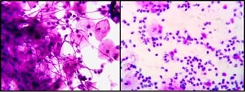
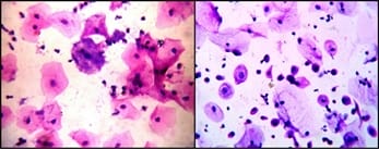
Introduction: Cervical cancer is a major health problem. It ranks second in mortality affecting the world population. Cervical cancer has the advantage that it takes a long time for it to develop. During this time it can be diagnosed and treated properly. For this purpose pap smears (cervical smears) are routinely screened for pre-malignant lesions using Liquid Based Cytology. Purpose: The purpose is to study the prevalence of cervical lesions by EZI PREP method and to determine the prevalence of cervical lesions in various age groups. Material and Methods: This was a prospective study done in the Department of Pathology. All the data regarding the patient was recorded in a predefined proforma. Results: A total of 1500 cervical smears were taken into the study. The age group of females ranged from 20-80 years. All the satisfactory smears were evaluated. Inflammatory smears, infectious smears, and smears with epithelial cell abnormalities were identified in the present study. Conclusion: Out of the 1500 LBC smears studied, the majority (1134) of the smears studied were inflammatory. Epithelial cell abnormalities were found in 21 cases and the most common age group of epithelial abnormalities was the fifth decade.

Keywords: Epithelial cell abnormalities, EZI PREP, Liquid-based cytology, Pap smear

Corresponding Author How to Cite this Article To Browse
Chitturi Ramya, Assistant Professor, Department of Pathology, NRI Medical College and Hospital, Vijayawada, Andhra Pradesh, India.
Email:
Meghana BP, Renuka IV, Ramya C, Baddam DP, Kolla S. A study of liquid-based cytology in cervical smears. Trop J Pathol Microbiol. 2020;6(3):238-244.
Available From
https://pathology.medresearch.in/index.php/jopm/article/view/422

Introduction

Cervical cancer is a severe health care problem. According to global statistics, cervical cancer is in second place by the frequency and on the third by mortality among the cancers of the reproductive system

[1].

Invasive cancer of the uterine cervix is preventable when its precursor lesions are detected and treated early. Cervical cytology has been in use now for more than 50 years and has proven itself to be the main weapon of defense against this disease [2].


Manuscript Received Review Round 1 Review Round 2 Review Round 3 Accepted
04-02-2020 15-02-2020 20-02-2020 24-02-2020
Conflict of Interest Funding Ethical Approval Plagiarism X-checker Note
No Nil Yes 19%

© 2020 by Bolla Phani Meghana, Inuganti Venkata Renuka, Chitturi Ramya, Baddam Durga Prasad, Kolla Sireesha and Published by Siddharth Health Research and Social Welfare Society. This is an Open Access article licensed under a Creative Commons Attribution 4.0 International License https://creativecommons.org/licenses/by/4.0/ unported [CC BY 4.0].

Invasive cervical cancer is the end result of a long pathological process that begins with precursor lesions called cervical dysplasia or squamous intraepithelial lesions (SIL) [3].

Cervical cancer is a preventable disease due to the long preinvasive stage. Early detection and appropriate treatment are possible if robust screening is implemented [4].

For the accurate diagnosis of these preventable diseases, pap screening is routinely employed. Liquid-based, thin layer technology was later developed in order to address the limitations of Pap smear [5].

The timely introduction of Liquid-based cytology (LBC) aimed to improve the efficiency of gynecological cytology through procedure standardization, sample quality improvement, screening support, and speedup.

Nowadays two methodologies and solutions of LBC are widely available: ThinPrep (Hologic, Marlborough, MA, USA) and BD SurePath (BD Diagnostics – TriPath, Burlington, NC, USA) [6].

Material and methods

Setting: Cytopathology, Department of Pathology

Duration of study: 1 year, July 2018 to May 2019

Type of study: Prospective study

Sampling methods: All the LBC smears sent from Department of Gynaecology and Obstetrics

Sample size: 1500 cervical smears

Inclusion criteria: All the satisfactory LBC smears

Exclusion criteria: Unsatisfactory LBC smears, Conventional smears, Hormonal cytology

Data collection procedure: All the staff in the Department of Obstetrics and Gynaecology were instructed to collect cervical samples in LBC vials using the brush supplied. The vials containing preservatives were received in our cytopathology laboratory. The samples were processed as per the prescribed protocol for Eazy PREP (EP) technique. 7ml of the sample with the preservative solution was added to 5ml of separator solution followed by centrifugation at 1500 rpm for 5 min.

The supernatant was discarded and the sediment was loaded in the Nanocytchamber followed by centrifugation in the Nanocyt Neo processor

at 1500 rpm for 2 min with a pre-coated slide. The slides were stained by routine Haematoxylin and Eosin (H and E) staining method. The 2014 Bethesda system was employed for reporting of cervical smears.

Data analysis: The data is analyzed in the form of descriptive statistics like frequencies and percentages.

Ethical clearance: Institutional ethical committee clearance was taken.

Results

1500 LBC smears were analyzed in this study. EziPrep technique yielded monolayer smears without nuclear overlapping and spread over a diameter of 16 mm. Cellular overlapping and crowding were minimal. The background of smears was clear and mucus was also significantly less in these smears. The time taken to screen was less compared to conventional smears.

The predominant smears were inflammatory followed by normal smears. Atrophic smears accounted for 3.5% of the cases (Table 1).

Table-1: Incidence of various cervical lesions in the present study.

Cervical lesions n=1500 Percent
Inflammatory 1134 75.6%
Normal 182 12.2%
Atrophic 53 3.5%
Infections 110 7.3%
Epithelial cell abnormalities 21 1.4%

Infections accounted for 7.3% of the cases in the present study. Bacterial and viral etiologies were identified. Bacterial Vaginosis (Figure 2a) was the most common infection followed by Candidiasis (Figure1a) and Trichomonas (Figure 1b). Herpes simplex virus infection accounted for 2 cases (Table 2).

patho_422_01.jpg

Fig-1a) Candidiasis, H and E400X 1b) Trichomonas, H and E 400X.


patho_422_02.jpg

Fig-2 a) Clue cell - Bacterial Vaginosis, H and E 400X 2b) LSIL, H and E 400X.

Table-2: Various infectious organisms encountered in the present study.

Infectious organisms Total (110)
Bacterial Vaginosis 47
Candidiasis 46
Trichomoniasis 15
Herpes Simplex Virus 2

patho_422_03.jpg

Fig-3 a) HSIL, 400X 3b) Squamous cell carcinoma 400X.

Epithelial cell abnormalities accounted for 1.4% of the cases in the present study. Among epithelial cell abnormalities, the low-grade intraepithelial lesion (LSIL) in Figure 2b, the high-grade intraepithelial lesion(HSIL) in Figure 3a, Atypical squamous cells of undetermined significance (ASC-US), Atypical glandular cells (AGC) and squamous cell carcinoma (SCC) in Figure 3b were noted. The most common epithelial cell abnormality noted was HSIL followed by LSIL. Their incidence is shown in Table 3.

Table-3: Incidence of smears with epithelial cell abnormalities.

Epithelial cell abnormality (n=21)
LSIL 5
HSIL 8
ASC-US 3
AGC 1
SCC 4

Among all the age groups, inflammatory smears and normal smears were more common compared to other lesions except for age group 71-80yrs, where atrophic smears predominated. Infectious lesions were predominant in 31-40 years age group.

The most common age group for epithelial cell abnormalities was 41-50 years (Table 4).

Table-4: Distribution of cervical lesions according to age group.

LBC Findings 11-20 years 21-30 years 31-40 years 41-50 years 51-60 years 61-70 years 71-80 years
Inflammatory 29 340 393 336 30 5 1
Normal - 36 62 66 16 1 1
Atrophic - - 10 24 12 3 4
Candidiasis 1 3 39 2 1 - -
Trichomoniasis - - 9 5 1 - -
Bacterial Vaginosis 1 6 28 10 2 - -
HSV - - 1 1 - - -
LSIL - - 1 2 1 1 -
HSIL - - - 2 3 3 -
ASC-US - - - 2 1 - -
AGC - - - 1 - - -
SCC - - - 1 1 2 -

Discussion

Pap smear is a very simple, useful and safe tool for detecting precancerous cervical epithelial lesions. It is regarded as the gold standard for cervical screening methods. Due to a lack of proper guidance to the people of India, screening for cancer especially cervical cancer is minimal. Many studies have proven increased detection rate of epithelial cell abnormalities on LBC compared to conventional smears. Liquid-based cytology is a technique that enables cells to be suspended in a monolayer and thus better morphological assessment is possible [7]. The present study compared the present study with similar other studies (Table 5).

Table-5: Comparison of cervical lesions with other studies.

Cervical lesions Present Study, 2019 (n=1500) Pushpa Lata Sachan et al [8], 2018 (n=1650) Sherwani et al [9], 2007 (n=160) Vikrant et al [10], 2015 (n=1000)
Inflammation 75.6% 42.6% - 0.011%
Infection 7.3% - 8.75% 0.164%
ASC-US 0.2% 2.9% - 0.012%
AGC 0.07% - - 0.001%
LSIL 0.3% 5.89% 18.1% 0.004%
HSIL 0.6% 0.48% 4.3% 0.002%
SCC 0.2% - 3.7% 0.005%

Inflammatory smears accounted for the majority of the cases in the present study, which is similar to other studies by Pushpa Lata Sachan et al., but Vikrant et al. reported infectious smears as the predominant ones [8,10]. The percent of inflammatory smears in the present study was more compared to the other studies accounting to 75.66% [8,10]. Since the present study was conducted in a medical college and many of the patients undergo pap smear as a part of the routine gynecological examination, most of the smears were inflammatory.

The predominant infectious condition in the present study was bacterial vaginosis which is similar to Vikrant et al [10]. Normal cervical smears show rod-shaped lactobacilli. In bacterial vaginosis, clue cells were found, which were the squamous cells covered by a layer of coccobacillus obscuring the cellular outlines. There is an associated absence of lactobacilli and scant inflammatory infiltrate. Moderate to severe inflammatory infiltrate should exclude the diagnosis of bacterial vaginosis. Bacterial vaginosis is associated with pelvic inflammatory disease, preterm birth, postoperative gynecological infections and abnormal cervical cytology [11, 12].

Trichomonas appeared as pear-shaped oval organisms with pale vesicular nuclei. Flagellae were seen in few cases. Collections of neutrophils known as “poly balls? were also seen in the background. In candidiasis, budding yeast or pseudohyphae were seen. These pseudohyphae lack true septations but show constrictions, which indicates the formation of new cells. In the present study, there were 47 cases of bacterial vaginosis, 46 cases of candidiasis and 15 cases of trichomonas. In a study by Vikrant et al., there were 112 cases of bacterial vaginosis, 24 cases of candida, and 6 cases of trichomonas [10]. The predominant organism in a study by Sherwani et al. is Candida and trichomonas by Singh et al. [9,13]. Herpes simplex infection was not reported in all the other three studies. Table 6 shows a comparison of the present study with various other studies.

Table-6: Infectious conditions in comparison to other studies

Organisms Present study, 2019 (n=1500) Sherwani et al [9], 2007 (n=160) Vikrant et al [10], 2015 (n=1000) Singh et al [13], 2015 (n=160)
Candida 46 (3.06%) 7 (4.3%) 24 (0.024%) 5 (0.03%)
Trichomonas 15 (0.09%) 4 (0.02%) 16 (0.016%) 6 (0.03%)
Bacterial vaginosis 47 (3.13%) - 112 (0.112%) 3 (0.017%)
Herpes simplex 2 (0.13%) - - -

In a study by Siebers et al., HSIL in LBC smears accounted for 6.7% of the total cases, whereas in the present study, HSIL accounted for 0.6% of cases [14]. The number of squamous cell carcinoma cases encountered in the present study was 0.27% which was more than that of Vikrant et al., but less than that of Sherwani et al. [9,10]. In a study by Vikrant et al., epithelial cell abnormalities were found in 25 cases [10]. Atypical squamous cells (ASC) refer to cytologic changes suggestive of SIL, but which are qualitatively or quantitatively insufficient for a definitive interpretation. The interpretation of ASC requires that the cells demonstrate three essential features i.e., squamous differentiation, increased nuclear to cytoplasmic (N/C) ratio and minimal nuclear changes which may include hyperchromasia, chromatin clumping, irregularity, smudging and or multinucleation [15]. Abnormal appearing nuclei are prerequisite for the interpretation of ASC. Incomplete changes suggestive of koilocytosis or poorly preserved cells with features suggestive of LSIL were designated as ASC-US [15]. LSIL cases had Squamous cells with abundant cytoplasm, nuclear enlargement more than three times the area of normal intermediate nuclei with anisonucleosis and inconspicuous nucleoli. Multinucleation and koilocytic change were seen in some cases. HSIL cases showed smaller dysplastic cells with a high N/C ratio, nuclear hyperchromatism, and irregular nuclear contours. Squamous cell carcinoma cases showed singly scattered and clusters of squamous cells with poorly defined cell borders, high N/C ratio, irregular distribution of chromatin, prominent nucleoli and tumor diathesis. Smears from AGC’s revealed sheets of cells with crowding, nuclear overlapping, nuclear enlargement up to 3-5 times with variation in nuclear size and shape.

Table-7: Table depicting the most common age group for epithelial cell abnormalities

Studies Decade
Present Study Fifth decade
Zhu et al [16] Fourth decade
Ilter et al [17] Fourth decade
Khamankar et al [18] Fourth decade
Macharid et al [19] Fourth decade
Almonte et al [20] Fourth decade
Chinaka et al [21] Fifth decade
Nigerio et al [22] Fifth decade
Sharma et al [ 23] Third decade

In the present study, the most common age group for epithelial cell abnormalities was 41-50 years that is the fifth decade which correlates with two other studies by Chinaka et al. and Nigerio et al. as shown in table 7 [21, 22].

These results were in contrast with studies done by Zhu et al., Ilter et al., Khamankar et al., Macharid et al., and Almonte et al. in which the most common age group was fourth decade [16-23].

Atrophic smears were found in 53 cases in the present study, whereas Vikrant et al. found in 43 cases [10]. Flat monolayered sheets or discreetly scattered parabasal cells with uniformly distributed chromatin were found in our smears. Occasional histiocytic collections were also seen.

Limitations of the study: The limitations of the study are control population of conventional smears has not been taken and since mucous is removed during processing, infectious organisms might not have been diagnosed effectively.

Conclusion

The majority of the smears in the present study were inflammatory smears. Among infectious organisms, Bacterial Vaginosis was most commonly seen followed by candida. 21 cases showed epithelial cell abnormalities of which, HSIL has the highest incidence. The most common age group of epithelial cell abnormalities was 41-50yrs. Epithelial cell abnormalities were easily identified in LBC smears, as LBC provides a clear background for making the diagnosis of epithelial cell abnormalities. This study emphasizes the importance of routine cervical screening.

What does this study add to the existing knowledge?

LBC is a simple and rapid screening method for the diagnosis of intraepithelial lesions of the cervix. Since epithelial cell abnormalities are common after 40yrs, women should be routinely screened.

Author’s contributions

Dr. B. Phani Meghana: Concepts, Design, Definition of intellectual content, Literature search, Clinical studies, Experimental studies, Data acquisition, Data analysis, Statistical analysis, Manuscript preparation, Manuscript editing

Dr. I.V Renuka: Concepts, Design, Definition of intellectual content, Manuscript editing, Manuscript review

Dr. Ch. Ramya: Concepts, Design, Definition of intellectual content, Literature search, Clinical studies, Experimental studies, Data acquisition, Data analysis, Statistical analysis, Manuscript preparation, Manuscript editing

Dr. B. Durga Prasad: Data acquisition

Dr. K. Sireesha: Data acquisition

Reference

  1. Costa MP, Heráclio SA, Coelho AVC, Acioly VL, Souza PE, et al. Comparison of conventional papanicolaou cytology samples with liquid-based cervical cytology samples from women in Pernambuco, Brazil. Braz J Med Biol Res. 2015;48(9)831-838.
  2. Monsonego J, Autillo-Touati A, Bergeron C, Dachez R, Liaras J, Saurel J, et al. Liquid-based cytology for primary cervical cancer screening- a multi-centre study. British J Cancer. 2001;84(3)360-366.
  3. Singh VB, Gupta N, Nijhawan R, Srinivasan R, Suri V, Rajwanshi A. Liquid-based cytology versus conventional cytology for evaluation of cervical Pap smears- Experience from the first 1000 split samples. Indian J Pathol Microbiol. 2015;58(1)17-21.
  4. Macharid HC, Chaseem EJ, Bukiri E, Muchiri L. A comparative analysis of conventional PAP smear cytology, liquid based cytology and colposcopy clinical impression with colposcopy biopsy histology as gold standard in women undergoing colposcopy in Kenyatta national hospital. Int J Reprod Contracept Obstet Gynecol. 2014;3(1):58-63.

  1. Richart RM, Barron BA. A follow up study of patients with cervical dysplasia. Am J Obstet Gynecol. 1969;105(3)386-393.
  2. Bal MS, Goyal R, Suri AK, Mohi MK. Detection of abnormal cervical cytology in papanicolaou smears. J Cytol. 2012;29(1)45‑47.
  3. Shobana R, Saranya B. Comparison of Conventional Papanicolaou Smear and Liquid-based Cytology for Cervical Cancer Screening. Int J Sci Stud. 2019;6(12)64-73.
  4. Kavatkar AN, Nagwanshi CA, Dabak SM. Study of a manual method of liquid-based cervical cytology. Indian J Pathol Microbiol. 2008;51(2)190-194.
  5. Sachan PL, Singh M, Patel ML, Sachan R. A study on cervical cancer screening using pap smear test and clinical correlation. Asia Pac J Oncol Nurs. 2018;5(3)337-341.
  6. Sherwani RK, Khan T, Akhtar K, Zeba A, Siddiqui FA, Rahman K, et al. Conventional Pap Smear and Liquid Based Cytology for Cervical Cancer Screening- A Comparative Study. J Cytol. 2007;24(4)167-172.
  7. Donders GG, Van Bulck B, Caudron J, Londers L, Vereecken A, Spitz B. Relationship of bacterial vaginosis and mycoplasmas to the risk of spontaneous abortion. Am J Obstet Gynecol. 2000;183(2)431-437.
  1. Bojgua S, Kldiashvili E. Liquid based cytology cervical cancer screening program–Georgian experience. Arch Cancer Res. 2016;4(3):89-95.
  2. Schwebke R. Bacterial vaginosis. Curr Infect Dis Rep. 2000;2;14-17.
  3. Singh A, Joshi C, Kujur P, Chandrakar K, Shrivastava S. Liquid-based Cytology versus Conventional Cytology for Evaluation of Cervical Cytology in a Tertiary Care Centre of Chhattisgarh. Int J Sci Stud. 2016;4(9)161-167.
  4. Siebers AG, Klinkhamer PJ, Grefte JM, Massuger LF, Vedder JE, Beijers-Broos A, et al. Comparison of liquid-based cytology with conventional cytology for detection of cervical cancer precursors- a randomized controlled trial. JAMA. 2009;302(16)1757-1764.
  5. Nayar R, Wilbur DC (eds). The Bethesda System for Reporting Cervical Cytology- Definitions, Criteria, and Explanatory Notes. New York, Springer, 3rd Ed. 2015;29-240.
  6. Zhu J, Norman I, Elfgren K, Gaberi V, Hagmar B, Hjerpe A, et al. A comparison of liquid-based cytology and pap smearas a screening method for cervical cancer. Oncol Rep. 2007;18(1)157-160.
  7. İlter E, Midi A, Haliloğlu B, Çelik A, Yener AN, Ulu İ, et al. Comparison of conventional and liquid-based cytology: Do the diagnostic benefits outweigh the financial aspect?. Turk J Med Sci. 2012;42(1):1200-1206.
  8. Khamankar ST, Belekar V, Bangal VM, Baniskar SR. Cervical cancer screening: Risk factors for cervical neoplasia among rural women of Nanded Maharashtra. Innov J Med Health Sci. 2014;4(1):312-316.

  1. Almonte M, Ferreccio C, Winkler JL, Cuzick J, Tsu V, Robles S, et al. Cervical cancer screening by visual inspection, HPV testing, liquid based and conventional cytology in Amazonian Peru. Int J Cancer. 2007;121(4):796-802.
  2. Sharma P, Rahi M, Lal P. A community-based cervical cancer screening program among women of Delhi using camp approach. Indian J Community Med. 2010;35(1):86-88.
  1. Chinaka CC, Abudullahi M, Mohammed OM. A comparative study on the use of liquid based cytology and conventional PAP smear in cervical screening. J Med Med Res. 2014;2(4):40-50.
  2. Nigerio SE, Eze JN, Emeke-Trem EN, Edegbe FO. A six-year study of the clinical presentation of cervical cancer and the management challenges encountered at a state teaching hospital. Clin Med Insights Oncol. 2013;7:151-158.