E-ISSN:2456-1487
P-ISSN:2456-9887
RNI:MPENG/2017/70771

Research Article

Tumor

Tropical Journal of Pathology and Microbiology

2020 Volume 6 Number 1 January
Publisherwww.medresearch.in

A rare mesentric tumor "paraganglioma" - a case report

Gidwani N.1*, Jain N.2, Agarwal S.3
DOI: https://doi.org/10.17511/jopm.2020.i01.16

1* Nikita Gidwani, Final Year Resident, Department of Pathology, Mahatma Gandhi Medical College and Hospital, Jaipur, Rajasthan, India.

2 Niharika Jain, Final Year Resident, Department of Pathology, Mahatma Gandhi Medical College and Hospital, Jaipur, Rajasthan, India.

3 Sonia Agarwal, Associate Professor, Department of Pathology, Mahatma Gandhi Medical College and Hospital, Jaipur, Rajasthan, India.

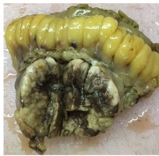
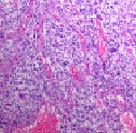
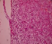
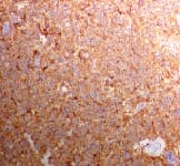
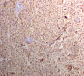
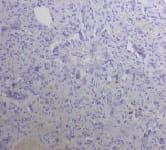
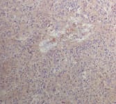
Paraganglioma are rare type of neuroendocrine tumors that arise from embryonal neural crest. Paraganglioma of adrenal medulla is known as Pheochromocytoma. Extra adrenal localization has incidence of 2-8 per million people per year, which can extend from upper cervical region to pelvis related to autonomic nervous system. Even rarer is occurrence of paragangliomas outside the usual distribution of paraganglionic tissue. Mesenteric paragangliomas are extremely rare. The present case was of a non-functional extra adrenal paraganglioma occurring in a 43-year-old male presented with complain of pain in abdomen and mild discomfort. Abdominal CECT revealed well defined enhancing solid mass with central necrosis in right side of mesentry. Explorative laparotomy and surgical excision were done, on histology typical "zellballen" pattern was found. Paragangliomas are rare neuroendocrine tumor known to store and secrete catecholamines and hence are known as functional paragangliomas and may present with symptoms like headache, sweating, palpitations and hypertension.

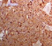
Keywords: Paraganglioma, Intra-Abdominal surgery, Abdominal surgery, Neuroendocrine cells, Immunohistochemistry

Corresponding Author How to Cite this Article To Browse
Nikita Gidwani, Final Year Resident, Department of Pathology, Mahatma Gandhi Medical College and Hospital, Jaipur, Rajasthan, India.
Email:
Gidwani N, Jain N, Agarwal S. A rare mesentric tumor "paraganglioma" - a case report. Trop J Pathol Microbiol. 2020;6(1):98-102.
Available From
https://pathology.medresearch.in/index.php/jopm/article/view/418

Introduction

Paraganglioma are rare type of neuroendocrine tumor that arise from embryonal neural crest [1]. Paraganglia contain tissues such as the adrenal medulla, carotid and aortic bodies, paraganglia in sympathetic and parasympathetic nerves and organs of Zuckerkandl. Paragangliomas are tumors originating from the neuroendocrine elements of the paraganglia. The most comm-

on localization of paragangliomas is the adrenal medulla and they are usually pheochromocytomas. Extra-adrenal localization is observed in 5-10% of all paragangliomas [2]. The most common extra-adrenal paragangliomas occur as carotid bodies. Paragangliomas have been seen in the gastrointestinal tract of the duodenum. Mesenteric paragangliomas are exceedingly infrequent [3]. With an annual incidence estimated at 1/100,000, paragangliomas re-


Manuscript Received Review Round 1 Review Round 2 Review Round 3 Accepted
05-01-2020 15-01-2020 25-01-2020 28-01-2020
Conflict of Interest Funding Ethical Approval Plagiarism X-checker Note
No Nil Yes 18%

© 2020 by Nikita Gidwani, Niharika Jain, Sonia Agarwal and Published by Siddharth Health Research and Social Welfare Society. This is an Open Access article licensed under a Creative Commons Attribution 4.0 International License https://creativecommons.org/licenses/by/4.0/ unported [CC BY 4.0].

present ten percent of catecholamine secreting tumors. Paragangliomas arise from chromaffin tissue most commonly found in the Zuckerkandl body, the sympathetic plexus of the urinary bladder, the kidneys, and the heart or in the sympathetic ganglia of the head or neck [1]. Some paragangliomas have been described in the Gastrointestinal System, the majority of which were associated with the duodenum. Only a very few were described to arise from the mesentery [2]. Given their catecholamine secreting properties, paragangliomas have the potential to present as a mass with paroxystic symptoms of palpitations, pallor, tremor, headache and diaphoresis as well as hypertension [3]. This however is only the case in 25% of the paragangliomas. The rest have presented as abdominal masses with or without hypertension [2]. Paraganglioma of adrenal medulla is called Pheochromocytoma while outside adrenal they are known as Paraganglioma. Extra adrenal localization is observed in 5-10% of all paragangliomas with incidence rate of 2-8cases per million people per year [2]. Mesenteric paraganglioma is extremely rare [3-6]. They are usually seen in 2-3 decades [7] and both sexes are equally affected [8]

Types of paraganglioma [9]

  1. Carotid Body Paraganglioma – They are located at bifurcation of carotid artery.
  2. Vagal Paraganglioma – They are located in the anterolateral portion of neck.
  3. Mediastinal Paraganglioma– They originate from aorticopulmonary portion, so are found in antero-superior part of mediastinum.
  4. Retroperitoneal Paraganglioma– They are located anywhere along the paravertebral chain.
  5. Zuckerkandl Body Paraganglioma– They are found close to the angle formed by anterior wall of aorta and origin of inferior mesenteric artery.
  6. Others – Thyroid, Uterus, Gall Bladder, Mesentery.

patho_418_01.jpg

Fig-1: Gross image of paraganglioma mesentery.

Case Report

A 43-year-old male patient came to Mahatma Gandhi Hospital for routine health check up. Patient had no complaints related to abdomen or any other chronic condition. No significant past or family history / No co-morbidities. On examination no abnormalities were detected.

Abdominal USG revealed a well-defined lesion with small cystic area measuring 36x42mm is seen in right lumbar region in peritoneum just posterior to anterior abdominal wall. Abdominal CT Scan revealed well defined enhancing solid mass lesion with central necrosis in right side of mesentery measuring 49x50x42mm.

Simultaneously plasma free metanephrine was done and it was 28.4pg/ml (normal < 65), its done to rule out presence of any other adrenaline secreting tumor present in body. Chromogranin A was 227.1ng/ml (normal <108). Exploratory laprotomy was done for excision of tumor under general anaesthesia.

A hard lump of size approx 5x6cm was present in mesentery of ileum (small bowel) around 1 feet proximal to ileo-caecal junction close to ileum wall but not invading the wall. The lump was excised from the mesentery in total but due to involvement of locally supplying mesenteric vessels, the involved segment of ileum was also resected. The excised lump along with ileum was sent for intra-operative frozen section. On frozen section, features were suggestive of paraganglioma. The entire specimen was sent for HPE.

Pathological findings

Gross: Intestinal segment with mesentery was received. Nodular grey white growth seen attached measuring 5x4.5x2cm. On cut section mass was grey white to grey brown and hemorrhagic.

Microscopy: Multiple sections showed well circumscribed neoplasm made up of nests of cells (Zell Ballen Pattern) separated by thin walled blood vessels. The cells are round to polyhedral with vesicular nuclei and prominent nucleoli. The cells have abundant eosinophilic, clear to granular cytoplasm. Significant mitotic activity is not seen. High cellularity and diffuse growth pattern is not seen. Necrosis is not seen. The cells show only mild nuclear pleomorphism. Occasional tiny focus of capsular invasion is seen with vascular invasion. Overall suggestive of Paraganglioma.


patho_418_02.jpg patho_418_03.jpg

Fig-2: Circumscribed neoplasm made up of nests of cells (Zell Ballen Pattern).

Immunohistochemistry findings

  • Synaptophysin (neuroendocrine marker) – Positive
  • CD 56 (neuroendocrine marker) – Positive
  • Ki 67 (proliferation index) - <1%
  • Pancytokeratin – Negative
  • S-100 – Negative
  • Diagnosis – consistent with paraganglioma

patho_418_04.jpg

Fig-3: Synaptophysin Positive

patho_418_05.jpg

Fig 4: CD-56 Positive

patho_418_06.jpg

Fig-4: KI-67 <1%

patho_418_07.jpg

Fig-5: Cytokeratin Negative

patho_418_08.jpg

Fig-6: S-100 (Negative).

Discussion

Neural crest cells give rise to the parenchymal cells of the paraganglia and other elements of the autonomic nervous system. These neural crest cells have the ability to migrate to various regions along the paravertebral and para-aortic axis, while remaining in close relation to the sympathetic nervous system. They extend to various places, anywhere from the neck to the base of the pelvis [4]. In rare occasions, paragangliomas have been identified in areas where chromaffin tissue has not yet been characterized, such as the genitourinary tract, spermatic cord, sacro-coccygeal area, anus, renal capsule, broad ligament, ovary and vaginal wall and can only be explained by the migratory property of the neural crest cells [5]. From the vast survey of the literature, there appears to be a marked predilection for females (9:3), which contrasts with the slight male predominance (1.3:1) reported for retroperitoneal paraganglioma [5,6]. At the time of diagnosis, most patients are older


(median, 57.5 years of age) than those with retroperitoneal paraganglioma (median, 39-43 years of age [4-6]). No significant difference was noted in the size of mesenteric (average, 9.3 cm) and retroperitoneal tumors (average, 7.4-10.5 cm [4-6]). The pathogenesis of paragangliomas is not fully understood. They may be either sporadic or hereditary. Overall, as many as 10%-50% of paragangliomas are considered to be hereditary [7]. Hereditary paragangliomas are multicentric in 20%-50% of cases [8,9], whereas sporadic paragangliomas are multicentric in 10% of cases. In hereditary cases, they may be associated with multiple endocrine neoplasia type 2, von Hippel-Lindau disease, familial paraganglioma, Carney triad and neurofibromatosis type 1 [10]. For this reason, especially in patients diagnosed before 50 years of age and in those who present with bilateral, multifocal, and malignant paragangliomas, genetic testing may be beneficial [11]. Diagnosing paragangliomas, and in particular those of the mesentery, can be achieved via biochemistry and/or imagery. Given the capacity of a paraganglioma to secrete catecholamines, plasma or urinary metanephrines have been described in the literature as a very sensitive technique. Unfortunately, the secreting property is only found in 25% of mesenteric paragangliomas [1]. Anatomical imagery with US/CT/MRI are equally as effective in identifying these abdominal masses. In addition, specific functional imaging with metaiodo-benzylguanidine scintigraphy or PET imaging with 6 [18F] fluoro-DOPA help identify and characterize the extent of the mass as well as the staging [6]. These techniques are then followed up with seemingly essential laparoscopic exploration and biopsy [7]. Finally, tumor resection is the form of treatment that has achieved the best results. Throughout all the cases described in the literature, none described any recurrence post-excision of the mass, but median follow-up was relatively short. Chemotherapy and radiotherapy have not demonstrated convincing results for patients with unresectable or metastatic disease. Its involvement remains palliative, as there is no current evidence of increased survival using these modalities [6]. Treatment with radiolabelled MIBG is gaining popularity given its avidity for the chromaffin cell tumors and in particular their metastases [8]. The literature stipulates that while 131I-MIBG is not a curative therapy, its involvement as an adjuvant to surgical resection as well as the possibility of a synergistic effect with chemotherapy seem

promising and are venues to be explored in the near future [9]. Radioactive somatostatin analogues is yet another radiopharmaceutical to be considered [6]. Focus has now shifted to specific molecular targets involved in the malignant transformation of chromaffin cell tumors, and its development has shown signs of promise, yet development in these areas is still necessary [10]. Paraganglioma are rare neuroendocrine tumors that arise from specialized cells referred to as chief cells of paraganglia which are present over entire body. The adrenal medulla has largest collection of receptor cells derived from neural crest which accounts for highest rate of occurrence of these tumors in this site [2]. Paraganglioma can be functional when they secrete catecholamines and their metabolites in blood and urine and may present with headache, sweating, palpitation and symptoms of hypertension [10-12]. Nonfunctional paragangliomas are mostly asymptomatic and found incidently or present as a mass with symptoms of surrounding organ compression [13]. 20-50% of extra adrenal paraganglioma are malignant in contrast to pheochromocytoma which is 10% malignant [14]. FOLLOW UP: Patient has been advised for Gallium 68 DOTANOC PET CT scan to scan the whole body for presence of any residual lesion or any undiagnosed lesions in the body [15-17]. Genetic screening if any positive family or genetic history.

Conclusion

Mesenteric paraganglioma is very rare tumor and its diagnosis is very difficult. It should be among the preoperative differential diagnosis of abdominal masses of unknown etiology. The diagnosis is best achieved by imagery and laproscopic exploration. Surgical excision is treatment of choice which can be performed successfully by laproscopy. Long term follow up is mandatory as tumor is prone to recur and to metastatise.

Reference

  1. Shah U, Giubellino A, Pecak K. Pheochromocytoma- complications in tumorigenesis and the actual management. Minerva Endocrinol. 2012;37(2)141-156.
  2. Sclafani LM, Woodruff JM, Brennan MF. Extraadrenal retroperitoneal paragangliomas- natural history and response to treatment. Surg. 1990;108(6)1124-1129.

  1. Wen J, LI HZ, Ji ZG, Mao QZ, Shi BB, Yan WG. A decade of clinical experience with extra-adrenal paragangliomas of retroperitoneuml Report of 67 cases and a literature review. Urol Ann. 2010;2(1)12-16.
  2. Cherit M, Dube P, Royal V, Leblanc G, Sideris L. Malignant paragangliomaof the mesentery- a case report and review of literature. Worls J Surg oncol. 2012;10;46.
  3. Lagorga JB, Vaquero M, Juanpere N. Paragastric Paraganglioma- A case report with Unusual Alveolar Pattern and Myoxid Component. Cytopathol. 2012;40(9)815-819.
  4. Pruiti V, Mazzeo F, Rossitto M, Foti A, Macri A, Cucinotta E. Gastric paraganglioma- A case report and a review of the literature. Ann Ital Chir. 2014;29;85.
  5. Crosbie L, Humphreys WG, Maxwell M, Maxwell P, Cameron CHs, Toner PG. Castric paraganglioma- an immunohistological and ultrastructural case study. J Submicrosc Cytol Pathol. 1990;22(3)401-408.
  6. Buonuomo V, Ruggiero A, Rando G, D’urzo C, Manzoni C, pintus C. Malignant retroperitoneal paraganglioma- case report and review of the literature. Pediatr Hematol Oncol. 2004;21(8)731-738.
  7. Belhamidi MS, Ratbi MB, Tarchouli M, Adioui T, Ali AA, Zentar A Sair K. An unusual localization of retroperitoneal paraganglioma- a case report. Pan Af Med J. 2015;22(1)12.
  8. Chrisoulidou A, Kaltsas G, Ilias I, Grossman AB. The diagnosis and management of malignant phaeochromocytoma and paraganglioma. Endocr Relat Cancer. 2007;14(3)569-585.
  1. Absher KJ, Witte DA, Trung LD, Ramzy I, Mody DR, Ostrowski ML. Aspiration biopsy of osseous metastasis of retroperitoneal paraganglioma- report of a case with cytologic features and differential diagnostic considerations. Actacytol. 2001;45(2)249-253.
  2. Offergeld C, Brase C, Yaremchuk S, Mader I, Rischke HC, Glasker S, et al. Head and neck paragangliomas- clinical and molecular genetic classification. Clinics. 2012;67(1)19-28.
  3. Perrot G, Pavic M, Mitou F, Crozes C, Faucompret S, Vincent E. diagnostic difficile d’un paragangliomepancreatuque. Rev Med Interne. 2007;28(10)701-704.
  4. Kinney Ma, Narr BJ, Warner MA. Perioperative management of pheochromocyta. J Cardiothorac Vasc Anesth. 2002;16(3)395-369.
  5. Vanderveen Ka, Thompsan SM, Callstrom Mr, Young WF, Jr, Grant CS, Farley DR, Richards ML, Thompson GB. Biopsy of pheochroocytomas and paraganglioma potential for disaster. Surg. 2009;146(6)1158-1166.
  6. Orkan Z, Ozdemir CS, Yasar G, Altas O, Koc M, Gul Y, Durdag E. An unusual mesenteric tumor ‘paraganglioma’- a case report. Iran Red Crescent Med J. 2014;16(12)e16837.
  7. Hemalatha AL, Avadhani Geeta K, Anoosha K, Ashok KP. Extra-adrenal silent retroperitoneal paraganglioma- Report of a rare case. Journal of clinical and diagnostic research- JCDR. 2014;8(11)FD06-FD07.