E-ISSN:2456-1487
P-ISSN:2456-9887
RNI:MPENG/2017/70771

Research Article

Syndrome

Tropical Journal of Pathology and Microbiology

2020 Volume 6 Number 2 February
Publisherwww.medresearch.in

Testicular regression syndrome: A series of 22 cases at a tertiary care hospital

Sharma D.1, Sagar N.2*, Khurana N.3
DOI: https://doi.org/10.17511/jopm.2020.i02.12

1 Divya Sharma, Dr. Baba Saheb Ambedkar Medical College and Hospital, Delhi, India.

2* Nishant Sagar, Maulana Azad medical College and Hospital, Delhi, India.

3 Nita Khurana, Maulana Azad medical College and Hospital, Delhi, India.

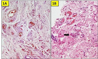
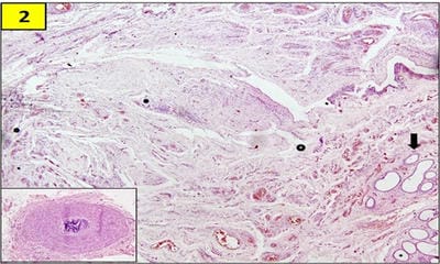
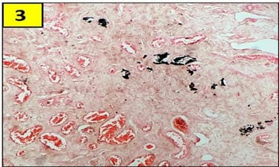
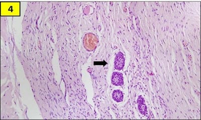
Testicular regression syndrome (TRS) represents a congenital condition in which no normal testicular tissue can be identified following exploration for a clinically impalpable testis. To study the Clinicopathological spectrum of Testicular regression syndrome (TRS) and review the literature. Study design: The study included 22 patients with nonpalpable testis, who had undergone resection of testicular nubbins. Original diagnosis was studied in context of pathological recognition of TRS and additional sections and stains were examined. Pathological assessment included identification of epididymis and vas deferens, vascularised fibrous nodule (VFN), dystrophic calcification, hemosiderin and pampiniform plexus like veins. Stain for iron and calcium were performed. On microscopy, VFN was observed in 17 (77.3%), calcification in 4 (18.2%), hemosiderin in 15 (68.2%), vas deferens in 13 (59%), epididymis in 11 (50%), prominent vessels in 21 (95.4%) and seminiferous tubules in 6 (27.3%) cases. The presence of dystrophic calcification and hemosiderin deposition with absent viable tissue points to the hypoxic injury to the testis. TRS theoretically carries a long-term risk for malignant degeneration therefore In the typical situation in which a blind-ending spermatic cord without viable testis is submitted for tissue analysis, it is imperative to characterize such cases as consistent with regressed testis thus eliminating the need for further surgical intervention.

Keywords: Impalpable, Regression, Vanishing testis, Nubbin

Corresponding Author How to Cite this Article To Browse
Nishant Sagar, Maulana Azad medical College and Hospital, Delhi, India.
Email:
Sharma D, Sagar N, Khurana N. Testicular regression syndrome: A series of 22 cases at a tertiary care hospital. Trop J Pathol Microbiol. 2020;6(2):191-197.
Available From
https://pathology.medresearch.in/index.php/jopm/article/view/406

Introduction

Clinically impalpable testis is a common problem encountered by urologists and paediatric surgeons. This may be due to undescended testis regarded as crypto-

orchidism. Testicular descent occurs in two phases in the intrauterine life: between 8–15 weeks and 25–35 weeks gestation.

This descent is under the influence of hormones like testosterone, human chorionic gonadotrophin (hCG) and the protein hormone


Manuscript Received Review Round 1 Review Round 2 Review Round 3 Accepted
30-01-2020 07-02-2020 14-02-2020 18-02-2020
Conflict of Interest Funding Ethical Approval Plagiarism X-checker Note
No Nil Yes 11%

© 2020 by Divya Sharma, Nishant Sagar, Nita Khurana and Published by Siddharth Health Research and Social Welfare Society. This is an Open Access article licensed under a Creative Commons Attribution 4.0 International License https://creativecommons.org/licenses/by/4.0/ unported [CC BY 4.0].

Insulin-Like 3 (INSL3) [1].

Around 5% of undescended testicles have failed to form completely, manifesting clinically as an "empty" scrotum. The local examination of scrotum demonstrates tissue nubbins or tiny nodules corresponding to the vestigial remnants of the vanishing testis.

The presence of spermatic cord structure is evidence of presence of testis in the early intrauterine life which if associated with a blind ending spermatic cord, is named as testicular regression syndrome (TRS) in the pathology literature [1].

TRS is seen in less than 5% of cryptorchidism cases however it accounts for 35-60% of clinically impalpable testis [2]. This absence of a testis in an otherwise normal 46XY male is assumed to be a consequence of intrauterine or perinatal torsion or infarction [2].

Although this entity has been well-described in the surgical literature, few pathological studies have been performed.

The histopathological features are distinct and include a fibrovascular nodule, areas of dystrophic calcification and hemosiderin deposition at the end of the spermatic cord. Presence of seminiferous tubules with remnants of germ cell has also been seen in early stages of TRS.

The association of malignancy arising from cryptorchid testis is well known occurrence. However the malignant change of residual germ cells in testicular regression syndrome is not yet established.

Materials and Methods

Setting and type of study: The study was conducted at a Tertiary care hospital on all patients undergoing orchidectomy for undescended testis, over a period of 9 years.

Type of study: Retrospective Observational study

Inclusion criteria: Patients with clinical diagnosis of UDT, atrophic testis or histopathological diagnosis consistent with cryptorchidism were selected in the study.

Exclusion criteria: Cases showing infarction of testicular parenchyma, torsion of testis and ambiguous genitalia.

Sample size: The clinicopathological study was done of 22 patients with nonpalpable testis, who had undergone resection of testicular nubbins.

Data collection and analysis: Original hematoxylin and eosin stained microscopic slides of each case was examined and deeper sections were obtained as necessary. Iron stain was performed in which there was prominent brown pigment deposition or was inadequate in amount. Special stain like Von kossa was done to demonstrate small foci of calcification.

Pathological assessment included presence of vascularised fibrous nodule (VFN), identification of vas deferens, epididymis, hemosiderin, calcification and pampniform plexus like veins. The presence of seminiferous tubules, germ cells and leydig cells was also documented in each case.

Malignancy potential was defined as the presence of germ cells in the seminiferous tubules. The gross description of each case was reviewed for identification of a fibrous nodule prospectively. The specimen was whole processed in all the cases to detect any residual testicular remnants.

The original diagnosis was compared with the secondary diagnosis and TRS was determined by the presence of a) a vascularised fibrous nodule with hemosiderin and or calcification; b) seminiferous tubules with cord elements in proximity.

Results

A total of 22 patients met the criteria for inclusion in the study. The clinical information provided in the cases was "impalpable testis", "testicular nubbin" or "vanished testis".

The age of the patients ranged from 7 month to 14 years with a mean of 5.6 years and median age of 3 years. Majority (68.1%) of excisions were done on the left side.

The original diagnosis in 6 of 22 cases was that of TRS. Remaining of the cases were designated as TRS on secondary review. The concurrence for primary and secondary diagnosis was 27.2% (6/22).

Of the 22 cases, 17 cases contained a VFN (Figure 1), 15 cases showed intranodular hemosiderin, 4 cases showed calcification (Figure 1,3), 11 cases demonstrated epididymis, 13 cases showed vas deferens (Fig 2) and prominent vessels was noted in


21 cases.

In only five of these 13 cases with vas deferens, was there no accompanying epididymis. There was no correlation between nodule size and degree of

calcification or hemosiderin. The nodule size ranged from 0.9 to 3.6 cm, with a mean size of 1.8 cm. Residual seminiferous tubules were observed in 6 cases (27.2%) (Figure 4).

patho_406_01.jpg

Fig-1A : Vascularised fibrotic nodule (H/E, 200x).

Fig-1B: Focal calcification (arrow) and hemosiderin deposition (H and E, 200x).

patho_406_02.jpg

Fig-2: Vascularised fibrous nodule with residual seminiferous tubules (arrow)

(H/E, 200x) (Inset: vas deferens).

patho_406_03.jpg

Fig-3: Areas of calcification (Von kossa, 100x).

patho_406_04.jpg

Fig-4: Residual testicular elements (H and E, 400x).

Discussion

TRS is a condition which is considered to be due to subsequent atrophy and disappearance in fetal life of an initially normal testis [3]. There is a spectrum of syndromes characterized as TRS, the exact categorisation depending on the stage of fetal or early neonatal life at which testicular function ceased [4].

Such individuals are genetically male (46, XY), presenting with unilateral or bilateral absence of recognizable testis structures and absence of the mullerian duct system [1]. The degree of masculinization of the internal and external genitalia depends on the duration of testicular function prior to its loss [2].

Absence of testis, intra-abdominal testis, inguinal or intra-abdominal vanishing testis are reasons for nonpalpable testis [5,6]. Vanishing testis is more common than testicular agenesis in patients with nonpalpable testis [7].

One fifth of the 0.7 to 1% of the cryptorchid males presenting at age 1 year or older are found to have a non palpable gonad. Forty percent of this subpopulation is thought to harbour a vanished testis [8] Bader et al reviewed testicular remnants in 208 cases and observed evidence of TRS histologically in 180 cases (87%) [9].

Yet this entity is infrequently addressed in the pathology literature.

Nonpalpable testis is a heterogenous group; with two of the possible situations: (1) regression of the gonad; or (2) failure to localize the testis by surgeon. The pathologist may play a pivotal role in the management of these patients. As 95%


of the testis are located at or below the internal inguinal ring, therefore, pathologic confirmation reassure the patient and the surgeon of the correctness of the diagnosis and avoid further surgical intervention.

Diagnosis of TRS is based on a clinically nonpalpable testis located within the retroperitoneum or exiting a closed internal inguinal ring. Typical gross descriptions include several cm of spermatic cord with a small mass of firm, fibrotic tissue at one end;

elements of the vas deferens, spermatic artery and venous plexuses are usually present [10].

Histological features include dense vascularised fibrous nodule, in the absence of seminiferous tubules or normal testicular elements; scattered foci of calcification and hemosiderin pigment deposition. The combined histological features from the current data and previous series of testicular regression syndrome are provided in Table 1.


Table-1: Histopathological features of TRS in case series reporting more than 10 cases in literature

Study Year No. VFN (%) Vas (%) Epididymis (%) Hemosiderin (%) Calcification (%) Tubules (%) Germ cell (%)
Present 2020 22 77.3 59 50 68.2 18.2 27.3 0
Md Shakir 2018 19 10.5 - 100 68.4 73.7 21.1 0
Mizuno11 2012 88 85.2 54 27.3 23.9 29.5 12.5 3.4
Bader9 2011 180 81 92 59 48 49 15 11
Emir24 2007 44 - 52 - 27 32 11.3 4.5
Mushtaq16 2006 117 44 61 33 73 44 10 0
Nishizaw25 2000 43 63 72 26 - - 5 0
Spires 1 2000 13 85 69 38 69 62 0 0
Grady26 1998 14 - - - 93 - 7 0
Cendro22 1998 29 100 72 28 86 83 0 0
Merry19 1997 47 23 68 - - 15 9 -
Turek27 1994 110 - 81 36 30 35.5 6.4 -
Smith 10 1991 77 - 79 36 42 42 4 -

Hormonal and mechanical factors have been implicated in the descent of testis but no specific factor has been identified for the pathogenesis of nonpalpable or undescended testis. Two possibilities can be speculated for the formation of testicular nubbins.

First, the testis develops in the genital ridge and starts to descend toward the scrotum, but undergoes regression due to vascular accident or endocrinopathy during testicular descent. Second, testicular development in the genital ridge by itself might be insufficient [11].

TRS is thought to be the result of vascular compromise due to antenatal or perinatal thrombosis or torsion, kinking of vasculature or direct trauma [10,12-14].

It has been suggested that the preterm testis is particularly susceptible to haemorrhagic infarction once the fibrous tunica albuginea has formed, due to the numerous thin walled vessels and relatively sparse loose stromal tissue. Therefore, the fetal response to infarction may include fibrosis and

calcification. If haemorrhage is extensive at the time of infarction, accumulation of iron containing blood pigment may also occur and be reflected in post-natal life as haemosiderin pigment in the infarcted tissue [10].

Secondary fibrous replacement of the testicular parenchyma may then lead to the formation of the characteristic vascularised fibrous nodule. Similar to other studies, 80.9% of testicular remnants had calcification and fibrosis in our series which also supports the vascular accident theory as a cause of vanishing testis.

Cryptorchidism is more common on the right, but Honore and Smith et al found that the left testis was more susceptible to regression, and the present study concurs with this impression, with 68% of cases being left-sided [10,15].

It has been suggested that the anatomical arrangement of the left spermatic vein, draining into the left renal vein, may predispose to kinking, due to an unusually mobile left kidney. As there is no venous anastomosis across the midline until after


16 weeks, venous infarction may result [9].

Residual Seminiferous tubules are rarely encountered and where present, appear predominantly composed of Sertoli cells [16]. The literature reports that viable germ cells or seminiferous tubules are present in 0 to 16% of excised testicular remnants [17-20].

The precise fate of these remnants though not clear, viable germ cells are theoretically at risk for degeneration and intratubular germ cell neoplasia is a known precursor of invasive germ cell tumors [21].

Cendron et al presented a histologic evaluation of 25 vanishing testis specimens and noted no identifiable testicular elements in any of these cases [22].

However, Mizuno et al detected seminiferous tubules in 12% of cases with 3% cases demonstrating germ cells [11].

In addition, intratubular germ-cell neoplasia in a testicular remnant has been reported in 1 case [21].

Although the risk of subsequent development of testicular germ cell neoplasia is significantly increased in the presence of cryptorchidism, the risk associated with residual testicular tubules as a component of TRS remains unknown [16].

In the majority of cases, TRS appears to be a sporadic occurrence, and the patients are otherwise normal with no significant family history.

However, there are now several reports of TRS occurring in association with other defects, including severe mental retardation in chromosomally normal siblings [14], or occurring in family members, suggesting a possible genetic basis in some subjects [23].

Controversy remains among paediatric urologists regarding the appropriate management of such cases, in particular the extent of surgical exploration and need for surgical removal if TRS is suspected [24].

The variable reports of viable germ cell elements found within the testicular remnants may be the reason for the differences in the opinion regarding the proper management [2].

The lack of association of frequency of residual testicular tubules with either patient’s age or macroscopically identifiable nodule indicates that

surgical removal of tissue at the end of the spermatic cord is warranted to ensure that no residual tubules remain in an intra-abdominal location [16].

Although this study was spanning the time period of 9 years, a limitation of this study was the rarity of the condition resulting in a small sample size.

Furthermore, due to unawareness of this entity by the pathologist, a definitive diagnosis was not rendered on the initial reporting in the appropriate clinical settings.

Despite these limitations, our findings highlight that definitive pathological diagnosis of regressed testis is desirable and achievable in majority of the cases of nubbin testis.

Pathologic confirmation of a regressed testis reassures the surgeon and the patient of accuracy of the diagnosis and removes the need for further surgical intervention.

What does this study add to the existing knowledge?

Therefore, this study aims to delineate the histopathological features of this entity to facilitate the definitive diagnosis, in a typical clinical setting of impalpable testis.

Authors contributions

Dr. Divya Sharma: Conception of the work with analysis of the data and literature

Dr. Nishant Sagar: Acquisition of images and clinical data

Dr. Nita Khurana: Final draft and revision of the manuscript with critical inputs

Reference

  1. Spires SE, Woolums CS, Pulito AR, Spires SM. Testicular regression syndrome- a clinical and pathologic study of 11 cases. Arch Pathol Lab Med. 2000;124(5)694-698.
  2. Desai A, Verma R, Parab M. A Case of Testicular Regression Syndrome. Int J Surg. 2008;21:2.

  1. Redman F. Impalpable testis- observations based on 208 consecutive operations for undescended testis. J Urol. 1980;124(3)379-381.
  2. Hegarty PK, Mushtaq I, Sebire NJ. Natural history of testicular regression syndrome and consequences for clinical management. J Pediatr Urol. 2007;3(3)206-208.
  3. Selby DM. Sexual maldevelopment syndromes, In- Stocker JT, Dehner LP, eds. Pediatric Pathology Philadelphia, Pa- JB Lippincott Co. 1992;117-159.
  4. Pirgon Ö, Dündar BN. Vanishing testes- a literature review. J Clin Res Pediatr Endocrinol. 2012;4(3)116-120.
  5. Smolko MJ, Kaplan GW, Brock WA. Location and fate of the nonpalpable testis in children. J Urol. 1983;129(6)1204-1206.
  6. Law H, Mushtaq I, Wingrove K, Malone M, Sebire NJ. Histopathological features of testicular regression syndrome- relation to patient age and implications for management. Fetal Pediatr Pathol. 2006;25(2)119-129.
  7. Ferro F, Lais A, Bagolan P, Talamo M, Caterino S. Impact of primary surgical approach in the management of impalpable testis. Eur Urol. 1992;22(2)142-146.
  8. Smith NN, Byard RW, Bourne AJ. Testicular regression syndrome- a pathological study of 77 cases. Histopathol. 1991;19(3)269–272.
  1. Mizuno K, Kojima Y, Kamisawa H, Kurokawa S, Moritoki Y, Nishio H, et al. Feasible etiology of vanishing testis regarding disturbance of testicular development- histopathological andimmunohistochemical evaluation of testicular nubbins. Int J Urol. 2012;19(5)450-456.
  2. Wright JE. The ‘‘atrophic’’ testicular remnant- atrophy or agenesis?. Pediatr Surg Int. 1986;1;229-231.
  3. Bader MI, Peeraully R, Ba'ath M, McPartland J, Baillie C. The testicular regression syndrome--do remnants require routine excision?. J Pediatr Surg. 2011;46(2)384-386.
  4. Sutcliffe JR, Wilson-Storey D, Smith NM. Ante-natal testicular torsion- only one cause of the testicular regression syndrome?. J R Coll Surg Edinb. 1996;41(2)99-101.
  5. Honore LH. Unilateral anorchism, Report of 11 cases with discussion of etiology and pathogenesis. Urol. 1978;11(3)251-254.
  6. Van Savage JG. Avoidance of inguinal incision in laparoscopically confirmed vanishing testis syndrome. J Urol. 2001;166(4)1421-1424.
  7. Storm D, Redden T, Aguiar M, Wilkerson M, Jordan G, Sumfest J. Histologic evaluation of the testicular remnant associated with the vanishing testes syndrome- is surgical management necessary?. Urol. 2007; 70(6)1204-1206.
  8. Rozanski TA, Wojno KJ, Bloom DA. The remnant orchiectomy. J Urol. 1996;155(2)712-714.

  1. Merry C, Sweeney B, Puri P. The vanishing testis- anatomical and histological findings. Eur Urol. 1997; 31(1)65-67.
  2. Emir H, Ayik B, Eliçevik M, Buyukunal C, Danişmend N, Dervişoglu S et al. Histological evaluation ofthe testicular nubbins in patients with nonpalpable testis- assessment of etiology andsurgical approach. Pediatr Surg Int. 2007;23(1)41-44.
  3. Belman AB, Rusthon HG. Is the vanished testis always a scrotal event?. Br J Urol Int. 2001;87(6)480-483.
  4. Cendron M, Schned AR, Ellsworth PI. Histological evaluation of the testicular nubbin in the vanishing testis syndrome. J Urol. 1998;160(3 Pt 2)1161-1162.
  5. Marcantonio SM, Fechner PY, Migeon CJ, Perlman EJ, Berkovitz GD. Embryonic testicular regression sequence- a part of the clinical spectrum of 46, XY gonadal dysgenesis. Am J Med Genet. 1994;49(1)1-5.
  1. Williams EV, Appanna T, Foster ME. Management of the impalpable testis- a six year review together with a national experience. Postgrad Med J. 2001;77;320-322.
  2. Schned AR, Cendron M. Pathologic findings in the vanishing testes syndrome. J Urol Pathol. 1997;6;95-107.
  3. Nishizawa S, Suzuki K, Tachikawa N, Nukui A, Kumamaru T, Shioji Y, et al. The vanishing testis- diagnosis and histological findings. Nippon Hinyokika Gakkai Zasshi. 2000;91(6)537-541.
  4. Grady RW, Mitchell ME, Carr MC. Laparoscopic and histologic evaluation of the inguinal vanishing testis. Urol. 1998;52(5)866-869.
  5. Turek PJ, Ewalt DH, Snyder HM, Stampfers D, Blyth B, Huff DS et al. The absent cryptorchid testis- The absent cryptorchid testis- surgical findings and their implications for diagnosis and etiology. J Urol. 1994;151(3)718-720.