E-ISSN:2456-1487
P-ISSN:2456-9887
RNI:MPENG/2017/70771

Research Article

Meningioma

Tropical Journal of Pathology and Microbiology

2019 Volume 6 Number 1 January
Publisherwww.medresearch.in

Clinicopathological study of Meningioma

Patel JP.1, Jansari TR.2*, Chaudhari VV.3
DOI: https://doi.org/10.17511/jopm.2020.i01.02

1 Jigna Prakashbhai Patel, Assistant Professor, Department of Pathology, SBKS MI & RC, Sumandeep Vidyapeeth, Vadodara, Gujarat, India.

2* Trupti Rajeshbhai Jansari, Assistant Professor, Department of Pathology, SBKS MI & RC, Sumandeep Vidyapeeth, Vadodara, Gujarat, India.

3 Vaibhavi Vinodbhai Chaudhari, Resident Doctor, Department of Pathology, SBKS MI & RC, Sumandeep Vidyapeeth, Vadodara, Gujarat, India.

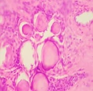
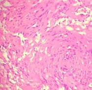
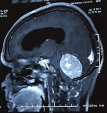
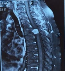
Introduction: Meningiomas are tumors that arise from the meningothelial cells. They are commonly located at intracranial, intraspinal or occasionally ectopic site. They show histological diversity and are categorized into three grades. Aims and Objectives: To study the incidence, anatomical location, sex and age Predilection, histological variants and grading of meningiomas based on WHO 2016 classification. To correlate clinical features and radiological findings with those of histopathological findings. Materials and Methods: The study is carried out in the Department of Pathology, Dhiraj General Hospital, Piparia from November 2016 to July 2018. 30 tumors specimen diagnosed as meningioma by radiology and neurosurgery department, sent to department of pathology were included in the study. Results: Total 30 meningioma tumors were included in the study. Most of them were intracranial, predominantly involving the posterior fossa of brain, females and the 41-60 age group. The most common histological subtype was psammomatous followed by meningothelial. Majority (93.33%) were benign grade I tumors. Conclusion: Meningiomas are slow growing tumors arising from the meningothelial cells accounting for 15-30 % of all CNS neoplasms showing a variety of histological patterns, more common in women, predominantly Grade I tumors.

Keywords: Intracranial, Meningioma, Meningothelial cells, WHO grade

Corresponding Author How to Cite this Article To Browse
Trupti Rajeshbhai Jansari, Assistant Professor, Department of Pathology, SBKS MI & RC, Sumandeep Vidyapeeth, Vadodara, Gujarat, India.
Email:
Patel PJ, Trupti RJ, Chaudhari VV. Clinicopathological study of Meningioma. Trop J Pathol Microbiol. 2019;6(1):9-17.
Available From
https://pathology.medresearch.in/index.php/jopm/article/view/403

Introduction

Meningiomas are the most common primary non-glial intracranial brain tumours arising from the meninges [1]. Harvey Cushing coined the name "MENINGIOMA", in1922 for the most common dural based tumor, accounting for 15-30% of all primary

intracranial tumors [2]. The meningiomas arise from the arachnoid cap cells of the arachnoid villi in the meninges. These tumours usually are benign in nature, however, a small percentage are cancerous. More than 90% of all meningiomas are solitary. Exact etiology is unknown. Ionizing radiation is the only established environmental risk factor for


Manuscript Received Review Round 1 Review Round 2 Review Round 3 Accepted
28-11-2019 08-12-2019 14-12-2019 18-12-2019
Conflict of Interest Funding Ethical Approval Plagiarism X-checker Note
No Nil Yes 12%

© 2019 by Jigna Prakashbhai Patel, Trupti Rajeshbhai Jansari, Vaibhavi Vinodbhai Chaudhari and Published by Siddharth Health Research and Social Welfare Society. This is an Open Access article licensed under a Creative Commons Attribution 4.0 International License https://creativecommons.org/licenses/by/4.0/ unported [CC BY 4.0].

meningioma, with higher risk among people who were exposed in childhood than as adults. They can occur at any age, median age being 65 years, with risk increasing with increasing age. It has a female preponderance, with a female/male ratio of approximately 2:1 and intracranial/spine ratio approximately 10:1.3. Meningiomas produce neurological signs and symptoms due to compression of adjacent structures; the specific deficits depend on tumor location. Headache and seizures are common nonspecific presentations [3].

Meningiomas are regarded as a heterogeneous group of tumors and are histologically categorized into 14 distinct subgroups with three grades of malignancy. The current grading system is essentially based on histological features such as small cell changes, hypercellularity, sheeting, necrosis, mitotic count and brain invasion. These features have been found to be of prognostic importance by several clinicopathological studies [4,5]. However, the assessment of grading is subjective. Moreover, there is no precise definition of what constitutes a particular grade. This makes practical application of the grading rather difficult. The lack of reproducibility necessitates constant revision of histopathologic criteria [6,7,8,9]. Based on histology and clinical behavior, WHO classification categorizes meningiomas into three grades: Grade I (benign), Grade II (atypical) and Grade III (malignant).

Grade I meningiomas are characterized by their distinct histological type and absence of anaplastic features. Grade II meningiomas (atypical) are defined by one or more of the following four criteria: 1) chordoid or clear cell histologic subtype, 2) 4 to 19 mitoses per ten high-power field (HPFs), 3) brain infiltration, and 4) three or more of the following five histologic features: small cell change, increased cellularity, prominent nucleoli, sheet-like growth, or necrosis. Grade III meningiomas (anaplastic/malignant) are defined by rhabdoid or papillary subtypes, a histological picture of frank malignancy resembling that of carcinomas, melanomas, or high-grade sarcomas, or 20 or more mitosis per ten HPFs. Grade II and Grade III meningiomas recur with greater frequency [10]. Surgery is the treatment of choice for Grade I tumors, whereas Grade II and grade III tumors require both surgery and radiotherapy. Histological grade and extent of surgical resection are very important parameters to predict recurrence of tumors [11].

Materials and Methods

Place of the study: This study was conducted at Dhiraj Hospital and Shrimati Bhikiben Kanjibhai Shah Medical Institute & Research Centre, Sumandeep Vidyapeeth, Vadodara, Gujarat, India

Duration: November 2016 to July 2018

Type of the study: Observational

Sampling Methods: A total of 30 cases were included in the study. The specimens received were fixed in 10% buffer formalin to achieve optimum fixation, the tumor tissue was then immersed in 10-20 times more formalin. The representative sections were taken from the specimen. They were then embedded in paraffin wax. Multiple serial sections of 4-5 microns thickness were taken from the paraffin block and then stained with H &E (Hematoxyline and Eosin). Immunohistochemistry was also performed wherever required. Radiological imaging and operative findings were scrutinized to know the anatomical location of the tumor. Based on Histological features and typing grading of meningiomas was done as per the WHO 2016 classification of Meningiomas (Table 1) [12,13].

Table-1: WHO 2016 grading of meningioma and its variants

Grade Variants
Grade I Histological subtypes without anaplasia Meningioma
Meningothelial meningioma
Fibrous meningioma
Transitional (mixed) meningioma
Microcystic meningioma
Psammomatous meningioma
Angiomatous meningioma
Metaplastic meningioma
Secretory meningioma
Lymphoplasmacyte-rich Meningioma
Grade II Histological subtypes with 4-19 Mitotic figures per 10 HPF Atypical meningioma
Clear cell meningioma
Chordoid meningioma
Grade III Histological subtypes with >20Mitotic figures per 10 HPF. Anaplastic meningioma (malignant)
Papillary meningioma
Rhabdoid meningioma

Inclusion criteria: All the enrolling patients diagnosed with meningioma tumors by the neurosurgeons and radiologists, biopsies sent to our department over a period of November 2016 to July 2018 were included.

Exclusion criteria: 1) Autolysed specimen, 2) Inadequate biopsies.

Ethical consideration & permission: The study was conducted as per Ethical guidelines for biomedical research on human participants and ICMR (Indian Council of Medical Research (2006)].

Statistical analysis: Statistical analysis was done by calculating number and percentage for computing the incidence in various age groups, in sexes, location and also comparison with other studies.

Result

A total of 30 cases of meningiomas were enrolled in the study. Of all the patients enrolled 30% (n=9) were male patients and 70% (n=21) were females. The male to female ratio being 1:3.33. The most clinical symptoms were headache (11, 36.7%), lower limb paralysis (8, 26.7%) and seizures (4, 13.33%).

The mean age of the patients was 44.53 years. The median age was 45 years. The youngest patient was of 17 years while the eldest was of 70 years. Majority of the patients (50%), were in the age group of 41-60 years, this was followed by 30% patients in the age group of 21-40 years (Table 2).

Table-2: Age wise distribution of patients

Age distribution Number Percentage
10-20 2 6.66
21-40 9 30
41-60 15 50
61-80 4 13.33
Total 30 100

In the current study psammomatous meningioma (14, 46.66%) was the most common histological subtype reported followed by meningothelial meningioma (8, 26.66%) (Table 3, Figure 1).

Table-3: Histological distribution of meningioma subtypes.

Histopathological diagnosis Number Percentage
Meningioma 4 13.33
Meningothelial meningioma 8 26.66
Fibrous meningioma 1 3.33
Microcystic meningioma 0 0
Psammomatous meningioma 14 46.66
Angiomatous meningioma 0 0
Metaplastic meningioma 0 0
Secretory meningioma 1 3.33
Lymphoplasmacyte-rich Meningioma 0 0
Atypical meningioma 2 6.66
Clear cell meningioma 0 0
Chordoid meningioma 0 0
Anaplastic meningioma (malignant) 0 0
Papillary meningioma 0 0
Rhabdoid meningioma 0 0
Total 30 100%

patho_403_01.jpg

patho_403_02.jpg

Fig-1: (A) Psammomatous meningioma, WHO grade I (x400); (B) Meningothelial meningioma, WHO grade I (x100).

In this study, according to WHO grading system of meningioma tumors, out of 30 cases 28 were stated as Grade I and 2 meningiomas were stated as Grade II (Table 4).

Table-4: WHO Grading.

Grade Number Percentage
Grade I 28 93.33%
Grade II 02 6.66%
Grade III 00 0%

Most of the meningiomas were found in the intracranial region (20, 66.7%) (Table 5, Figure 2). Posterior fossa was the most favoured site (5,25%)


followed by olfactory groove (3,15 %).

Table-5: Location wise distribution of Meningioma

Location Number of cases Percentage
Intracranial 20 66.7%
Spinal 10 33.3%
Total 30 100%

patho_403_03.jpgpatho_403_04.jpg

Fig-2: MRI brain showing lesion at (A) Right sided posterior fossa; (B)Spinal meningioma at D4 level Of these 30 cases, histopathological diagnosis of meningioma was consistent with the radiological diagnosis in 27 cases (90%) and 3 were diagnosed as schwannoma on radiology (Table 6).

Table-6: Comparison of histopathology and radiology impression

Impression Histopathology Radiology
Meningioma 30 27
Schwannoma 0 3

Discussion

Meningiomas are slow growing intradural extramedullary tumors accounting for 15 - 30% of all CNS tumors and are the most common tumors arising from the meninges [1,11,14]. These are the most common extra-axial neoplasms. Most benign meningiomas occur in adult women, but atypical and anaplastic forms seem to be more common in men and the younger age group. Childhood meningiomas are less common [15,16].

Most meningiomas are intracranial. Most of the intracranial tumors occur in the convexities. Intraspinal meningiomas constitute 25-46% of all tumors occurring in the spinal cord and are more common in the thoracic region [17,18]. Extracranial location is rare.

Ionizing radiation increase the risk of intracranial tumours particularly meningiomas, probably by damaging the DNA [19]. Commonly meningiomas occur after radiation therapy for pituitary adenoma, glial tumours and scalp abnormalities like tineacapitis. Dental radiographs are another important causative factors of meningiomas [20].

Irrespective of the sex of the patient progesterone receptors are expressed by many and lack of its expression is associated with poor outcome [1,11,14]. The concept of genetic contribution to meningiomas has been derived from associated familial syndromes. The first and most widely described of these syndromes is neurofibromatosis 2 (NF2), in which 50 to 75% of patients develop one or more meningiomas [21].

In about 20% of the meningiomas defined mutations, epigenomic alterations may play an important role in tumor development and progression. For instance, RIZ1 expression negatively correlates with tumor grade: grade I, II, and III meningiomas [20]. Neuroimaging plays a critical role in the diagnosis of meningiomas and their therapeutic planning.

Meningiomas are commonly isodense to gray matter on noncontrast computed tomography (CT) and T1-weighted magnetic resonance imaging (MRI) studies, making it difficult to appreciate their borders. However, they are avidly contrast enhancing, such that meningiomas as small as 3 mm are often detectable. Radiologists use the "dural tail" sign as a helpful feature to diagnose meningioma [22,23].


Meningiomas unveil a heterogeneous histopathology, which may explain the repeated revisions of classification schemes. Histologically meningiomas are of three grades. Grade I meningiomas comprise 90%, Grade II atypical meningiomas comprise between 4.7% to 7.2% of meningiomas, whereas Grade III malignant meningiomas comprise between 1.0% to 2.8% [1,11,14].

Majority are positive for EMA and 100% for Vimentin. High grade types maybe negative or weakly reactive for both [1]. The treatment in grade I tumors is total resection [14,24].

Surgery and adjuvant radiotherapy are the treatment of choice in grade II and grade III meningiomas [24,25]. Extent of surgical resection is one of the most important factor in predicting recurrence along with histological grading. Subtotal resections were associated with more recurrence or re - growth.

Targeting of telomerases has been an area of research in regulating cancer senescence. Telomerase activation has been demonstrated in 10% of grade I, 50% of grade II, and 95% of grade III meningiomas [21].

Hence, targeted telomerase inhibitors may have greater potential in treating meningiomas. Recurrence is not limited to meningiomas with malignant histological features.

Benign meningiomas can also recur following incomplete resection, if large and associated with monosomy14 and del (1p36) [26]. The extent of surgical resection depends on the site, size of the tumor and its relation to vital structures.

Higher rates of recurrence are seen in younger age, male sex, parasagittal location and an aggressive histologic type.

Reported recurrence rates of grade I, II, and III meningiomas are 7-25%, 29-52%, 50-94%, respectively [24,25].

Metastasis is an exceedingly rare event, estimated to involve only 0.1% of meningiomas. Forms of spread include both systemic and CSF (e.g., drop metastases). Most metastatic meningiomas are histologically malignant.

Benign metastasizing meningiomas spread to the lungs and pleura most often, followed in frequency by the musculoskeletal system, liver, reticuloendothelial system, and kidneys; the prognosis for such patients can be surprisingly favorable.

In general, factors associated with increased risk of metastasis include prior craniotomy, venous sinus invasion, local recurrence, WHO grade III, and papillary or rhabdoid variant morphology.

For reasons that remain enigmatic, meningioma is one of the most common receptor neoplasms for systemic metastases. Breast and lung carcinomas are particularly common primaries to metastasize to meningioma [27,28].

In the present study of a total of 30 meningiomas cases, 30% (n=9) were males while 70% (n=21) were females; male to female ratio being 1:3.33, thus there was female preponderance, similar to studies done by Shrilakshmi et al (73.44%) [29], E. Fonkem et al (70.4%) [30], John Varlotto et al (68%) [31], Abu Khalid Muhammad Maruf Raza et al (26.2%) [32], Shah et al (67%) [33], Joseph Wanjeri et al (69.2%) [34], Thomas Backer

et al (75%) [24] and Nath HD et al (52%) [35] (Table 7). Female dominance of meningiomas was confirmed in the present study which could be explained due to progesterone dependent tumor growth.


Table-7: Comparison of sex distribution in relation to meningioma

Sex Current study Shrilakshmi et al [29] E. Fonkem et al [30] John Varlotto et al [31] Abu Khalid Muhammad Maruf Raza et al [32] Shah et al [33] Joseph Wanjeri et al [34] Thomas Backer et al [24] Nath HD et al [35]
Male 30% 26.56% 29.6% 32% 26.2% 33% 30.8% 25% 48%
Female 70% 73.44% 70.4% 68% 73.8% 67% 69.2% 75% 52%

Majority of the cases diagnosed were in the age group of 41-60 years. This amounted to a total of 50% cases. This was in concordance with a

literature by Shrilakshmi et al [29], Gadgil NM et al [36], Patil, P. R et al [37], Raza AKMM et al [32], Jat KC et al [38], Narmadha R et al [39] and Samadi N.


et al [40]. The next age group was 21-40 years. In this age group meningiomas constituted 30% of cases.

Grading of meningiomas is relatively easy when compared to other important CNS tumours like gliomas. Meningiomas are graded into Grade I, Grade II and Grade III with incidence in a ratio of 93.33%, 6.66% , 0% in this study similar to a studies done by Shrilakshmi et al (90.63%, 7.03%, 2.34%) [29], John Varlotto et al (64%, 34%, 2%) [31], Abu Khalid Muhammad Maruf Raza (94.1%,1.9%,3.9%) [32], Shah et al (92%, 8%, 5.9%) [33], Gadgil et al (11.5%, 2.9%,2.9%) [36], Nasrin Samadi et al (86.1%: 8%: 5.9%) [40] and Konstantinos Violaris et al (89.82%:5.82%:4.36%) [26]. Grade I meningiomas are benign and rarely recur.

Grade II and Grade III meningiomas tend to recur more frequently. In all the reference studies Grade I tumors were more common. Higher incidence of Grade II tumors was noted in the studies done by S Babu et al (26%) [41] and grondahl TB et al (30.1%) [24]. Grade III tumors were less common in all the studies. In the present study, the most common histologic type diagnosed was psammomatous meningioma.

It alone accounted for 46.66% of all meningiomas followed by meningothelial meningiomas which was 26.66%. In the study by Shrilakshmi et al, the most common type of meningioma was psammomatous meningioma comprising of 25.69% of all meningiomas followed by meningothelial and fibroblastic meningiomas each comprising of 22.22% [29].

Psammomatous meningioma is characterized by presence numerous psammoma bodies which outnumbered the meningothelial component. In some cases, these formed irregular calcified bodies. Some studies have found psammoma bodies to have a protective role [13].

In meningothelial meningioma, the meningothelial cells were arranged in syncytium and lobules. These lobules were separated by thin collagenous septae. Most of these cases showed oval nuclei with delicate nuclear chromatin. Some cases showed rounded eosinophilic cytoplasmic invaginations, and some cases demonstrated central nuclear clearing. There were 4 cases (13.33%) of meningioma, 2 cases (6.66%) of atypical meningioma ,1 case (3.33%) of fibrous meningioma and 1 case (3.33%) of secretory meningioma in the present study.

Histologically, fibrous meningioma consisted of spindle cells arranged in storiform pattern and interlacing bundles. Collagen rich matrix was also seen. Secretory meningioma demonstrated intracellular lumina with presence of eosinophilic secretions within showing positivity with PAS staining.

Atypical meningiomas include types having certain histological parameters that are associated with increased risk of recurrence and more aggressive behaviour than benign forms. These include meningioma with (a) mitotic index more than 4 per 10 high power fields OR (b) 3 of the 5 features which include- pattern less growth, hypercellularity, high nucleus: cytoplasm ratio, macronucleoli and geographic areas of necrosis OR (c) Brain invasion [29].

Immunohistochemistry (IHC) mainly has a role in differential diagnosis, for example, when distinguishing meningioma from hemangiopericytoma or other mesenchymal tumours. Majority of the meningiomas stains positive for epithelial membrane antigen (EMA). Vimentin positivity is also seen in all meningiomas. The intermediate filaments in meningiomas that are composed of vimentin give a consistent positive immunostaining with antibodies to this protein. Variable positivity is seen for S-100.

Secretory meningiomas demonstrate a characteristic positivity for cytokeratin and carcinoembryonic antigen. Some of these tumours also show positive staining for other IHC markers including claudin-1, CD99, bcl-2 and Factor XIIIa have also been noted in some of these tumours [42].

An important role for immunohistochemistry in meningioma diagnostics lies in the assessment of the proliferative index, which in clinical pathology is usually measured with the antibody MIB-1. MIB-1 is the clone that targets the proliferation marker Ki-67 in paraffin embedded tissue.

Raised MIB-1 labelling indices are associated with increased risk of recurrence. Demonstration of progesterone receptors also have a role in meningiomas [4]. Many studies have demonstrated higher PR expression in benign compared to aggressive forms. It was also reported that positive progesterone receptors are associated with less recurrence rate. These have inverse relationship with Ki-67 proliferative index, and thus associated with better prognosis [43].


Limitations to the study is, it’s a single institution-based study restricted by small sample size.

Conclusion

Meningiomas are slow growing neoplasm that exhibits a remarkably wide range of clinical spectrum and histological appearances with female preponderance. They usually present with headache and vomiting and are frequently located intracranially. Majority are grade I meningiomas with psammomatous meningioma as commonest variant, belonging to WHO grade I meningiomas are readily curable by resection. Few histological features and variants are associated with aggressive behaviour and high risk of recurrence. Thus, accurate histopathological diagnosis and grading of these tumors is essential to improve the accuracy and reproducibility.

What the study adds to the existing knowledge?

The present distribution of histomorphologic spectrum of meningioma is similar to most of the other studies worldwide as stated in discussion. However, the present study had only few cases of atypical meningioma as compared to others. Immunohistochemistry in prospective study may provide more details and elaborate the facts.

Author contribution

Dr. Jigna Prakashbhai Patel contributed to study designing, literature search and review, data acquisition and analysis, statistical analysis, manuscript preparation and editing.

Dr. Trupti Rajeshbhai Jansari contributed to study designing, literature review, data analysis, manuscript preparation and editing.

Dr. Vaibhavi Vinodbhai Chaudhari contributed to study designing, data analysis.Consensus of all authors was reached in finalization of draft for publication.

Acknowledgement

This article could not have been compiled without the help of Dr. Vaibhavi Chaudhari, resident doctor, Department of Pathology, Shrimati Bhikiben Kanjibhai Shah Medical Institute & Research Centre, Sumandeep Vidyapeeth.

Reference

  1. Perry A, Louis DN, Scheithauer BW, Budka H. von Deimling A- Meningiomas in WHO Classification of Tumours of the Central Nervous System. 4th Edition, IARC press, Lyon. 2007;1;164-172.
  2. Lamszus K. Meningioma pathology, genetics, and biology. J Neuropathol Exp Neurol. 2004;63(4)275–286.
  3. Gattu V. Histopathological analysis of meningiomas- A retrospective study. SAS J Surg. 2017;3(1)25-29.
  4. Louis DN, Ohgaki H, Wiestler OD, Cavenee WK, Burger PC, Jouvet A, et al. The 2007 WHO Classification of Tumours of the Central Nervous System. Acta Neuropathol. 2007;114(2)97-109.
  5. Perry A, Stafford SL, Scheithauer BW, Suman VJ, Lohse CM. Meningioma grading- an analysis of histologic parameters. Am J Surg Pathol. 1997;21(12)1455-1465.
  6. Willis J, Smith C, Ironside JW, Erridge S, Whittle IR, Everington D. The accuracy of meningioma grading- a 10-year retrospective audit. Neuropathol Appl Neurobiol. 2005;31(5)141-149.
  7. Perry A, Scheithauer BW, Stafford SL. "Malignancy" in Meningiomas A clinicopathological Study of 116 patient with Grading Implications. Cancer. 1999;85(9)2046-2056.
  8. Yoon SH, Chung CK, Jahng TA. Surgical Outcome of Spinal Canal Meningiomas. J Korean Neurosurg Soc. 2007;42(4)300-304.

  1. Louis DN, Perry A, Reifenberger G, von Diemling A, Figarella-Branger D, Cavenee WK, et al. The 2016 World Health Organization Classification of Tumors of the Central Nervous System: a summary. Acta Neuropathol. 2016;131(6):803-820. Epub 2016 May 9
  2. Commins DL, Atkinson RD, Burnett ME. Review of meningioma histopathology. Neurosurg Focus. 2007;23(4)E3.
  3. Miller DC. Meningiomas In- Miller DC, editor, Modern surgical pathology. Cambridge University Press. 2009, pp 217–236.
  4. Rogers L, Gilbert M, Vogelbaum MA. Intracranial meningiomas of atypical (WHO grade II) histology. J Neurooncol. 2010;99(3)393–405.
  5. Deborah L, Commins S, Roscoe D, Atkinson Margaret E, Burnett. Review of meningioma histopathology. Neurosurg Focus. 2007;23(4)E3.
  6. Reddy R. Histopathological spectrum of meningioma and its variants. Asian Pac J Health Sci. 2016;3(1)151-155.
  7. Wang DJ, Zheng MZ, Gong Y, Xie Q, Wang Y, Cheng HX, et al. Papillary meningioma- clinical and histopathological observations. Int J Clinic Experiment Pathol. 2013;6(5)878-888.
  8. Juong lee, Meningiomas. Diagnosis and treatment and outcome of meningiomas. 8th Ed. 2008.
  9. Perry A, Gutmann DH, Reifenberger G. Molecular pathogenesis of meningiomas. J Neuro Oncol. 2004; 70(2)183-202.
  10. Engelhard HH. Progress in the diagnosis and treatment of patients with meningiomas, Part I- diagnostic imaging, preoperative embolization. Surg Neurol. 2001;55(2)89-101.
  1. Germano IM, Michael S, Edwards B, Davis RL, Schiffer D. Intracranial meningiomas of the first two decades of life. J Neurosurg. 1994;80(3)447-453.
  2. Adlakha A, Rao K, Adlakha H. Meningioma metastatic to the lung. Mayo Clin Proc. 1999;74;1129-1133.
  3. Mehta N, Bhagwati S, Parulekar G. Meningiomas in children- A study of 18 Cases. J Pediatric Neurosci. 2009;4(2)61-65.
  4. Gottfried ON, Gluf W, Hinojosa AQ, Kan P, Schmidt MH. Spinal meningiomas- Surgical management and outcome. Neurosurg Focus. 2003;14(6)e2.
  5. Elizabeth B, Clauis MD, Melissa L, Bondy MD, Black PM. Epidemiology of Intra cranial meningiomas. Neurosurg. 2005;57(6)1088-1095.
  6. Longstreth WT. Dental X rays and the risk of intracranial meningiomas. Cancer. 2004;100(5)1026-1034. Crossref][PubMed][Google Scholar]
  7. Bi WL, Abedalthagafi M, Horowitz P, Agarwalla PK, Mei Y, Aizer AA et al. Genomic landscape of intracranial meningiomas. J Neurosurg. 2016; 125(3)525-535.
  8. Samadi N, Ahmadi SA. Meningioma- A clinicopathological evaluation. Malays J Med Sci. 2007;14(1)46-52.
  9. Babu S, Uppin SG, Uppin MS. Meningiomas- Correlation of Ki67 with histological grade. Neurol India. 2011;59(2)204-207.

  1. Grondahl TB, Moen BH, Torp SH. The histopathological spectrum of human meningiomas. Int J Clin Exp Pathol. 2012;5(3)231-242.
  2. Violaris K, Katsarides V, Sakellariou P. The recurrence rate in meningiomas- Analysis of tumor location,histological grading, and extent of resection Open. J Mod Neurosurg. 2012:2(1)6-10.
  3. Takei H, Powell SZ. Tumor-to-tumor metastasis to the central nervous system. Neuropathol. 2009;29(3)303-308.
  4. Lakshmi SS. Meningiomas- A Clinicopathological study. Int J Med Res Heal Sci. 2015;4(4)827-831.
  5. Varlotto J, Flickinger J, Pavelic MT. Distinguishing grade I meningioma from higher grade meningiomas without biopsy. Oncotarget. 2015;6(35)38421-38428.
  6. Jat KC, Vyas SP, Bihari NA, Mehra K. Central nervous system tumors- a histopathological study. Int J Res Med Sci. 2016;4(5)1539-1545.
  7. Raza AKMM, Ahmed F, Munni TA. Histomorphological spectrum of meningioma with variants and grading. Adv Surg Res. 2017;1(1)15-17.
  8. Shah S, Gonsai RN, Makwana R. Histopathological study of meningioma in civil hospital Ahmedabad. Int J Cur Res Rev. 2013;5(3)76-82.
  1. Joseph, Wanjeri et al. Histology and clinical pattern of meningiomas at the Kenyatta National Hospital Nairobi, Kenya. A thesis submitted for the award of the degree of master of medicine in neurosurgery, University of Nairobi. 2011.
  2. Nath HD, Mainuddin MD, Ehsammahmood KU. Surgical outcome of supratentorial meningioma, A study of 25 cases. JCMCTA. 2009;20(1)41-44.
  3. Narmadha R, Dhanalakshmi S, Priyadharshini M. Histomorphological spectrum of central nervous system tumours- A three-year retrospective descriptive study in atertiary care centre. J Evolution Med Dent Sci. 2017;6(43)3362-3366.
  4. Gadgil NM, Margam SR, Chaudhuri CS, Kumavat PV. The histopathological spectrum of meningeal neoplasms. Indian J Pathol Oncol. 2016;3(3)432-436.
  5. Patil PR, Sondankar D. Clinicopathological Study of Meningioma. Int J Med Res Rev. 2016;4(4)592-601.
  6. Fonkem E, Dandashi JA, Stroberg E, Garrett D, Harris FS, Nihum IM, et al. A retrospective analysis of meningioma in Central Texas. J Epidemiol Glob Health. 2016;6(2)87-93.
  7. Prayson RA. Pathology of Meningiomas, In Lee JH(eds) Meningiomas. Springer, London. (2009).
  8. Taghipour M, Rakei SM, Monabati A. The role of estrogen and progesterone receptors in grading of the malignancy of meningioma. Iran Red Crescent Med J. 2007;9(1)17-21.