E-ISSN:2456-1487
P-ISSN:2456-9887
RNI:MPENG/2017/70771

Research Article

Gastrointestinal

Tropical Journal of Pathology and Microbiology

2019 Volume 6 Number 1 January
Publisherwww.medresearch.in

Evaluating the spectrum of histomorphological patterns on endoscopic biopsy in patients with upper gastrointestinal tract disorders

Theresa J.1, Lavanya M.2, Rakesh J.3*, Basha K.4
DOI: https://doi.org/10.17511/jopm.2020.i01.01

1 J. Margaret Theresa, Assistant Professor, Department of Pathology, Sri Venkateshwaraa Medical College Hospital and Research Centre, Pondicherry, Puducherry, India.

2 Lavanya M., Associate Professor, Department of Pathology, Sri Venkateshwaraa Medical College Hospital and Research Centre, Pondicherry, Puducherry, India.

3* J. Gerard Rakesh, Associate Professor, Department of Pathology, Sri Venkateshwaraa Medical College Hospital and Research Centre, Pondicherry, Puducherry, India.

4 K. Sultan Basha, 2nd year MBBS Student, Department of Microbiology, Sri Venkateshwaraa Medical College Hospital and Research Centre, Pondicherry, Puducherry, India.

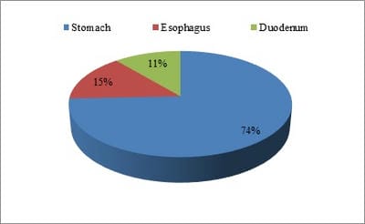
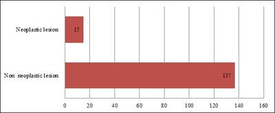
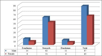
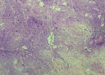
Introduction: The upper gastrointestinal tract (UGT) disorders are more common complaints in clinical practice and have got high degree of mortality and morbidity. Many different types of lesions can affect the upper gastrointestinal tract and can be classified as congenital anomalies, infections, inflammation and neoplastic lesions. Material and Methods: A total of 152 cases of upper gastrointestinal tract biopsies are included in this study, out of which 113 cases were gastric biopsies, 22 cases were esophageal biopsies and the remaining 17 cases were duodenal biopsies. Present study was carried out in the Department of Pathology at Sri Venkateshwaraa Medical College Hospital and Research Centre, Puducherry for a period of one year between March 2018 and February 2019. All the biopsies were performed using fiberoptic endoscopy. Results: It is reported among 152 cases of UGT biopsies 137 were non-neoplastic lesions and 15 were neoplastic. Commonly affected age group was 31-40 years followed by 41 to 50 years. As per the present study, males were affected more predominantly than females. Out of the 22 cases from esophageal biopsies, 16 cases showed non-neoplastic lesions and 6 were neoplastic.

Keywords: Gastrointestinal tract disorders, Endoscopic Biopsy, Histomorphological examination, Helicobacter pylori, Malignant neoplasms and Squamous cell carcinoma

Corresponding Author How to Cite this Article To Browse
J. Gerard Rakesh, Associate Professor, Department of Pathology, Sri Venkateshwaraa Medical College Hospital and Research Centre, Pondicherry, Puducherry, India.
Email:
Theresa MJ, Lavanya M, Gerard RJ, Sultan BK. Evaluating the spectrum of histomorphological patterns on endoscopic biopsy in patients with upper gastrointestinal tract disorders. Trop J Pathol Microbiol. 2019;6(1):1-8.
Available From
https://pathology.medresearch.in/index.php/jopm/article/view/401

Manuscript Received Review Round 1 Review Round 2 Review Round 3 Accepted
10-12-2019 20-12-2019 24-12-2019 28-12-2019
Conflict of Interest Funding Ethical Approval Plagiarism X-checker Note
No Nil Yes 14%

© 2019 by J. Margaret Theresa, Lavanya M., J. Gerard Rakesh, K. Sultan Basha and Published by Siddharth Health Research and Social Welfare Society. This is an Open Access article licensed under a Creative Commons Attribution 4.0 International License https://creativecommons.org/licenses/by/4.0/ unported [CC BY 4.0].

Introduction

The upper gastrointestinal tract disorders are the common complaints in the clinical practice and has got high degree of mortality and morbidity [1]. The upper gastrointestinal tract comprises of oral cavity, esophagus, stomach and duodenum. Several lesions can affect the UGT which are broadly classified into congenital anomalies, infections, inflammation, benign and malignant neoplasms [2].

To visualize and to get the specimen from the gastrointestinal tract a flexible, fiberoptic endoscopy is used. Endoscopic biopsy in combination with histopathological examination plays an important role in the early diagnosis of esophago-gastro-duodenal lesions [3].

Though endoscopic biopsies are invasive procedures, it is considered to be the gold standard for determining UGT lesions. It also helps the clinician and pathologist to correlate with the clinical data [4].

Endoscopic biopsy coupled with histopathological examination in patients with gastrointestinal disorders help us to provide early detection of pre-cancerous and cancerous lesions and timely management of the disease [5].

Endoscopic biopsies are not only used for the early diagnosis of lesion but also for monitoring the course of disease, its extent, response to therapy and early detection of complications [6,7].

Classification of gastritis based on Sydney system, emphasizes the importance of morphological, etiological and topographical informations. This system was revised in the year 1994 at Houston gastritis workshop [8].

The updated Sydney system explains the chronic inflammation, activity, atrophy, intestinal metaplasia and histological severity of H. Pylori density [9].

H. Pylori has been confirmed to be the causative organism for the development of gastric carcinoma. It is more frequently found within the mucous layer and in the gastric pits, which is often visible in Hematoxylin and Eosin stain. Special stains like Giemsa, Warthin starry stain can help us in easy demonstration of H.pylori [10,11].

According to National Cancer Registry - Carcinoma of stomach and esophagus are seen commonly in males. Early detection of these cancers through endoscopic biopsies, can provide appropriate

treatment and have great impact on the survival of the patients [12,13].

Therefore, this study is planned to evaluate the histomorphological patterns of upper gastrointestinal disorders on endoscopic biopsy and to correlate various histomorphological lesions of upper gastrointestinal tract with respect to age, sex and with other clinical details.

Materials and Methods

Type of study: Prospective, observational and descriptive.

Place of study: Department of Pathology at Sri Venkateshwaraa Medical College Hospital and Research Centre, Pondicherry.

Duration of study: One year between March 2018 and February 2019.

Sample collection: The present study included upper gastrointestinal tract biopsies received in the department of pathology. The biopsies were processed and section cutting were carried out. Those sections were stained by Hematoxylin and Eosin. Special stains such as Giemsa, Warthin starry stains were also done whenever indicated. The stained slides were examined and histomorphological patterns were studied.

All the relevant data such as age, gender, duration of symptoms of the patients were also obtained and documented. Other routine investigation results and radiological examination like X–ray and ultrasound reports of the patients were also recorded.

Inclusion criteria: All the upper gastrointestinal endoscopic biopsies, which included the lesions from esophagus, stomach and up to second part of duodenum.

Exclusion criteria: Patients presenting lesion in the oral cavity, pharynx and beyond the second part of duodenum.

Results

In the present study, the frequency of distribution of histomorphological patterns on endoscopic biopsy in patients with upper gastrointestinal tract disorders was analyzed.

Among the 152 cases of UGT biopsies,113 biopsies were from stomach (74%). A total of 22 biopsies were from esophagus which showed (15%) and the


remaining 17 biopsies were from duodenum (11%) showed in Figure 1.

patho_401_O.jpg

Fig-1: Site wise distribution of UGT biopsies.

Among the 152 cases of upper endoscopic biopsies 137were non-neoplastic (90%) and the other15were neoplastic lesions (10%) as represented in Figure 2.

patho_401_02.jpg

Fig-2: Frequency of distribution of neoplastic and non-neoplastic lesions.

As per the gender concern, out of the total 152 cases of UGT biopsies 87 were from males and the remaining 65 were from females. Distribution of UGT lesions in esophagus, stomach and duodenum among males and females as represented in Figure 3.

patho_401_03.jpg

Fig-3: Frequency of distribution of lesions in male and female.

The study result in relevance to the age groups revealed that, among the 152 cases of upper gastrointestinal biopsies, the commonly affected age group was 31-40 years followed by 41 to 50 years.

The youngest age included in the study was 16 years female with esophagitis. The elderly age included in the study was 72 years male with gastric carcinoma as represented in Table 1.

Table-1: Frequency of distribution of lesion according to age.

Age Esophagus Stomach Duodenum
<20 1 - -
21-30 5 6 5
31-40 6 56 10
41-50 4 32 2
51-60 4 8 -
61-70 2 3 -
71-80 - 8 -
Total 22 113 17

Common non-neoplastic lesion among the esophageal biopsies were chronic non-specific esophagitis, followed by esophageal ulcer.

patho_401_04.jpg

Fig-4: Microscopic feature of squamous cell carcinoma of esophagus, Haematoxylin and Eosin stain (10X).

Table-2: Frequency of distribution of esophageal lesion

Histopathological Diagnosis No.of Cases Percentage
Non-neoplastic lesions
Chronic non-specific esophagitis 9 40.9
Esophageal ulcer 3 13.63
Esophageal candidiasis 1 4.54
Barrets esophagus 3 13.63
Neoplastic lesions
Carcinoma in situ 2 9.09
Squamous cell carcinoma 4 18.18
Total 22 100

The common neoplastic lesion observed was squamous cell carcinoma (Figure 4) and there are three cases of barrets esophagus was also observed


in this study as represented in Table 2.

Among 113 cases of gastric biopsies 104 were non-neoplastic and 9 were neoplastic lesions. Similarly, among the non-neoplastic lesions chronic superficial gastritis observed in 24 cases followed by chronic active gastritis in 22 cases and an equal number of cases in chronic superficial gastritis with intestinal metaplasia (Figure 5). Among the neoplastic lesion’s adenocarcinoma of stomach (Figure 6) was the commonly observed lesions shown in Table 3.

patho_401_05.jpg

Fig-5: Chronic superficial gastritis with intestinal metaplasia, Haematoxylin and Eosin stain (10X).

patho_401_06.jpg

Fig -6: Adenocarcinoma of stomach, Haematoxylin and Eosin stain(40X).

Table-3: Frequency of distribution of gastric lesion

Histopathological diagnosis No. of Cases %
Non- neoplastic lesions
Chronic superficial gastritis 24 21.23
Chronic active gastritis 22 19.43
Chronic superficial gastritis with intestinal metaplasia 22 19.46
H.pylori associated gastritis 21 18.58
Eosinophilic gastritis 3 2.65
Gastric ulcer 8 7.17
Gastric polyp 4 3.53
Neoplastic lesions
Gastricintraepithelial neoplasm 3 2.65
Adenocarcinoma 6 5.30
Total 113 100

Among the17 duodenal biopsies obtained, on analysis, all of them were found as non-neoplastic. The lesion observed was chronic nonspecific duodenitis as represented in Table 4.

Table-4: Frequency of distribution of duodenal lesion

Histopathological diagnosis No. of cases Percentage
Non-neoplastic lesions
Chronic non-specific duodenitis 8 47.06
Duodenal ulcer 6 35.29
Duodenal polyp 3 17.65
Total 17 100

Discussion

Various lesions can affect the upper gastrointestinal tract and may due to congenital anomalies, infections, inflammation, polyps, benign and malignant neoplasms. According to national cancer registry the incidence of gastric and esophageal cancers is gradually increasing according to the Globocan [14,15].

Its worldwide incidence is believed to be increasing and are associated with poor prognosis. Hence there is an essential need to detect the lesion at an early stage.

Endoscopy helps in visualizing the gastrointestinal mucosa. The biopsies taken during the procedure help in confirming the diagnosis and to categorize them. Before the advent of endoscopic biopsy direct access to the lesions of gastrointestinal tract for the confirmation of the diagnosis was difficult, this resulted in difficulty in contemplating adequate and appropriate surgery [16].

Endoscopic biopsy also played an important role in the early detection of malignancies. Biopsies obtained under direct vision, through a working endoscopic channel in greater number of biopsies from a suspected malignancy increases the diagnostic yield [17] and may be useful in making therapeutic decisions to improve the prognosis of the patients.


H.pylori are found to be the frequent organism within the muscular layer and within the gastric pits. The organisms are visible in Hematoxylin and Eosin stain. In Hematoxylin and eosin staining sensitivity and specificity as 69-93% and 87 -90 respectively which are also more easily visualized in special stains like Giemsa and Warthins starry stains.

Particularly it is useful in detecting the bacilli when they are in small number. The specificity can be improved 90-100% by using special stains such as modified Giemsa stain, Warthin –Starry silver stain [18,19]. Electron microscopic examination showed that the bacilli are adherent to the surface epithelium [20,21]. It has been confirmed that the development of gastric cancer arises from background of H. pylori infection [22,23].

Sydney system of classification for gastritis emphasizes the importance of morphological, topographical and etiological information about the lesion. The revised Sydney system which was held in Houston gastric workshop also defined the histological severity of H. Pylori density, inflammation, activity, atrophy and intestinal metaplasia.

The introduction of the Sydney system made it possible to grade histological parameters, identify topographical distribution and etio-pathogenesis of the gastritis. However still non-standard histological reporting formats are widely used for gastritis which make it difficult to identify the patients for clinico-endoscopic surveillance. Additionally, there is no validated test to quantify the density of H. Pylori infection [24].

In the present study, among 152 cases of upper gastrointestinal tract biopsies, more number of cases were received from stomach 113(74.34%), followed by esophagus 22 (14.47%) and duodenum 17 (11.19%). Similar result was observed in a study conducted by Bilal A Sheikh [25], wherein the site wise distribution of endoscopic biopsies showed that in stomach 127 (64.8%), oesophagus 50 (25.51%), gastro-esophageal junction 15 (7.65%) and duodenum 4 (2.04%).

In the case of neoplastic verses non neoplastic lesions in the present study report showed that out of 152 cases 137 (90.13%) were non neoplastic lesions and 15 (9.87%) were neoplastic lesions.

However, yet another study by Khatib W M et al [26] showed a slightly altered report in which the non-neoplastic lesions observed in 75.29% of case

cases and the neoplastic lesions were seen in 24.71% of cases. The variation in the results may due to personal, nutritional and environmental factors of the individuals.

Age and sex wise distribution of lesions of the present study reports showed that the most commonly affected age group was 31-40 years followed by 41 -50 years. Our result is slightly contradict with the studies carried out by Shepherd NA [27].

Shennak M M et al [28] and Islam SM et al [29], Wherein they studied the endoscopic biopsy for gastrointestinal tract lesions in which the commonly affected age group was between fifth and sixth decades of life.

As per the present study, among the 152 cases of upper endoscopic biopsies, 87(57.20 %) were males and 65 (42.80%) were female. Males were the predominant population affected with UGT disorders when compared to female [30].

Similar results were observed by Nwafor CC et al [30], who studied on 190 case of gastrointestinal biopsies showed the result 58% with male predominance and remaining 41% females were affected. The age related difference could be due to varied exposure to the risk factors among the different age groups.

Among the 22 esophageal biopsies received 16 (72%) of them showed non neoplastic lesions and the remaining 6 (28%) were neoplastic lesions. The present study report showed that the common non neoplastic lesion was chronic nonspecific esophagitis, followed by esophageal ulcer. It was also observed that squamous cell carcinoma was the most frequently reported neoplastic lesion.

A study conducted by Rosy Khandelia et al [31] also showed similar findings in that it was observed out of 115 upper gastrointestinal biopsies, 48 (41.74%) were from esophagus in origin and the most common non neoplastic lesion was Chronic nonspecific esophagitis. The study also confirmed that the squamous cell carcinoma of esophagus was the common neoplastic lesion.

In the case of gastric biopsies 113 as per the present study 104 (92%) were non neoplastic and only 9 (8%) cases were neoplastic, which is similar to that of the study result of Syed Imtiyaz et al [32] report showed that 57.4% of cases were non neoplastic and 42.6% were neoplastic.


In the present study, among the non-neoplastic lesions it was observed that chronic superficial gastritis, 24 (21.23%) followed by chronic active gastritis 22 (19.43%), chronic superficial gastritis with intestinal metaplasia 22 (19.43%) and 21 cases (18.58%) were H. Pylori associated gastritis. The common neoplastic lesion observed in the present study is adenocarcinoma of stomach.

The study by Rupendra Thapa et al [33] stated that the common non-neoplastic lesion observed was mild chronic gastritis 22 (27.5%) followed by

chronic active gastritis 15 (18.75%) while H.pylori was present in 13(16.25%) and common neoplastic lesion were adenocarcinoma which correlated with the present study report.

Among the 17 cases of duodenal biopsies in the present study result showed that all 17 were non neoplastic and the commonly observed condition was chronic nonspecific duodenitis.

This result is in correlation with the study result of previous study [34].

Conclusion

The study report concluded that the upper gastrointestinal biopsies, on examination in combination with histopathological studies provides a useful information in the determination of overall extent of mucosal architecture and the use of special stains like Giemsa, Warthin starry stains made it easy to demonstrate H. Pylori in the gastric pits and mucous layer.

All the biopsies were performed using fiberoptic endoscopy which plays an important role in the improved visualization and accuracy in the UGT lesions could be directly inspected and helps to evaluate the various clinical conditions at the early stage.

Further the early result favors to correlate various disorders in correspondence to the clinical parameters and also the neoplastic and non-neoplastic lesions. Use of fiberoptic endoscopic biopsy helps to detect the site and distribution of the benign and malignant lesions.

Early and more specific diagnosis help for the management of disease which intern reduces the mortality and morbidity of patients. For better understanding of disease, its progression and associated risk factors, further studies are essential

to describe the biological behavior of non-neoplastic and neoplastic lesions.

What this study adds to the existing knowledge?

The present study emphasis the usefulness of fiberoptic endoscopic biopsies along with histopathological examination helps in identifying the histomorphological patterns in upper gastrointestinal tract at an early stage. Early detection of these gastrointestinal tract diseases results in timely management, which also decreases the mortality and morbidity of patients.

Author’s contributions

Dr. J. Margaret Theresa: Designed the study, protocol writing, manuscript preparation.

Dr. M. Lavanya: Manuscript review and guided the research work.

Dr. J. Gerard Rakesh: Manuscript review and guided the research work.

Mr. K. Sultan Basha: Data acquisition and statistical analysis.

Reference

  1. Blackstone MO. Endoscopic interpretation- Normal and pathologic appearances of Gastrointestinal tract. New York- Raven Press. 1984, P 13-15.
  2. Gulia SP, Chaudhury M, Noorunnisa N, Balakrishnan CD, Balagurunathan K. Interpretation of Upper GastroIntestinal Tract Endoscopic Mucosal Biopsies–A Study Conducted In Teaching Hospital In Puducherry, India. Int J Med Health Sci. 2012;1(3)17-24.
  3. Memon F, Baloch K, Memon AA. Upper gastrointestinal endoscopic biopsy; Morphological spectrum of lesions. Professional Med J. 2015;22(12)1574-1579.
  4. Krishnappa R, Horakerappa MS, Mangala AK, Gouri M. A study onhistopathologic spectrum of upper gastrointestinal tract endoscopic biopsies. Int J Medical Res Health Sci. 2013;2(3)418-424.

  1. Afzal S, Ahmad M, Mubarik A, Saeed F, Rafi S, Saleem N, Qur AH. Morphological spectrum of gastric lesions-Endoscopic biopsy findings. Pak Armed Forces Med J. 2006;56(2)143-149.
  2. Hirachand S, Sthapit RR, Gurung P, Pradhanang S, Thapa R, Sedhai M, RegmiS. Histopathological spectrum of upper gastrointestinal endoscopic biopsies. J BP Koirala Inst Health Sci. 2018;1(1)67-74.
  3. Saha. Studies on Helicobacter pylori- National Institute of Cholera and Enteric Diseases. Annual Report. 2004-2005.
  4. Teriaky A, Al Nasser A, McLean C, Gregor J, Yan B. The utility of endoscopic biopsies in patients with normal upper endoscopy. Can J Gastroenterol Hepatol. 2016;2016.
  5. Sgambato D, Visciola G, Ferrante E, Miranda A, Romano L, Tuccillo C, et al. Prevalence of H Pylori infection in sexual partners of H pylori-infected subjects- Role of gastroesophageal reflux. United Europe Gastroenterol J. 2018;6(10)1470-1476.
  6. Kobayashi O, Eishi Y, Ohkusa T, Ishige. Gastric mucosal density of H- Pylori estimated by real-time PCR compared with results of urea breath test and histological grading. J Med Microbiol. 2002;51(4)305-311.
  7. Shennak MM, Tarawneh MS, Al Sheik. Upper gastrointestinal diseases in symptomatic Jordanians- A prospective study. Ann Saudi Med. 1997;17(4)471-474.
  8. Nafees A Qureshi, Michael T Hallissey, John W. Fielding Outcome of indexupper gastrointestinal endoscopy in patients presenting with dysphagia in atertiary care hospital - A 10 years review. BMC Gastroenterol. 2007;7;43.
  1. National Cancer Registry Programme. First All India Report 2001-2002. Vol 1. Indian Council of Medical Research- Bangalore, India. 2004
  2. International Agency for Research on Cancer. Latest world cancer statistics Global cancer burden rises to 14. 1million new cases in 2012- Marked increase in breast cancers must be addressed. World Health Organization.
  3. Shin A, Won YJ, Jung HK, Kong HJ, Jung KW, Oh CM, et al. Trends in incidence and survival of esophageal cancer in Korea- Analysis of the Korea Central Cancer Registry Database. J Gastroenterol Hepatol. 2018;33(12)1961-1968.
  4. Khandelia R, Saikia M. Histopathologic Spectrum of Upper Gastrointestinal Tract Mucosal Biopsies- A Prospective Study. Int J Med Sci Clinic Invent. 2017;4(11)3314-3316.
  5. Ozturk S, Serinsoz E, Kuzu I, Ensari A, Erden E, Kansu A, et al. The Sydney System in the assessment of gastritis- Inter-observer agreement. The Turkish J Gastroenterol. 2001;12(1)36-39.
  6. Fallone CA, Loo VG, Lough J, Barkun AN. Hematoxylin and eosin staining of gastric tissue for the detection of Helicobacter pylori. Helicobacter. 1997;2(1)32-35.
  7. Pandya HB, Agravat HH, Patel JS, Sodagar NR. Emerging antimicrobial resistance pattern of H Pylori in central Gujarat. Indian J Med Microbiol. 2014;32(4)408-413.
  8. Singh V, Trikha B, Vaiphei K, Nain CK, Thennarasu K, Singh K. Helicobacter pylori- Evidence for spouse-to-spouse transmission. J Gastroenterol Hepatol. 1999;14(6)519-522.
  9. Kadish SL, Kochman ML. Endoscopic diagnosis and management of gastrointestinal malignancy. Oncol. 1995;9(10)967-983.

  1. Laine L, Lewin DN, Naritoku W, Cohen H. Prospective comparison of H&E, Giemsa, and Genta stains for the diagnosis of Helicobacter pylori. Gastrointest endos. 1997;45(6)463-467.
  2. Morilla A, Melón S, ?lvarez-Argüelles ME, Armesto E, Villar H, de Oña M. Utility of normalized genome quantification of H Pylori in gastric mucosa using an in-house real-time polymerase chain reaction. PloS one. 2017;12(6)e0178674.
  3. Hassan TM, Al-Najjar SI, Al-Zahrani IH, Alanazi FI, Alotibi MG. H Pylori chronic gastritis updated Sydney grading in relation to endoscopic findings and H pylori IgG antibody-diagnostic methods. J Microscop Ultrastruct. 2016;4(4)167-174.
  4. Sheikh BA, Hamdani SM, Malik R. Histopathological spectrum of lesions of upper Gastrointestinal tract-A study of endoscopic biopsies. GJMEDPH. 2015;4(4)1-8.
  5. Shepherd NA, Valori RM. The effective use of gastrointestinal histopathology-guidance for endoscopic biopsy in the gastrointestinal tract. Front Gastroenterol. 2014;5(2)84-7.
  6. Islam SM, Ahmed AM, Ahmad MS, Hafiz SA. Endoscopic and histologicDiagnosis of upper gastrointestinal lesions, Experience in a port city of Bangladesh. Chattagram Maa-O-Shishu Hospital Med College J. 2014;13(3)11-14.
  1. Khatib WM, Demde RB, Aher VC, Patel PM. Histopathological Spectrum of Non-Malignant Lesions of Gastrointestinal Tract-An Institutional Stud. IOSR J Dent Med Sci. 2016;15(10)113-116.
  2. Nwafor CC, Nwafor NN, Etuk EB, Kanu O. Histopathological spectrum of gastrointestinal lesions seen in university of uyo teaching hospital, South–South Nigeria. Ann Trop Pathol. 2019;10(1)27-33.
  3. Khandelia R, Saikia M. Histopathologic Spectrum of Upper GastrointestinalTract Mucosal Biopsies- A Prospective Study. Int J Med Sci Clinic Invent. 2017;4(11)3314-3316.
  4. Hussain SI, Reshi R, Akhter G, Beigh A. Clinico histopathological study ofupper gastrointestinal tract endoscopic biopsies. Int J Curr Res Rev. 2015;7(16)78-85.
  5. Thapa R, Lakhey M, Yadav PK, Kandel P, Aryal C, Subba K. Histopathologicalstudy of endoscopic biopsies. J Nepal Med Assoc. 2013;52(190)354-356.
  6. Prasaad PR, Rao B. Histopathological spectrum of gastrointestinal lesions-anexperience in a tertiary care centre in South India. Int J Res Med Sci. 2016;4(8)3407-3412.
  7. Shennak MM, Tarawneh MS, Al Sheikh TM. Pattern of upper gastrointestinal disease in symptomatic Jordanians- a prospective endoscopic study of 5657 patients. Dirasat. 1998;25(2)69-81.