E-ISSN:2456-1487
P-ISSN:2456-9887
RNI:MPENG/2017/70771

Research Article

Granulomatous

Tropical Journal of Pathology and Microbiology

2020 Volume 6 Number 2 February
Publisherwww.medresearch.in

Granulomatous pathology in salivary glands: a secondary health care center experience

Malhotra P.1, Saraf A.2*, Bansal N.3
DOI: https://doi.org/10.17511/jopm.2020.i02.02

1 Pooja Malhotra, Specialist Pathology Grade I, Department of Pathology, ESIC Model Hospital, Baddi, Himachal Pradesh, India.

2* Ashish Saraf, Specialist Pathology, Department of Pathology, ESIC Model Hospital, Baddi, Himachal Pradesh, India.

3 Naveen Bansal, Medical Officer, Department of Pathology, ESIC Model Hospital, Baddi, Himachal Pradesh, India.

Introduction: The diagnosis of parotid tuberculosis requires a high degree of clinical suspicion, so as to avoid unnecessary surgery, and fine-needle aspiration cytology is an easy and reliable investigation for the same. Salivary glands are suitable for unguided fine needle aspiration cytology (FNAC) because of their superficial location. Methods: The present study involved a total of 42 cases of salivary gland swellings in which fine needle aspiration cytology was performed in the department of pathology of our hospital from December 2011 to August 2019. Results: The prevalence of granulomatous inflammation among salivary gland swellings was 38.1 % (n=16). Anti-tubercular treatment was given in 5 patients, all of whom showed improvement after anti-tubercular treatment. Therefore, 5 out of the total of 16 cases were a case of tuberculous parotitis (31.2 %). Conclusion: Although tuberculous parotitis is a rare entity, it should be always kept in mind as a differential during the evaluation of solitary parotid mass lesion. Fine needle aspiration cytology of all mass lesions of salivary glands must be done before doing excision of salivary gland so as to avoid unnecessary surgery.

Keywords: Salivary gland, Tuberculous parotitis, Fine needle aspiration cytology

Corresponding Author How to Cite this Article To Browse
Ashish Saraf, Specialist Pathology, Department of Pathology, ESIC Model Hospital, Baddi, Himachal Pradesh, India.
Email:
Malhotra P, Saraf A, Bansal N. Granulomatous pathology in salivary glands: a secondary health care center experience. Trop J Pathol Microbiol. 2020;6(2):118-123.
Available From
https://pathology.medresearch.in/index.php/jopm/article/view/394

Manuscript Received Review Round 1 Review Round 2 Review Round 3 Accepted
14-01-2020 24-01-2020 30-01-2020 03-02-2020
Conflict of Interest Funding Ethical Approval Plagiarism X-checker Note
No Nil Yes 13%

© 2020 by Pooja Malhotra, Ashish Saraf, Naveen Bansal and Published by Siddharth Health Research and Social Welfare Society. This is an Open Access article licensed under a Creative Commons Attribution 4.0 International License https://creativecommons.org/licenses/by/4.0/ unported [CC BY 4.0].

Introduction

Tuberculosis is a chronic illness that is more common in developing countries such as India. It mainly involves lungs and is characterized by granulomatous inflammation with or without caseous necrosis. Extrapulmonary forms of the disease account for 20 % of overall active tuberculosis and can affect any organ of the body. However, the involvement of salivary glands is a very rare event [1]. Tuberculosis of salivary glands is rare because of the bactericidal effect of salivary enzymes [2]. Only about less than 200 cases are reported in the literature [1,2].

It is difficult to distinguish tuberculous parotitis from other inflammatory conditions and neoplastic lesions of the salivary glands. Tuberculosis of the salivary gland should be considered especially when there is a family history of tuberculosis. The parotid gland and its lymph nodes can be involved by two mechanisms. Firstly, as a result of a focus of mycobacterial infection in the oral cavity (infected tooth, tonsil or sputum). Secondly, as a result of hematogenous or lymphatic spread from a distant primary lung focus. Tuberculous parotitis may mimic neoplasm even on imaging studies. Most of the reported parotid swelling in tuberculous parotitis is unilateral.

Clinically it presents as a slow-growing, painless and non-tender salivary gland swelling. However, the swelling can be a painful and tender lump in case of abscess or fistula formation. Salivary glands are suitable for unguided fine needle aspiration cytology (FNAC) because of their superficial location. The diagnosis of tuberculous parotitis requires a high degree of clinical suspicion, so as to avoid unnecessary surgery, and FNAC is an easy and reliable investigation for its diagnosis [3]. Incision and drainage should not be performed until tuberculosis has been ruled out otherwise fistula or sinus formation may occur [1,2]. FNAC is ideally suited for use in resource-limited settings, especially in developing countries. Therefore, the present study was planned to assess the diagnostic accuracy of FNAC in salivary gland swellings.

Materials and Methods

Type of study- The present study was a retrospective observational study that involved a total of 42 cases of salivary gland swellings in which FNAC was performed in the department of

pathology of our hospital from December 2011 to August 2019.

Inclusion criteria- Cases of salivary gland swellings which showed granulomatous inflammation on FNAC were included in the study.

Exclusion criteria- Inadequate aspirates in which cytological diagnosis was not possible, were excluded from the study.

Procedure- All the cases of parotid swellings were analyzed by taking a detailed history, clinical findings, and investigation findings (radiological and laboratory findings). FNAC of the salivary glands was performed under aseptic conditions using a 21-gauge needle attached to an FNAC gun with aspiration using a 20 ml syringe. Air-dried smears were stained with Giemsa stain. Alcohol fixed smears were stained with Papanicolaou stain, Haemotoxylin and Eosin stain. Acid-fast bacilli (AFB) staining was done in all the patients.

Ethical Consideration- The study was conducted in accordance with the ethical standards of the institute and with the 1964 Helsinki declaration and its later amendments.

Results

Out of these 42 cases of salivary gland swelling, 14 cases of granulomatous inflammation of the parotid gland and 2 cases of granulomatous inflammation of the submandibular salivary gland were included in the study. The detailed clinical and investigative findings are shown in Table 1. The prevalence of granulomatous inflammation among salivary gland swellings was 38.1 % (n=16). Out of these 16 cases, 11 were males and 5 were females. Male: Female ratio was 2.2:1. The duration of illness was 1-12 months in 11 patients (68.75%), less than 1 month in 3 patients (18.75%) and more than 12 months in 2 patients (12.5%).

The parotid gland was involved in 14 patients (87.5%), whereas the submandibular gland was involved in 2 patients (12.5%). A family history of tuberculosis was present in 2 patients (12.5%) while a history of subjective weight loss was present in 1 patient (6.2%). One patient presented with recurrent parotid swelling, 9 months after an operation for parotid swelling. The size of predominant salivary gland swelling was less than 3 cm in 12 patients (75%) and ≥ 3 cm in 4 patients (25%). In 3 patients adjacent lymph nodes were also palpable.


Table-1: Clinical data of patients with granulomatous salivary gland swellings.

Age/Sex Site Duration Description Material aspirated ZN stain for AFB Other associated findings
40/F Left parotid 1 year 2 x 1.4 cm Whitish particulate Negative Submental and submandibular lymph nodes; Culture-Mycobacterial species;AFB of the lymph node FNAC is negative
27/M Left Parotid 1.5 years 3 cm, firm to hard, non-tender Particulate Negative Family history of TB present; previous FNAC -Occasional granuloma; AFB-negative
28/M Right parotid 15 days-newer swelling, operated 9 months back 2.1 x 1.3 cm, non-tender, firm to hard Particulate, mucoid Negative Operated 9 months back, new swelling since 15 days, USG-Right parotid mass- 2.8 x 1.8 x 2 cm, mixed echogenicity, septations; Colour ppler- increased vascularity
38/M Right parotid 0.25 years 1 cm, soft, mobile Particulate Negative FNAC was repeated
30/M Left submandibular 2.5 years 1.2 x 1.1 cm, Blood mixed Negative Previous FNAC -Reactive hyperplasia
32/M Right parotid 1 year 1.3 x 1.2 cm, firm Particulate blood mixed Negative  
37/F Left parotid   2 x 1.5 cm, firm, mildly tender Pus aspirated Negative  
55/F Left parotid Few months 1 cm, mobile, firm, tender Scanty Negative  
27/M Left parotid 1 month 2 cm, painless Blood mixed particulate Negative No H/O past TB, ATT taken, symptoms relieved with treatment
45/M Left parotid 1 week 4 x 3 cm, painful Pus aspirated Negative Painful, HIV, HBsAg, HCV negative, no H/O fever, not resolving to treatment
21/M Right parotid 3 months 1.3 cm, firm Blood mixed particulate Positive Firm, no other complaints, HIV, HBsAg, HCV –negative, ATT taken
39/M Left parotid 15 days 4 x 3 cm Blood mixed Negative HIV, HBsAg, HCV –negative; random blood glucose - 500 mg/dL, ATT taken; symptoms relieved with treatment
37/F Left parotid 1 month 1.2 cm Blood mixed Negative  
40/M Left parotid 1 week 1 cm, soft to firm, non-tender Bloody aspirate Negative No H/O fever, cough; Left submandibular lymph node 1 cm; FNAC was repeated, FNAC of submandibular lymph node showed granulomatous ymphadenitis
22/M Left submandibular 1 month 1 cm, mobile, non-tender Scanty blood mixed Negative  
32/F Right parotid 1.5 months 4 x 3 cm, painless Blood mixed Negative H/O subjective weight loss, H/O contacts with T.B., USG-Right parotid gland-enlarged with a large cystic lesion; ATT taken, symptoms elieved on treatment; FNAC of right cervical lymph node shows reactive lymphadenitis

ZN: ZiehlNeelsen; USG: Ultrasonography; H/O: History of; ATT: Antitubercular treatment

The largest lymph node measured 1.3 cm approximately in maximum dimension. A complete blood count report was available in 7 out of 16 patients. Out of these 7 patients, the differential

monocyte count was more than 10 % in only 2 patients. Absolute monocyte count was less than the upper normal limit of 1000/mm3 in all the patients. Upon FNAC of the swelling, blood mixed


material was aspirated in 9 patients (56.2 %), pus was aspirated in 2 patients (12.5%), while whitish material and mucoid material were aspirated in one patient each (6.2 %). Both patients in whom pus was aspirated had a tender swelling. FNAC was repeated in 3 patients.

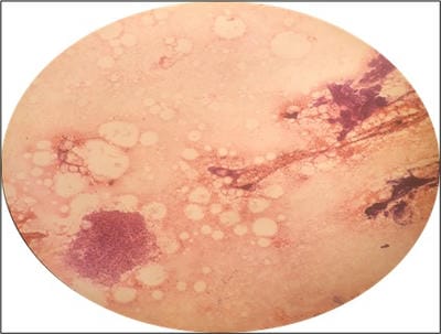
The microscopic findings of FNAC are shown in Table 2. Ziehl-Neelsen (ZN) staining was done in all patients and only 1 patient was positive for AFB. In one patient culture report from aspirated material showed mycobacterium species.

Table-2: Microscopic findings upon FNAC.

Microscopy of salivary gland shows No. of cases (% of cases)
Epithelioid cell granuloma 16 (100%)
iant cells 8 (50 %)
Necrosis 3 (18.8 %)
Neutrophils 3 (18.8 %)
Acinar cells 6 (37.5 %)
Ductal cells 2 (12.5 %)

Anti-tubercular treatment (ATT) was given in 5 patients, all of whom showed improvement after the treatment. Therefore, 5 out of the total of 16 cases were most likely to be tuberculous parotitis (31.2 %), while 12 cases had granulomatous diseases other than tuberculosis. In 1 patient, the parotid swelling reduced completely after antibiotic treatment for 5 days.

Discussion

Tuberculosis is a major health problem, especially in developing countries. However, tuberculosis of salivary glands is a rare variety of extrapulmonary tuberculosis. Tuberculosis of salivary glands is rare because of the bactericidal effect of salivary enzymes [2]. The pathogenesis of parotid tuberculosis remains unclear. The parotid gland and its lymph nodes can be involved by two mechanisms. Firstly, as a result of a focus of mycobacterial infection in the oral cavity (infected tooth, tonsil or sputum). Secondly as a result of hematogenous or lymphatic spread from a distant primary lung focus [1,4-6]. In the present study, swellings in 3 patients were associated with lymphadenopathy. The most commonly implicated agent is Mycobacterium Bovis and rarely by atypical mycobacteria [6].

Lee and Liu described the clinicopathological features of tubercular parotitis based on a study of 49 cases of tubercular parotitis [7]. Two clinical forms of tubercular parotitis have been described.

Acute tuberculous sialadenitis usually presents with diffuse glandular enlargement and is often painful. In the present study, 3 cases had painful enlargement of the parotid gland. Chronic sialadenitis manifests as an asymptomatic localized lesion within the parotid gland, which slowly keeps growing in size for many years [4]. This mimics a neoplasm [2]. It may also present as pre-auricular fistula and in advanced cases, facial nerve involvement [2,8]. The differential diagnosis includes malignancy of the parotid gland, mumps, sarcoidosis, and actinomycosis. A total of 25 % of patients with parotid tuberculosis have a concomitant pulmonary infection [1,2].

Parotid tuberculosis becomes a real diagnostic problem in the absence of clinical lung disease and without any systemic symptoms or signs of tuberculosis [2]. A chest radiograph may be helpful in cases of associated pulmonary tuberculosis. The use of erythrocyte sedimentation rate and tuberculin skin testing (PPD) can provide valuable information but requires an initial suspicion [1]. One of our cases had slightly raised ESR. Similarly, ESR was raised in the case report by Sharma T et al. [9]. Apart from the presence of non-caseating granulomas, elevated angiotensin-converting enzyme levels and a negative Mantoux test favor a diagnosis of sarcoidosis over other granulomatous lesions [9].

FNAC is an easy and reliable outpatient procedure for the diagnosis of tubercular parotitis, and it is ideally suited for use in resource-limited settings, especially in developing countries. FNAC has a sensitivity of 81-100 % and specificity of 94-100 % in the diagnosis of parotid tuberculosis [10]. A total of 20.6 % of FNAC are non-diagnostic; which could be due to the complexity of tissue architecture. However, this can be overcome by doing a repeat FNAC. Also, in the case of active infection, the FNAC report may be inconclusive [11]. FNAC was repeated in 3 out of 16 patients in the present study.

In the current study, FNACs of 38.1 % of the patients showed granulomatous inflammation (Figure 1) and at least 11.9% of the swellings were likely due to tuberculosis. This is more as compared to the study by Van Der Walt et al in which 12.1% of the swellings showed granulomatous inflammation and only 1.9% of these swellings were due to tuberculosis [11]. This difference could be due to a lesser prevalence of tuberculosis in western countries.


patho_394_01.jpg

Figure-1: Fine needle aspiration smear from parotid gland showing a cluster of epithelioid histiocytes and cluster of acinar cells (H and E smear, 10X).

In the present study, 5 out of a total of 37 parotid FNACs (13.5%) were cases of parotid tuberculosis. In different studies, tuberculous parotitis accounts for 2.5 % to 10 % of parotid pathologies [12]. However, the incidence rate of parotid tuberculosis in the present study was 0.6 cases/ year which was less as compared to that reported by Sengupta A et al (2 cases/year) [8]. The incidence of parotid tuberculosis could be falsely low in the present study because nucleic acid amplification testing was not performed because of the cost factor.

In the present study, all cases were unilateral. Most of the reported parotid swelling in tuberculous parotitis is unilateral but bilateral parotitis has also been described [1,4]. Parotid glands are the most common salivary glands involved in primary tuberculosis, whereas submandibular glands are most commonly involved in systemic tuberculosis [5].

If the diagnosis of tuberculosis of the salivary gland is made pre-operatively, then the patient is spared of surgery and can be cured by ATT under Directly observed treatment short course [5]. When imaging and FNAC are inconclusive, then excision biopsy of the parotid becomes necessary [2,6,13]. Incision and drainage should not be performed until tuberculosis has been ruled out otherwise fistula or sinus formation may occur [1,2].

Limitations- The incidence of parotid tuberculosis could be falsely low in the present study because nucleic acid amplification testing was not performed on the aspirate sample.

Conclusion

Although tuberculous parotitis is a rare entity, it should be always kept in mind as a differential during the evaluation of solitary parotid mass lesion. History of contact with tuberculosis, the clinical appearance of abscess in the salivary gland, elevated ESR, positive Mantoux test and ultrasound examination can help diagnose tuberculous parotitis in a particular patient. FNAC of all mass lesions of salivary glands must be done before doing excision of salivary gland so as to avoid unnecessary surgery.

What does this study add to the existing knowledge?

The present study emphasizes that FNAC is an easy and reliable investigation, which can help in the diagnosis of tubercular parotitis. As compared to the western data, our data shows that tuberculous parotitis constitutes a significant proportion of parotid swellings in India. Therefore, FNAC must be performed in all cases of salivary gland swellings for accurate diagnosis and treatment.

Author’s contributions

Dr. Pooja Malhotra conceived and designed the study. Dr. Pooja Malhotra and Dr. Ashish Saraf performed the study. Dr. Ashish Saraf and Dr. Naveen Bansal performed the statistical analysis. Dr. Naveen Bansal prepared the final manuscript. All authors read and approved the final manuscript.

Reference

  1. Maurya MK, Kumar S, Singh HP, Verma A. Tuberculous parotitis- A series of eight cases and review of literature. Natl J Maxillofac Surg. 2019;10(1)118-122.
  2. Satarkar RN, Sangwaiya A, Singh P, Kalhan S, Sharma P, Saini N. Fine needle aspiration cytology-based spectrum of salivary gland lesions at a teaching institute in north India. J Pathol Microbiol. 2020;5(12)976-982.

  1. Jain A, Meher R, Wadhwa V, Raj A, Tandon S. Parotid tuberculosis- A diagnostic dilemma. Acta Oto-Laryngologica Case Reports. 2017;2(1)73-76.
  2. Poorey VK, Tyagi A. Accuracy of fine needle aspiration cytology in head and neck masses. Indian J Otolaryngol Head Neck Surg. 2014;66(2)182-186.
  3. Bhattacharya G, Khadanga S, Dugar D, Kumar Thatoi P, Prasad Satapathy S. Tubercular parotitis- a forgotten entity. Int J Med Res Rev. 2016;4(7)1108-1110.
  4. Thakur JS, Thakur A, Mohindroo NK, Mohindroo S, Sharma DR. Bilateral parotid tuberculosis. J Global Infect Dis. 2011;3(3)296-299.
  5. Bakshi GD, Shaikh AS, Borisa AD, Jamadar NM, Abhishek G Singh, Sameer H Vora. Primary tuberculosis of parotid gland mimicking parotid tumour. Bombay Hosp J. 2009;51(2)14-16.
  6. Gupa V, Patankar K, Shinde A, Bhosale C, Tamhane A. Tuberculosis of the parotid gland. Case Rep Radiol. 2012; 2012;278793.
  1. Lee IK, Liu JW. Tuberculous parotitis- Case report and literature review. Ann Otol Rhinol Laryngol. 2005;114(7)547-551.
  2. Sengupta A, Paul SS, Majumder L, Jyoti. Tuberculosis of the parotid gland-How we diagnose and manage it?. J Evidence Based Med Healthcare. 2016;3(19)798-802.
  3. Sharma T, Joshi D, Khurana A, Gupta V, Kapoor N. Bilaterally enlarged parotids and sicca symptoms as a presentation of sarcoidosis- Pivotal role of aspiration cytology in diagnosis. J Cytol. 2015;32(4)281-283.
  4. Gayathri B, Kalyani R, Manjula K. Primary tuberculous parotitis. J Cytol. 2011;28(3)144-145.
  5. Van Der Walt JD, Leake J. Granulomatous sialadenitis of the major salivary glands, A clinicopathological study of 57 cases. Histopathol. 1987;11(2)131-144.