E-ISSN:2456-1487
P-ISSN:2456-9887
RNI:MPENG/2017/70771

Case Report

Leiomyosarcoma

Tropical Journal of Pathology and Microbiology

2020 Volume 6 Number 3 March
Publisherwww.medresearch.in

Leiomyosarcoma of the tongue: A case report

Gahlot A.1*, Zaheer S.2
DOI: https://doi.org/10.17511/jopm.2020.i03.08

1* Adwaita Gahlot, Department of Pathology, Vardhman Mahavir Medical College, New Delhi, India.

2 Sufian Zaheer, Department of Pathology, Vardhman Mahavir Medical College, New Delhi, India.

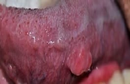
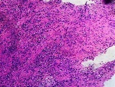
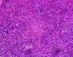
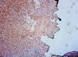
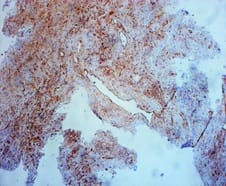
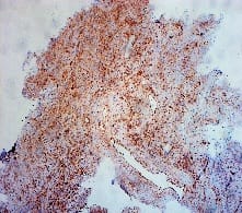
Leiomyosarcoma (LMS) of the tongue is an extremely rare malignant mesenchymal tumor. Visceral lesions are the most common and account for about 7% of all soft-tissue sarcomas. The lesion is rare in the head and neck, making up 3%–10% of all leiomyosarcomas. Primary leiomyosarcoma of the tongue is extremely rare leiomyosarcomas andrepresents 5–6% of all soft tissue sarcomas and usually, they arise in the uterus, gastrointestinal tract, and retroperitoneum. The most common sites of head and neck regions are the oral cavity (22%), the sinonasal tract (19%), and the facial skin-subcutis (17%). Smooth muscle tumors in the oral cavity usually arise from the buccal mucosa, the gingiva, and the oral floor,but only a few numbers of cases arise in the tongue.The present case shows the tumor cells arranged in fascicles and interlacing bundles. The nuclei of individual tumor cells are elongated and hyperchromatic with a moderate amount of eosinophilic cytoplasm. The tumor cells were strongly positive for immunoreactivity with Vimentin, and SMA. However, CK, P63, EMA, desmin, CD99, and S100 were negativefrom these immunohistochemical results, the pathological diagnosis was leiomyosarcoma at the tip of the tongue.The tumor cells were strongly positive vimentin and SMA. However, CK, P63, EMA, desmin, S100, and CD99 were negative. From these immunohistochemical results, the pathological diagnosis was leiomyosarcoma, left side of the tongue.

Keywords: Tongue, leiomyosarcoma, SMA

Corresponding Author How to Cite this Article To Browse
Adwaita Gahlot, Department of Pathology, Vardhman Mahavir Medical College, New Delhi, India.
Email:
Gahlot A, Zaheer S. Leiomyosarcoma of the tongue: A case report. Trop J Pathol Microbiol. 2020;6(3):261-264.
Available From
https://pathology.medresearch.in/index.php/jopm/article/view/391

Introduction

Leiomyosarcoma is a malignant mesenchymal tumor. It is an aggressivetumor and tends to recur. These are mainlytumors of adults, as are liposarcomas and malignant fibrous histiocytomas. Leiomyosarcomas represent 5–6% of all soft tissue sarcomas [1,2] and usually they arise in the uterus, gastrointest

-inal tract, skin, andretroperitoneum. Leiomyosarcoma is a rare neoplasm in the head and neck, and is particularly rare in the oral cavity, with only 71 previously reported cases of primary oral leiomyosarcoma. LMSs usually show typical histologic and architectural features: they are composed of cells which are spindle-shaped with elongated, 'cigar-shaped' nuclei and eosinophilic


Manuscript Received Review Round 1 Review Round 2 Review Round 3 Accepted
05-03-2020 16-03-2020 22-03-2020 28-03-2020
Conflict of Interest Funding Ethical Approval Plagiarism X-checker Note
No Nil Yes 18%

© 2020 by Adwaita Gahlot, Sufian Zaheer and Published by Siddharth Health Research and Social Welfare Society. This is an Open Access article licensed under a Creative Commons Attribution 4.0 International License https://creativecommons.org/licenses/by/4.0/ unported [CC BY 4.0].

cytoplasm, sometimes showing longitudinal striation. More rarely, these tumors consist of rounded epithelioid cells with eosinophilic or clear cytoplasm (epithelioid leiomyosarcoma). Frequent and atypical mitotic gures and necrotic foci, more than hypercellularity and nuclear pleomorphism suggest the malignant nature of the tumor. The differential diagnosis includes several tumor types characterized by prominent spindle-cell features and may be extremely difficult when these tumors display atypical morpho-architectural features and/or arise at uncommon sites. In such instances, ancillary techniques, such as immunohistochemistry or electron microscopy are widely accepted as useful tools for confirming the diagnosis. [3] The treatment of choice is complete resection. Although radical resection alone results in a high rate of local control, achieving complete compartmental resection in the head and neck especially the oral cavity is usually not feasible. The local recurrence rate for high-grade soft tissue sarcoma after a wide local excision may be as high as 50%. Thus, most patients with soft tissue sarcomas of head and neck receive postoperative adjuvant radiotherapy (RT), including those with low-grade lesions. The 5-year local control rates of the combination of surgery and RT in soft tissue sarcomas arising in the head and neck range 60-70%, which are similar to the 5-year cause-specific and overall survival rates [1].

Case Report

A 50-year-old male came to Safdarjung hospital ENT OPD with the chief complaint of a painless mass,1.5 x 1cm in size, at the left lateral border of his tongue since 1year (Fig. 1). The mass was soft and discrete, palpable, and covered by healthy oral mucosa. The biopsy was performed. The sample was fixed with 10% neutral buffered formalin and dehydrated with a graded ethanol series before being embedded in paraffin. Sections, approximately 5 mm in thickness, were cut and stained with hematoxylin and eosin.

patho_391_01.jpg

Fig-1: Soft discrete painless mass 1.5*1cm palpable on the left lateral border of the tongue.

Pathological findings:Section showed tumor cells arranged in fascicles and interlacing bundles. The nuclei of individual tumor cells are elongated and hyperchromatic with a moderate amount of eosinophilic cytoplasm. Further Immunohistochemical staining; smooth muscle actin(SMA), desmin, vimentin, cytokeratin,CD99, S-100, P63, CD34, and epithelial membrane antigen(EMA) were performed.The tumor cells were strongly positive vimentin and SMA. However, CK, P63, EMA, desmin, S100and CD99 were negative. From these immunohistochemical results, the pathological diagnosis was leiomyosarcoma, left side of the tongue.

patho_391_02.jpg patho_391_03.jpg

Fig-2: Hyperchromaticspindle-shaped cells arranged in fascicles and interlacing bundles.

patho_391_04.jpg


patho_391_05.jpg

Fig-3: VIMENTIN Positive.

patho_391_06.jpg

Fig-4: Positive smooth muscle actin (SMA).

Discussion

LMS is an uncommon malignant mesenchymal neoplasm originating from smooth muscle. It occurs frequently in the gastrointestinal tract and female genital tract [1]. Due to the paucity of smooth muscle in the head and neck region, only 3–7% of LMS cases occur in the head and neck region. However, when present in the head and neck region, they are usually localized on the tongue, lips, and palate [3]. The cause of LMS is still uncertain, although the association with trauma, estrogen therapy, ionizing radiation, and Epstein–Barr virus has been documented in the literature [4].There is a variable male preponderance; the median age is approximately 50 to 55 years. Most patients are initially seen with a painless mass. The median interval between onset and diagnosis is 5.5 months. Most head and neck soft tissue sarcomas are high grades. A wide spectrum of histologic subtypes is observed, but there is the preponderance of angiosarcoma in this site. The prognosis may vary depending on the histologic findings, and angiosarcoma is the worst one[5]. In a study, 103 known cases of soft tissue sarcoma of the head and neck were reviewed and were discovered that survival was dependent on recurrence, grade, the

extension of the primary tumor. The only independent prognostic factor in local control was the T stage stout reported that smooth muscle tumors of the tongue might develop from the walls of blood vessels since vessels are formed in bunches entangled in large areas in the smooth muscle of the tongue. Cases of big vessels with thick walls are particularly common [6]. Enzinger and Weiss reported that pathologically, leiomyosarcomas originate from vessels similar to other types of leiomyosarcomas. They concluded that the tumor cells grew as long spindle-shaped cells in bunches, [7] and Ishikawa and Enjouji suggested that the tumor cells have eosinophilic cytoplasm and pleomorphic nuclei. Enzinger and Weiss reported that more than 80% of leiomyosarcomas showed 5 out of 10 HPF and/or more. (7)Hajdu considers the standard diagnosis as leiomyosarcoma if the tumor size is larger than 50 mm and heterotypic fission is found in more than 5 out of 10 HPF [8]. Musio et al. stated that it is necessary to discriminate leiomyosarcoma from fibrosarcoma, myxofibrosarcoma, synovial sarcoma, malignant peripheral nervesheath tumor, solitary neurofibroma, rhabdomyosarcoma, liposarcoma, and malignant fibrous histiocytoma, and they concluded that the final diagnosis should not be made only from an H-E stain [9]. CT and/or MRI are used to evaluate the extent of the primary lesion and to assess the regional lymph nodes. The likelihood of regional lymph node metastases is low. In the reports, the incidence of positive regional nodes was about 2-5%Fine-needle aspiration (FNA) is the first step to obtain a histologic diagnosis of a neck mass. However, the sample is limited and may not be diagnostic, so a core needle biopsy or limited incisional biopsy may be necessary. Soft tissue sarcomas are prone to seeding, and care should be taken not to contaminate a potential future operative site [5]. Postoperative adjuvant RT is indicated for patients with low-grade tumors who have close (|as well as those with high-grade tumors. If surgery is not feasible, the tumor is treated with definitive RT to a high dose (74.4-81.6 Gy at 1.2 Gy per fraction twice daily in a continuous course). It is difficult to assess the efficacy of RT alone because it is usually used for large unresectable tumors that have a low chance of cure after any treatment modality. However, it is likely that surgery and RT are more efficacious than RT alone for resectable tumors [5]. Due to the rarity of this tumor, there is a limited number of leiomyosarcoma of the tongue reported in the


literature. A 48-yearold woman with leiomyosarcoma of the tongue was reported by Wollenberg et al. [10]. The tumor was surgically removed. The prognosis was not mentioned in the report. A case report of this disease was published by Mayall et al. in a 60-year-old man [11]. Partial glossectomy was done without further treatment, and in one year of follow up, there was no evidence of recurrence. Another report by Aydin et al. was a patient, a known case of the leiomyosarcoma of the base of the tongue, who was treated with radiation therapy, and at follow up of about 1.5 years he had no sign or symptom of local recurrences or distant metastasis [12]. Muzio et al. reported a case of leiomyosarcoma of the oral tongue in a 67-year-old man who had a wide local excision and after five years of follow up, he had no symptom of recurrence [9]. Appropriate treatment options, and accurate estimation of survival outcome, however, will not be conclusive until sufficient cases have been reported. With the rarity of this tumor, such knowledge may require more practice.

Conclusion

Although primary LMS of the tongue is a rare mesenchymal tumor. Wide local excision is the most appropriate primary treatment with, we believe, selective neck dissection of the ipsilateral neck. Adjuvant radiotherapy is indicated in cases where there is a residual disease. it’s critical that the ENT surgeon should be familiar with these unusual lesions because early diagnosis and aggressive initial management are the mainstays of the therapy.

Reference

  1. Azevedo RS, Pires FR, Gouvêa AF, Lopes MA, Jorge J. Leiomyosarcomas of the oral cavity- Report of a radiation-associated and a metastatic case. Oral Maxillofac Surg. 2012;16(2)227-232.
  2. Muzio LL, Favia G, Mignogna MD, Piattelli A, Maiorano E. Primary intraoral leiomyosarcoma of the tongue- an immunohistochemical study and review of the literature. Oral Oncol. 2000;36(6)519-524.
  1. Schenberg ME, Slootweg PJ, Koole R. Leiomyosarcomas of the oral cavity, Report of four cases and review of the literature. J Craniomaxillofac Surg. 1993;21(8)342-347.
  2. Ishikawa I, Enjouji M. Leiomyosarcoma cutaneous andsubcutaneous type. Areasof soft tissue tumors; Tokyo- Bunkodo. 1989;134-142.
  3. Barnes, L, Enjouji M. Tumor and tumor-like lesions of the soft tissues. Surgical pathology of the head and neck, New York. 1985;2(6)814-821.
  4. Mendenhall W, Mendenhall C, Werning J, Riggs C, Mendenhall N. Adult head and neck soft tissue sarcomas. Head & Neck. 2005;27(10)916-922.
  5. Stout AP. Leiomyoma of the oral cavity. Am J Cancer. 1938;34(9)31.
  6. Enzinger FM, Weiss SW. Chapter 19 leioyosarcoma, Soft tissue tumors. Missouri- Mosby. 1995;491-510.
  7. Hadju SI. Pathology of soft tissue tumors. Philadelphia, PA- Lea and Febiger. 1979;311.
  8. Aydin H, Dreyer T. Leiomyosarcoma of base of the tongue treated with radiotherapy- a case report. Eur J Cancer B Oral Oncol. 1994;30(B)351-355.
  9. Wollenberg B, Muller-Hocker J, Wustrow TP. Leiomyosarcoma of the tongue. Laryngorhinootologie. 1993;72;342-345.
  10. Mayall F, Hickman J, Bulman C, Blewitt R. Leiomyosarcoma of the tongue- a very rare tumor. J Laryngol Otol. 1994;108(7)617-618.