E-ISSN:2456-1487
P-ISSN:2456-9887
RNI:MPENG/2017/70771

Research Article

Spectrum

Tropical Journal of Pathology and Microbiology

2019 Volume 5 Number 8 August
Publisherwww.medresearch.in

Spectrum of IgA nephropathy in a tertiary care centre, Chennai

Gomathi R.1*
DOI: https://doi.org/10.17511/jopm.2019.i08.11

1* Gomathi R., Shri Sathya Sai Medical College Sri Balaji Vidhyapeeth University, Puducherry, Tamil Nadu, India.

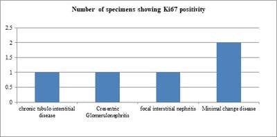
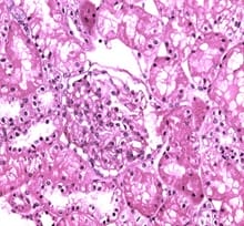
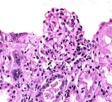
Background: IgA nephropathy is a common renal phenomenon which progresses to chronic kidney disease. It accounts for approximately one-third of the total renal biopsy diagnosis and nearly one half of all primary glomerular diseases diagnosed by renal biopsy. Ki67 functions as a nuclear marker and measures the proliferation index which is seen as increase in the proliferation in glomeruli and also in proximal tubules and renal interstitium. There is a great degree of histologic variability in this disease, varying from minimal histological lesion to a diffuse proliferative nephritis and glomerulosclerosis. This study was carried out to evaluate the spectrum of IgA nephropathy with Ki67 expression. Methods: This cross-sectional record-based study was carried out on 16 renal core biopsies. The cases were histologically diagnosed as IgA nephropathy. Hematoxylin and Eosin staining was done on the slides and immunohisto chemistry was done on paraffin blocks for evaluation of Ki67 using mouse antihuman Ki67 monoclonal antibody-Biogenex at the dilution of 1:50. Results: The most common histological variant was focal proliferative glomerulonephritis Haas Class III. Minimal change disease and focal segmental glomerulosclerosis were the most prevalent histological subtypes. It was observed that chronic tubulointerstitial disease, cresentric glomerulonephritis, Minimal Change Disease and focal interstitial nephritis were positive of Ki67 in 5 cases (31%). Conclusion: Although this study was undertaken on a relatively small number of specimens, the preliminary data obtained from this study could be the basis for a future prospective analysis in a larger number of cases of IgAN in order to be statistically significant.

Keywords: IgA nephropathy, Ki-67, Immunohistochemistry, Chronic kidney disease

Corresponding Author How to Cite this Article To Browse
Gomathi R., Shri Sathya Sai Medical College Sri Balaji Vidhyapeeth University, Puducherry, Tamil Nadu, India.
Email:
Gomathi R. Spectrum of IgA nephropathy in a tertiary care centre, Chennai. Trop J Pathol Microbiol. 2019;5(8):574-579.
Available From
https://pathology.medresearch.in/index.php/jopm/article/view/305

Manuscript Received Review Round 1 Review Round 2 Review Round 3 Accepted
2019-07-30 2019-08-10 2019-08-18 2019-08-22
Conflict of Interest Funding Ethical Approval Plagiarism X-checker Note
No Nil Yes 9%

© 2019 by Gomathi R. and Published by Siddharth Health Research and Social Welfare Society. This is an Open Access article licensed under a Creative Commons Attribution 4.0 International License https://creativecommons.org/licenses/by/4.0/ unported [CC BY 4.0].

Introduction

IgA nephropathy is of the most common type of glomerulonephritis encountered worldwide. It has a high prevalence in Asian subcontinent and is most often characterized by heterogenous clinical course. The prevalence of IgA nephropathy in India is approximately 7.8%. Although studies have documented that the 10-year renal survival rate is greater than 80%, it results in rapidly progressive kidney disease ending in chronic kidney disease and end stage renal failure [1]. However, in some proportion of the patients, it maintains a relatively stable clinical outcome.

An important morphological manifestation of IgA nephropathy is mesangial hypercellularity which is triggered by mesangial IgA deposits resulting in proliferation of the native mesangial cells as well as inflammatory infiltrate. The exact pathogenesis of IgA nephropathy (IgAN) is largely unknown. However, the final step in the process of IgA nephropathy appears to be the deposition of aberrantly glycosylated polymers of the IgA1 molecules and complexes in the glomerular mesangium. The deposition of these polymers (pIgA1) in the mesangium leads to the activation and proliferation of the mesangial cells resulting in proinflammatory cytokines, chemokines and growth factor which eventually causes tubulointerstitial damage and glomerulosclerosis [2, 3]

Considering heterogenous pattern of disease presentation, there is a compelling need to identify the high-risk groups based on the progression of the disease. Studies done earlier have identified several clinical predictors of progression including the degree of renal impairment at the time of diagnosis, histological grading and proteinuria [4].

There are certain studies which have developed risk scoring which reflects the cumulative effects of the individual predictors for progression of IgAN to CKD. However, they have been effectively used in patients with diabetes and nephropathy [5]. Since mesangial cells proliferation is a key morphological feature of IgAN, characterization of this proliferative activity within the glomerulus using antibody against Ki67 (a proliferative marker) may have a significant role to play in predicting progression of the disease.

Ki67 functions as a nuclear marker and measures the proliferation index which is seen as increase in the proliferation in glomeruli and also in proximal tubules and renal interstitium.

The evidence of IgA in terms of muscle layer thickening and increased matrix in presence or absence of hypertension could possibly indicate the intensity and magnitude of the risk of the patient progressing towards CKD.

Therefore, an in-depth evaluation of the role of Ki67 in understanding the spectrum of IgA nephropathy will go a long way in long identifying predictive accuracy for early evaluation of risk factors, and prevention of CKD.

Objectives

This study was carried to evaluate the spectrum of IgA nephropathy in a tertiary care center in Chennai.

Methodology

Study setting and participants: This study was carried out in the Department of Pathology in our tertiary teaching institution.

Study duration: This study was carried out for a period of 7 years. A total of 16 whole renal cell biopsy specimens were analyzed in this study.

Type of study: This study was carried out as a retrospective record based cross sectional study.

Sampling methods: The specimens for the study were chosen by convenient sampling method.

Sample size calculation: Based on the available literature, the incidence of IgA nephropathy in India varied from 7% to 16% [6]. At 95% confidence limits and 12.5% absolute precision, the sample size was calculated at 15.99 and rounded off to 16.

Inclusion criteria

  1. Age above 18 years, both sexes
  2. Renal biopsies diagnosed with IgA nephropathy

Exclusion criteria

  1. Absence of IgA nephropathy
  2. Pediatric population

Ethical Approval: Approval was obtained from the Institutional Ethics Committee prior to the commencement of the study.

Data collection: The samples included sixteen renal core biopsies. The cases were histologically diagnosed as various medical renal diseases and immuno-fluorescence proved IgA nephropathy.


Upon receiving the specimen the clinical data regarding the age, sex and medical history were documented.

Hematoxylin and Eosin staining was done on the slides and immunohisto chemistry was done on paraffin blocks for evaluation of Ki67 using mouse antihuman Ki67 monoclonal antibody-Biogenex at the dilution of 1:50.

Data Analysis: Data was entered and analyzed using EpiInfo version 6.04D. Chi-squared test was used for evaluating the role of Ki67 expression among various categories. A p value is less than 0.05 was considered statistically significant.

Results

This study was carried out among the patients who were diagnosed with IgA Nephropathy in our tertiary teaching institution. A total of 57 samples of native renal diseases of which 16 were diagnosed to have IgA Nephropathy (28%) were received.

The age range of the participants was 19 to 62 years with a mean age of 28.3 years. Majority of the participants were males (56.3%) while females were 43.7%. (Table 1) The pattern of glomerular involvement showing Ki67 expression is given in Table 2. Minimal change disease and focal segmental glomerulosclerosis were the most prevalent histological subtypes.

It was observed that chronic tubulointerstitial disease, cresentric glomerulonephritis, Minimal Change Disease and focal interstitial nephritis were positive of Ki67 (Figure 1). Ki67 expression was carried out in the study which showed focal positively in 5 cases (31%) (Figure 2, 3).

Table-1: Background characteristics of the study participants.

S. no. Characteristics Frequency N=16 Percentage (%)
1. Age (in years)
≤20 4 25
21-30 6 37.5
31-40 4 25
41-50 1 6.25
51-60 0 0
61-70 1 6.25
2. Gender
Males 9 56.3
Females 7 43.7

Table-2: The pattern of glomerular involvement showing Ki67 expression.

S No. Glomerular involvement Frequency N=16 (%)
1. Chronic tubulo interstitial disease 1 6.25
2. Mesangial proliferative glomerulonephritis 1 6.25
3. Cresentric glomerulonephritis 2 12.5
4. Chronic glomerulonephritis 1 6.25
5. Post infectious glomerulonephritis 2 12.5
6. Membranoproliferative glomerulonephritis 1 6.25
7. Focal segmental glomerulosclerosis 3 18.75
8. Minimal change disease 3 18.75
9. Chronic interstitial nephritis 1 6.25
10. Focal interstitial nephritis 1 6.25

patho_305_01.jpg

Figure-1: Ki67 positivity among the study samples:

patho_305_02.jpg

Figure 2: The expression of Ki67

patho_305_03.jpg


Discussion

IgAN is a primary glomerulopathy which encompasses a spectrum of histopathologic patterns ranging from minimal change disease to diffuse proliferative and sclerotic patterns. Granular, dominant or co-dominant deposits of IgA within glomeruli form the diagnostic characteristic of IgA nephropathy [7]. The diagnosis on renal biopsy is based on the morphological changes in conjunction with the immunofluorescence finding of IgA positivity in the mesangial region.

Before labelling the case as primary IgAN, it is important to rule out the secondary causes of IgA positivity like infections and malignancies [8]. In similar situations, it is often possible to detect the presence of IgG and IgM, in association with IgA [9].

This study was carried out among 16 specimens, with predominant male samples. Moreover, the age range of the present study specimens was 19-62 years. In a study by Hall YN et al, the male: female ratio was found to be 60:40, similar to the present study. The age group of study participants was also comparable to the present study (20-50 years) [10]. In a study done by Das U et al, 73% of the participants were males with a mean age of 29.2 years, similar to the present study [11]. In the present study, according to the morphological subclassification, Haas III classification was the most predominant subtype observed. In a study done by Das U et al, Class I Haas subclassification was the most predominant types.

Similarly, in a study done by Ghani AA et al, majority were males, similar to the present study, while the Haas classification I was the most predominant sub-type [12]. The spectrum of glomerular changes in IgAN ranges from minimal glomerular change, focal sclerosis, mesangial hypercellularity, diffuse proliferation with or without crescents and advanced sclerotic changes. In the present study Haas classification was used to classify the spectrum of IgAN [13]. Accordingly, Ki-67 was present only in one case of chronic tubulo interstitial disease, cresentric glomerulonephritis and focal interstitial nephritis. There are several other classifications, predominantly that of Lee et al, WHO and Oxford classifications, that are based on the severity and extent of mesangial proliferation, percentage of glomeruli in segmental lesions, crescents and presence of advanced sclerotic changes.

In some of the classifications, the extent of tubulo-interstitial damage is also included [14, 15]. In our series the renal biopsies were categorized by the Haas classification and the distribution of cases in this study were comparable to that of the Haas series of 244 cases with the majority of cases (38%) being class III [13]. This was in contrast to the study by Hall Y N who studied 124 native renal biopsies and they found the majority of cases belonging to Haas Class V (40%) followed by Class IV (25%) [10]. Clinically, proteinuria is one of the most predominant signs of IgA nephropathy. However, presence of macroscopic hematuria is also reported in several studies. In such cases, it is often associated with aggressive histologic findings and is often associated with poor prognosis [16]. In the present study did show moderate degree of positive expression of IgAN in 6 out of 16 cases (37.5%) and moderate Ki67 positivity in only two cases (12.5%). This data however did not have any statistical significance. Quantitative IHC expression of Ki67 has been studied by Groma V et al who showed both increase in glomerular cellularity and proliferative index [17]. Radford et al, in his study postulated that characterization of proliferative activity using Mib1 directed against Ki67 may be an additional marker predicting renal outcome in these patients [18].

Ki-67 antigen is expressed in the cell nucleus in all stages of cell cycle, thereby being indicative of proliferative stage [19]. The Ki-67 protein was originally defined by the prototype monoclonal antibody Ki-67, which was generated by immunizing mice with nuclei of the Hodgkin lymphoma cell line L428. The name is derived from the city of origin (Kiel, Germany) and the number of the original clone in the 96-well plate [20]. The Ki-67 is a commercially available monoclonal antibody that reacts with a nuclear antigen expressed in proliferating but not in quiescent cells.

Expression of this antigen occurs preferentially during late G1, S, G2, and M phases of the cell cycle, while in cells in G0 phase the antigen cannot be detected. Consequently, the antibody is used in tumor pathology to detect proliferating cells in neoplastic diseases. Ki-67 labeling index is estimated immunohistochemically, using the monoclonal antibody MIB-1. The Ki-67 protein (also known as MKI67) is a cellular marker for proliferation [21]. Assessment of cell proliferation in renal biopsy samples is a potentially promising analytical tool to evaluate disease activity.


So far, no information is available on the correlation between proliferative activity in different anatomic compartments of the kidney and clinical symptoms. Ki67 index was associated with renal outcome; it did not provide additional prognostic information, when compared to clinical and histologic features. Proliferation activity is correlated with glomerular and interstitial histopathologic scoring. The most common morphologic expression in IgA Nephropathy is mesangial hypercellularity with matrix expansion. So ki67 is used to detect the proliferative activity and can be utilized as a marker to predict progressive renal failure. Limitations- This study was carried out as a record-based study and therefore, the clinical risk factors and other histological parameters to evaluate the correlation with spectrum of IgA nephropathy could not be evaluated.

Conclusion

In the present study of 16 cases examined over seven years period, the most common histological variant was Focal Proliferative glomerulonephritis Haas Class III. Although this study was undertaken on a relatively small number of specimens, the preliminary data obtained from this study could be the basis for a future prospective analysis in a larger number of cases of IgAN in order to be statistically significant.

What the study adds to the existing knowledge?

There are very few studies done in South India that have evaluated the Ki-67 expression in the context of spectrum of IgA nephropathy. In the present study, one of the fore runners in establishing hypothesis for further evaluation through exploratory studies.

Author’s contribution

Dr. Gomathi R. was involved in conceptualization, data collection, manuscript writing and review.

Reference

01. SiddappaS, Kowsalya R, Mythri KM. IgA nephron-pathy in a tertiary care center from south India. Indian J Nephrol. 2011;21(4):230-234. doi: 10.4103/ 0971-4065. 82635 [Crossref][PubMed][Google Scholar]

02. Koyama A, Igarashi M, Kobayashi M. Natural history and risk factors for immunoglobulin A nephropathy in Japan, Research Group on Progressive Renal Diseases. Am J Kidney Dis. 1997;29(4):526-532. doi:10.1016/ s0272-6386(97)90333-4 [Crossref][PubMed][Google Scholar]

03. Lai FM, To KF, Choi PC, Li PK. Primary immuno-globulin A nephropathy through the 'retrospectroscope'. Hong Kong Med J. 1999;5(4):375-382. [Crossref][PubMed][Google Scholar]

04. Xie J, Kiryluk K, Wang W, Wang Z, Guo S, Shen P, et al. Predicting progression of IgA nephropathy: new clinical progression risk score. PLoS One. 2012; 7(6):e38904. doi: 10.1371/journal.pone.0038904. Epub 2012 Jun 14 [Crossref][PubMed][Google Scholar]

05. Keane WF, Zhang Z, Lyle PA, Cooper ME, de Zeeuw D, Grunfeld JP, et al. Risk scores for predicting outcomes in patients with type 2 diabetes and nephropathy: the renaal study. Clin J Am Soc Nephrol. 2006;1(4):761-767. doi: 10.2215/CJN. 01381005 [Crossref][PubMed][Google Scholar]

06. Chacko B. IgA nephropathy in India: what we do know. Ren Fail. 2011; 33(1):102-107. doi: 10.3109/ 0886022X. 2010.523486 [Crossref][PubMed][Google Scholar]

07. Bellur SS, Troyanov S, Cook HT, Roberts IS; Working Group of International IgA Nephropathy Network and Renal Pathology Society. Immunostaining findings in IgA nephropathy: correlation with histology and clinical outcome in the Oxford classification patient cohort. Nephrol Dial Transplant. 2011; 26(8):2533-2536. doi: 10.1093/ndt/ gfq812. Epub 2011 Jan 27 [Crossref][PubMed][Google Scholar]

08. Saha MK, Julian BA, Novak J, Rizk DV. Secondary IgA nephropathy. Kidney Int. 2018;94(4):674-681. doi: 10.1016/j.kint. 2018.02.030. [Crossref][PubMed][Google Scholar]

09. Galla JH. IgA nephropathy. Kidney Int. 1995;47(2): 377-387. doi:10.1038/ki.1995.50 [Crossref][PubMed][Google Scholar]

10. Hall YN, Fuentes EF, Chertow GM, Olson JL. Race/ethnicity and disease severity in IgA nephropathy. BMC Nephrol. 2004;5:10. doi:10.1186/1471-2369-5-10 [Crossref][PubMed][Google Scholar]


11. Das U, Dakshinamurty KV, Prayaga A, Uppin M. Spectrum of IgA nephropathy in a single center. Saudi J Kidney Dis Transpl. 2015; 26(5):1057-1063. doi: 10.4103/ 1319-2442.164612 [Crossref][PubMed][Google Scholar]

12. Ghani AA, Al Waheeb S, Al Homoud E, Al Helal B, Hussain N. Clinical and histopathological spectrum of IgA nephropathy in Kuwait. Ann Saudi Med. 2011;31(2):152-157. doi: 10.4103/0256-4947.77491 [Crossref][PubMed][Google Scholar]

13. Haas M. Histologic subclassification of IgA nephropathy: a clinicopathologic study of 244 cases. Am J Kidney Dis. 1997;29(6):829-842. doi:10.1016/ s0272-6386(97)90456-x [Crossref][PubMed][Google Scholar]

14. Lee SM, Rao VM, Franklin WA, Schiffer MS, Aronson AJ, Spargo BH, et al. IgA nephropathy: morphologic predictors of progressive renal disease. Hum Pathol. 1982;13(4):314-322. doi:10.1016/s0046-8177 (82)80221-9 [Crossref][PubMed][Google Scholar]

15. Trimarchi H, Barratt J, Cattran DC, Cook HT, Coppo R, Haas M, et al. Oxford Classification of IgA nephropathy 2016: an update from the IgA Nephropathy Classification Working Group. Kidney Int. 2017; 91(5): 1014-1021. doi: 10.1016/j.kint.2017.02. 003. Epub 2017 Mar 22 [Crossref][PubMed][Google Scholar]

16. Bennett WM, Kincaid-Smith P. Macroscopic hematuria in mesangial IgA nephropathy: correlation with glomerular crescents and renal dysfunction. Kidney Int. 1983;23(2):393-400. [Crossref][PubMed][Google Scholar]

17. Groma V, Marcussen N, Olsen S. A quantitative immunohistochemical study of the expression of mesangial alpha-smooth muscle actin and the proliferation marker Ki-67 in glomerulonephritis in man. Virchows Arch. 1997;431(5):345-350. [Crossref][PubMed][Google Scholar]

18. Radford MG Jr, Donadio JV Jr, Bergstralh EJ, Grande JP. Predicting renal outcome in IgA nephropathy. J Am Soc Nephrol. 1997;8(2):199-207. [Crossref][PubMed][Google Scholar]

19. Schlüter C, Duchrow M, Wohlenberg C, Becker MH, Key G, Flad HD, et al. The cell proliferation-associated antigen of antibody Ki-67: a very large, ubiquitous nuclear protein with numerous repeated elements, representing a new kind of cell cycle-maintaining proteins. J Cell Biol. 1993;123(3):513-522. doi:10.1083/jcb.123.3.513 [Crossref][PubMed][Google Scholar]

20. Klöppel G. On the value of Ki-67 in the prognostic grading of pancreatic neuroendocrine neoplasms: an update. 2017. [Crossref][PubMed][Google Scholar]

21. Inwald EC, Klinkhammer-Schalke M, Hofstädter F, Zeman F, Koller M, Gerstenhauer M, et al. Ki-67 is a prognostic parameter in breast cancer patients: results of a large population-based cohort of a cancer registry. Breast Cancer Res Treat. 2013;139(2):539-552. doi: 10. 1007/s10549-013-2560-8. Epub 2013 May 16 [Crossref][PubMed][Google Scholar]