E-ISSN:2456-1487
P-ISSN:2456-9887
RNI:MPENG/2017/70771

Research Article

Tumors

Tropical Journal of Pathology and Microbiology

2019 Volume 5 Number 8 August
Publisherwww.medresearch.in

Role of fine needle aspiration cytology in the diagnosis of soft tissue tumors - a prospective study

Saraswathi Boni L.1, Kasturi S.2*, Uma P.3, Atla B.4
DOI: https://doi.org/10.17511/jopm.2019.i08.05

1 Lakshmi Saraswathi Boni, Assistant Professor, Department of Pathology, Andhra Medical College, Vishakhapatnam, Andhra Pradesh, India.

2* Sumalatha Kasturi, Associate Professor, Department of Pathology, Chalmeda Anandrao Institute of Medical Sciences, Karimnagar, Telangana, India.

3 Prasad Uma, Associate Professor, Department of Pathology, Andhra Medical College, Vishakhapatnam, Andhra Pradesh, India.

4 Bhagyalakshmi Atla, Professor, Department of Pathology, Andhra Medical College, Vishakhapatnam, Andhra Pradesh, India.

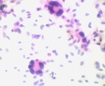
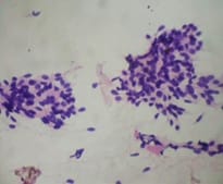
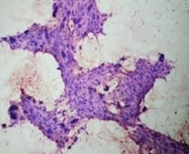
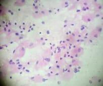
Background: Soft tissue can be defined as non- epithelial, extraskeletal tissues of the body exclusive of reticulo-endothelial system, glia and supporting tissues of various parenchymal organs. The annual incidence of soft tissue tumor is 1.4 per 1,00,000 population. FNAC is almost painless, easy to perform, safe and cost effective procedure without any anaesthesia and acts as a useful diagnostic technique in the initial diagnosis of tumors. Aims:To study the utility of fine needle aspiration cytology (FNAC) in the diagnosis of soft tissue tumours and in distinguishing benign or malignant tumors. Material and Methods: 105 cases of soft tissue tumors were included in this study for cytologic and histologic correlation. FNAC air dried smears were stained with Leishman stain and 95% ethanol fixed smears were stained with H&E. Smearswere categorised as benign, malignant, inconclusive or undetermined along with specific subtyping of the lesion. All diagnostic FNAC results were compared for diagnostic concordance using histology results as the gold standard. Results: The sensitivity, specificity, positive predictive value, negative predictive value and accuracy were 82.6%, 100%, 100%, 93.3% and 95% respectively. Conclusion: FNAC is an important preliminarydiagnostic tool in palpable soft tissue tumors with high degree of correlation with histopathology report.

Keywords: Benign, FNAC, Histopathology, Malignant, Soft tissue tumors

Corresponding Author How to Cite this Article To Browse
Sumalatha Kasturi, Associate Professor, Department of Pathology, Chalmeda Anandrao Institute of Medical Sciences, Karimnagar, Telangana, India.
Email:
Boni LS, Kasturi S, Uma P, Atla B. Role of fine needle aspiration cytology in the diagnosis of soft tissue tumors - a prospective study. Trop J Pathol Microbiol. 2019;5(8):535-541.
Available From
https://pathology.medresearch.in/index.php/jopm/article/view/301

Manuscript Received Review Round 1 Review Round 2 Review Round 3 Accepted
2019-07-25 2019-08-04 2019-08-10 2019-08-16
Conflict of Interest Funding Ethical Approval Plagiarism X-checker Note
No Nil Yes 9%

© 2019 by Lakshmi Saraswathi Boni, Sumalatha Kasturi, Prasad Uma, Bhagyalakshmi Atla and Published by Siddharth Health Research and Social Welfare Society. This is an Open Access article licensed under a Creative Commons Attribution 4.0 International License https://creativecommons.org/licenses/by/4.0/ unported [CC BY 4.0].

Introduction

Soft tissue tumors are defined as mesenchymal proliferations which occur in the extra-skeletal non epithelial tissues of the body, excluding the viscera, coverings of brain and lymphoreticular system [1]. Soft tissue tumours constitute a heterogeneous group of neoplasms in terms of clinical presentation, morphologic features and clinical behaviour [1]. The annual incidence of soft tissue tumor is 1.4 per 1,00,000 population[2]. Benign tumours outnumber malignant ones by 100:1[1]. Because of the rarity of primary tumors of soft tissue and large range of different types of tumours, the diagnosis and classification of soft tissue tumors becomes most difficult areas in surgical pathology and absence of recognisable tissue architectural patterns in cytological preparation makes diagnosis by fine needle aspiration cytology even more difficult [3].

However, FNAC is useful in distinguishing accurately between benign and malignant soft tissue tumours and also sub classify them into general, clinically relevant categories that permit initiation of therapy in many of the patients. In benign neoplasms, surgery can be avoided in the patients who are of poor surgical risk, and in malignant tumors, FNAC allows administration of a palliative treatment.

The main advantage of FNAC is its simplicity. It is a safe, cheap, relatively painless procedure, easy to perform without anaesthesiaand is acceptable to the patient [2]. It produces a speedy result and it acts as useful diagnostic technique in the initial diagnosis of tumours.

The present study was done to findout the efficacy of FNAC in the diagnosis of soft tissue tumors and in classifying benign and malignant tumours.

Materials and Methods

Type of study: This is a prospective cytohistological correlation study done at two tertiary care hospitalsin the Department of Pathology, Andhra Medical College and King George Hospital, Vishakhapatnam and Chalmeda Anand Rao Institute of Medical Sciences, Karimnagar.

Period of study: The present study was conducted over a period of one year between July 2018 and June 2019. A total number of 105 cases, 65 cases from King George Hospital and 40 cases from Chalmeda Anand Rao Institute of Medical Sciences were studied.

Complete clinical details, examination findings and radiological investigations were studied.

FNAC was done after taking consent with 23/24 G needle attached to 10 ml disposable plastic syringe and air dried smears were stained with Leishman stain and 95% ethanol fixed smears were stained with H & E stain. The cytosmears were studied and categorized into benign, malignant and inconclusive cases. The excised tissue specimens of all above cases were processed routienely and stained with H & E. Special stains were done as and when required. All diagnostic FNAC

results from patients who underwent a subsequent surgical excision were compared for diagnostic concordance using histology results as the gold standard. In addition FNAC results were analysed for ability to recognize malignancy using statistical parameters of sensitivity, specificity, positive predictive value and negative predictive value.

Histopathological diagnosis of soft tissue tumors were done based on degree of cellularity, cellular pleomorphism, mitotic activity, degree of necrosis, invasive growth, haemorrhage and inflammatory infiltrate. Grading of soft tissue sarcomas was done by Federation Nationale des Centres de Lutte Contre Le Cancer (FNCLCC) system.

Ethical clearance was taken from the Andhra medical college and Chalmeda Anandrao Medical College.

Inclusion criteria: Palpable soft tissue tumors of size more than 1 cm.

Exclusion criteria: Inflammatory swellings, autolysed biopsy specimens

Results

A total number of 105 cases clinically suspected as soft tissue tumors were subjected to FNAC and compared with histopathology. The observations of the study were as follows:

Table-1: Age and sex distribution of soft tissue tumors

Age (Years) Male Female Total
0 – 10 3 1 4
11 – 20 6 6 12
21 – 30 6 14 20
31 – 40 10 13 23
41 – 50 6 18 24
51 – 60 6 8 14

61 – 70 3 4 7
71 – 80 1 - 1
Total 41 64 105

Most of tumors were observed between the age group of 21-50 years. Male: female ratio was 1:1.5 (Table-1).

Table-2: Anatomical distribution of various soft tissue tumors.

Head & neck Upper extremity Lower extremity Trunk Retroperitoneum
20 25 30 21 9

Majority of the tumors were observed in the lower extremity, followed by upper extremity (Table -2)

Table-3:Tumor size of various soft tissue lesions

Tumor size (cm) Benign Intermediate Malignant Total
<5 36 5 7 48
6-10 24 4 8 36
11-15 7 2 11 20
>15 - - 1 1

Tumor size was highly variable irrespective of the grade of the lesion. Majority of benign tumors were <5 cm and malignant tumors were >10 cm in size (Table-3).

Table-4: Categorisation of soft tissue tumors on FNAC

Diagnosis on FNAC Number of cases (%)
Benign 60 (63%)
Malignant 19 (20%)
Inconclusive diagnosis 16 (17%)
Scanty material / only blood 10 (10%)
Total 105(100%)

Inadequate material was obtained in 10 (9.5%) cases. FNACsmears have been reported under three basic categories as benign, malignant and inconclusive or undetermined in this study. In 16/95 (17%) cases, the material yielded on FNAC was adequate but not sufficient to classify them as benign or malignant(Table-4).

Table-5: List of cases with inconclusive diagnosis

Diagnosis on FNAC Histopathological diagnosis No. of cases
Myxoidmesenchymaltumour Myxoidliposarcoma 1
Myxoid / chondroid lesion Extra skeletal chondroma 1
Granular cell tumor/ Alveolar soft part sarcoma Granular cell tumour 1
Spindle cell neoplasm Fibromatosis 6
Spindle cell neoplasm Dermatofibrosarcoma protuberance 2
Spindle cell neoplasm Hemangioendothelioma 2
Spindle cell neoplasm Nodular fasciitis 1
Spindle cell neoplasm Fibrosarcoma 1
Spindle cell neoplasm Infantile fibrosarcoma 1
Total   16

Most of the cases with inconclusive diagnosis on FNAC turned out to be intermediate grade lesions on histopathology (Table-5).

Table-6: Correlation between FNAC and histopathology–79 cases:

  Correlated Not Correlated Total
Benign 53 (94.64%) 3(5.3%) 56
Intermediate 2 - 2
Malignant 16 (76.19%) 5 (23.8%) 21
Total 71(90%) 8(10%) 79(100%)

Majority of benign tumors (94.64%) showed positive correlation on histopathology compared to malignant tumors (76.19%).

Table-7: List of cases with discordant results.

FNAC Diagnosis HPE Diagnosis Number of Cases
Lipoma Neurofibroma 1
Pleomorphic adenoma / Myoepithelioma Schwannoma 1
Benign vascular lesion Schwannoma 1
Lipoma Well differentiated liposarcoma 2
Benign nerve sheath tumour Malignant peripheral nerve sheath tumour 2
Round cell sarcoma Synovial sarcoma 1
Total   8

True positive cases (TP) = 19
True negative cases (TN) = 56
False positive cases (FP) = 0
False negative cases (FN) = 4
Sensitivity= 82.6%
Specificity= 100%
Predictive value of positive test= 100%
Predictive value of negative test= 93.3%
Accuracy = 95%

Discussion

Soft tissue can be defined as non- epithelial, extraskeletal tissues of the body exclusive of reticulo-endothelial system, glia and supporting tissues of various parenchymal organs. It is represented by voluntary muscles, fat and fibrous tissue along with the vessels serving these tissues.


By convention, it also includes peripheral nervous system [3]. The annual incidence of soft tissue tumor is 1.4 per 1,00,000 population[4].

Because of the rarity of primary tumors of soft tissue and large range of different types of tumors, the diagnosis and classification of soft tissue tumors become most difficult areas in surgical pathology and absence of recognizable tissue architectural patterns in cytological preparation makes diagnosis by fine needle aspiration cytology even more difficult[5]. Benign tumors are more common than malignant counterparts with a ratio of at least 100: 1[6].

FNAC is painless, easy to perform, safe and cost effective without any anaesthesia and acts as a useful diagnostic technique in the initial diagnosis of tumors [7].

For distinguishing benign tumors from malignant tumors, FNAC was very useful except for exact categorization of tumors [8].

However FNAC as a preliminary diagnostic tool offers several advantages as it can provide a predictive diagnosis of a benign or malignant neoplasm. In benign neoplasm, surgery can be avoided in the patients who are of poor surgical risk, and in malignant or recurrent cancers, FNAC allows the administration of a palliative treatment [9,10].

In the present study adequate material was obtained in 95/105(90%) cases. The possible causes for inadequacy of material in the 10 cases were either due to aspiration of blood (in vascular lesions), collagen (desmoid type fibromatosis) or due to pain (in neural lesions). Collagen rich intercellular stroma, areas of hyalinization, fibrosis and prominant vascularity are known to influence the yield[11].

Gonzalez – Campora R stated that the final cytology report should place the soft tissue tumors in one of the three basic categories – benign, malignant and inconclusive or undetermined [12].

Table-8: Comparison of results on FNAC with other studies

Maitra A et al [13] Benign on FNAC Malignant on FNAC Inconclusive on FNAC
42 (58%) 18 (25%) 12 (16%)
Benign on HPE Malignant on HPE Benign on HPE Malignant on HPE Benign on HPE Malignant on HPE
39(93%) 3 (7%) 1 (6%) 17(94%) 5(42%) 7(58%)
Present study Benign on FNAC Malignant on FNAC Inconclusive on FNAC
60 (63%) 19 (20%) 16 (17%)
Benign on HPE Malignant on HPE Benign on HPE Malignant on HPE Benign on HPE Malignant on HPE
56(93%) 4 (7%) 0 19(100%) 3 (28%) 13 (72%)

Correlation between cytological diagnosis on FNAC with histopathological diagnosis in the present study was comparable with other studies.

patho_301_01.jpg

Figure-1: PleomorphicLipoma- FNAC smears show floret like giant cells and spindle cells (H&E, x400)

patho_301_02.jpg

Figure-2: Cytosmears show bland looking round to spindle cells in synovial sarcoma (H&E, x400)


patho_301_03.jpg

Figure-3: Smears show syncytial sheet of pleomorphic spindle cells in MFH (H & E, x 100).

patho_301_04.jpg

Figure-4: FNAC smears show loose sheets of large polygonal cells with abundant granular eosinophilic cytoplasm in granular cell tumor (H&E,x400).

In the present study 16 of 95 (17%) cases were labeled under the group of inconclusive diagnosis- that is even though the material was adequate for interpretation, it was difficult to classify them as benign or malignant (Table-4).3 of 16 (72%) were subsequently diagnosed as benign and 13 of 16 (72%) malignant on histopathology. Neoplasms arising from fibrous tissue have varied patterns on morphology.

It is essentially a diagnosis by exclusion. Fibromatosis, dermatofibrosarcoma protuberance, nodular fasciitis and low-grade sarcomas have almost similar morphology on cytology.

It is only histopathology which can differentiate them, basing on infiltrative growth pattern, long fascicles, herringbone pattern, collagenous stroma and inflammatory cells at the border of infiltration [14]. Hence, 11/16 (68.75%) cases of fibrous tumors were given the diagnosis of spindle cell neoplasm on cytology.

Gonzalez – Compora et al, in their FNAB study of 16 myxoid soft tissue tumours opined that the only cells of diagnostic value in smears of lipomatoustumours were lipoblasts [14]. In the present study, a case of myxoidliposarcoma yielded only myxoid matrix with paucity of lipoblasts and hence the diagnosis was inconclusive (Table-5). Chondroid lesions are difficult to aspirate and usually they are hypocellular, as in our case of extraskeletalchondroma which yielded only amphophilic matrix with scattered round to oval cells showing minimal pleomophism. Vascular lesions usually yield blood and endothelial cells, which resemble spindle cells. Hence, 2 cases of hemangioendothelioma were given the diagnosis as spindle cell neoplasm. Granular cell tumour and alveolar soft part sarcoma show similar cytological features; hence the diagnosis was inconclusive in one of our case (Table-5).

Maximum correlation was seen in benign lesions (Table-6). A case of neurofibroma reported as lipoma on FNAC due to sampling error. A case of schwannoma in the parotid region was reported as pleomorphic adenoma / myoepithelioma due to similar morphology. Another case of schwannoma with high vascularity yielded blood and spindle cells on FNAC (Table-7).

5/21 (23.8%) malignant lesions were not correlated. Two cases of liposarcoma were under diagnosed as lipoma due to lack of typical lipoblasts on FNAC smears (Table-6). Two cases of low-grade malignant peripheral nerve sheath tumour were under diagnosed as benign nerve sheath tumours. A case of synovial sarcoma in a child was reported as round cell sarcoma on FNAC (Table-6). Smears of synovial sarcoma are highly cellular with both single cells and cohesive clusters. Single cells are often stripped of their cytoplasm and appear as round to oval naked nuclei. Keeping in view of the age of the patient, the diagnosis of round cell sarcoma was made. Of the 11/105 (10.47%) cases with intermediate grade malignancy, only two cases showed correlation, and nine were inconclusive (Table-5). The correlation rate was comparable with that of Keiko Nagira et al [15].


In the present study an accuracy of 95%, sensitivity of 82.6% and 100% specificity were observed which were similar to Maitra et al [13]. False negativity was seen in four cases, two liposarcomas, and two malignant peripheral nerve sheath tumors.

Limitations of the present study: Small sample size. The diagnosis on FNAC can be challenging because of interpretation and sampling issues.

Conclusion

From the present study, it was conclude that FNAC is an important tool in the preoperative diagnostic work up of soft tissue tumors with certain limitations and FNAC diagnosis is supplementary but not a substitute for histopathological examination.

What the study adds in the existing knowledge?

FNAC distinguishes benign and malignant tmors of soft tissue with few limitations.

Author’s contribution

Dr. Lakshmi Saraswathi Boni: Conception of idea, took lead in writing the manuscript, done investigations

Dr. Sumalatha Kasturi: Verified the analytical methods, done investigations, manuscript writing

Dr. Prasad Uma: Data analysis, analytical calculations, interpretation of results

Dr. BhagyalakshmiAtla: Supervising the project

All authors provided critical feedback and helped shape the research analysis and manuscript.

Reference

01. Jorge Albores S, Ruby Frank V. Soft tissue tumors; 4 thed, Steven G Silverberg, Baltimore, Maryland. Churchill Livingstone. 2006, pp 307-418. [Crossref][PubMed][Google Scholar]

02. Hajdu SI. Diagnosis of soft tissue sarcomas on aspiration smears, editorial. Acta Cytol. 1996;40;607-608. doi:10.1159/000333926 [Crossref][PubMed][Google Scholar]

03. Weiss SW, Goldblum JR. General Considerations, Ch-1 In- Enzinger and Weiss Soft tissue tumors. 4th edition, St Louis- Mosby. 2001;1-19. [Crossref][PubMed][Google Scholar]

04. Rydholm A, Berg NO. Epidemiology of Soft tissue sarcoma in locomotor system- A retrospective population based study of interrelationships between clinical and morphological variables. Acta Pathol Microbiol Immunol Scand. 1984;92(5)363-374. [Crossref][PubMed][Google Scholar]

05. Ackerman M, Domanski H. Soft tissues, In- Orell SR, Sterrett GF, editors, Orell and Sterrett’s Fine Needle Aspiration Cytology. 5th ed, Edinburgh, Scotland- Churchill Livingstone. 2012;387-400. [Crossref][PubMed][Google Scholar]

06. Rosenberg AE. Bones, joints and soft tissue tumors editors, In- Kumar V, Abbas AK, Fausto N, Aster JC, editors, Robbins and Cotran Pathologic Basis of Disease. 8th ed, Philadephia, Pa, USA- Saunders. 2010;235-249. [Crossref][PubMed][Google Scholar]

07. Rekhi B, Gorad BD, Kakade AC, Chinoy R. Scope of FNAC in the diagnosis of soft tissue tumors- a study from a tertiary cancer referral centre in India. Cyto J. 2007;4;20. doi: 10.1186/1742-6413-4-20 [Crossref][PubMed][Google Scholar]

08. Dey P, Mallik MK, Gupta SK, Vasishta RK. Role of fine needle aspiration cytology in the diagnosis of soft tissue tumors and tumor like lesions. Cytopathol. 2004;15(1)32-37. doi: 10.1046/j.0956-5507.2003.00102.x [Crossref][PubMed][Google Scholar]

09. Roy S, Manna AK, Pathak S, Guha D. Evaluation of fine needle aspiration cytology and its correlation with histopathological findings in soft tissue tumors. J Cytol. 2007;24(1)37-40. [Crossref][PubMed][Google Scholar]

10. Parajuli S, Lakhey M. Efficacy of fine needle aspiration cytology in diagnosing soft tissue tumors. J Pathol Nepal. 2012;2(4)305-308. doi: 10.3126/jpn.v2i4.6884 [Crossref][PubMed][Google Scholar]

11. Orell SR, Sterrett GF, Walters M, Whitaker D. Manual and Atlas Of Fine Needle Aspiration Cytology. Churchill Livingstone, London, fourth ed. 2005;409-432. [Crossref][PubMed][Google Scholar]

12. Gonzalez – Campora R. Fine needle aspiration cytology of soft tissue tumours. Acta Cytol. 2000;44(3)337–343. doi: 10.1159/000328475 [Crossref][PubMed][Google Scholar]


13. Maitra A, Ashfaq R, Saboorian MH, Lindberg G, Gokaslan ST. The role of fine – needle aspiration biopsy in the primary diagnosis of mesenchymal lesions- a community hospital – based experience. Cancer. 2000;90(3)178–185. [Crossref][PubMed][Google Scholar]

14. Enzinger and Weiss’s Soft Tissue Tumours, ed. SW Weiss and JR Goldblum, Mosby Co. St Louis, fourth ed. 2001;pp-147-188. [Crossref][PubMed][Google Scholar]

15. Keiko Nagira, Tetsuji Yamamoto, Toshihiro Akisue. Reliability of fine – needle aspiration biopsy in the initial diagnosis of soft–tissue lesions. Diagn Cytopathol. 2002;27(6)354–361. doi: 10.1002/dc.10200 [Crossref][PubMed][Google Scholar]