E-ISSN:2456-1487
P-ISSN:2456-9887
RNI:MPENG/2017/70771

Research Article

Lung

Tropical Journal of Pathology and Microbiology

2019 Volume 5 Number 7 July
Publisherwww.medresearch.in

Expression of EGFR in non small cell lung carcinoma

Devi E. P.1*, Sai Shalini C.N.2, Prathiba D.3
DOI: https://doi.org/10.17511/jopm.2019.i07.12

1* Prema Devi E., Assistant Professor, Sri Ramachandra Medical College and Research Institute, Chennai, Tamil Nadu, India.

2 Sai Shalini C.N., Associate Professor, Sri Ramachandra Medical College and Research Institute, Chennai, Tamil Nadu, India.

3 Prathiba D., Professor, Sri Ramachandra Medical College and Research Institute, Chennai, Tamil Nadu, India.

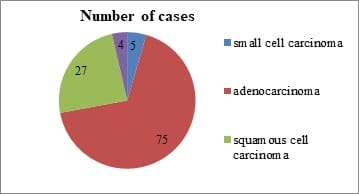
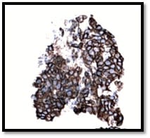
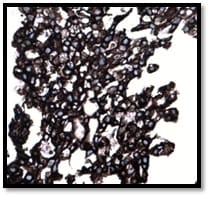
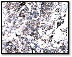
Background: The incidence and mortality associated with lung cancers are increasing at an alarming rate. Studies have shown that activation of Epidermal Growth Factor Receptor (EGFR) triggers the tumorigenesis in these cancers. The potential role of analyzing EGFR expression in these carcinomas could go a long way in devising screening tools for early detection of lung carcinoma. This study was carried out to evaluate the prevalence and factors associated with EGFR expression. Methods: This cross sectional study was carried out among 75 paraffin block specimens of lung carcinoma received in our tertiary care center for a period of five years. Three micron thick paraffin sections were cut and Hemotoxylin & Eosin staining was done. All the adenocarcinoma cases were immunostained with EGFR antibody and the results were analyzed. Results: Majority of the tumors were moderately differentiated (46%) and were negative for lymph node metastasis (69%). With regards to EGFR positivity, majority of the tumors showed EGFR expression 3+ (57.3%) flowed by 2+ (16%). Among the EGFR negative cases, ALK expression was positive in 5% of the cases. Conclusion: The present study has reinforced the fact that Indian patients have high expression of EGFR and therefore can be benefitted by targeted therapy. IHC coupled with molecular analysis would be of maximum benefit to patients with EGFR & ALK mutations.

Keywords: EGFR, Immunohistochemistry, Lung cancer, Tyrosine kinase

Corresponding Author How to Cite this Article To Browse
Prema Devi E., Assistant Professor, , Sri Ramachandra Medical College and Research Institute, Chennai, Tamil Nadu, India.
Email:
Devi EP, Sai Shalini CN, Prathiba D. Expression of EGFR in non small cell lung carcinoma. Trop J Pathol Microbiol. 2019;5(7):484-492.
Available From
https://pathology.medresearch.in/index.php/jopm/article/view/292

Manuscript Received Review Round 1 Review Round 2 Review Round 3 Accepted
2019-06-20 2019-06-30 2019-07-06 2019-07-11
Conflict of Interest Funding Ethical Approval Plagiarism X-checker Note
No Nil Yes 8%

© 2019 by Prema Devi E., Sai Shalini C.N., Prathiba D. and Published by Siddharth Health Research and Social Welfare Society. This is an Open Access article licensed under a Creative Commons Attribution 4.0 International License https://creativecommons.org/licenses/by/4.0/ unported [CC BY 4.0].

Introduction

Lung carcinoma is the leading cause of cancer deaths in the world [1]. It is the most common carcinoma among males and is associated with a high mortality rate.The five year survival rate for male patients with lung carcinoma is around 6% to 14% and for female patients with lung carcinoma it ranges between 7% and 18%.

The number of estimated deaths due to lung carcinoma is 1.38 million per year [2]. The carcinoma of lung is broadly classified into Non-Small Cell Lung Carcinoma (NSCLC) which accounts for 85% and Small Cell Lung Carcinomas (SCLC) which accounts for about 15% [3].

Extensive research on the pathophysiology of lung carcinoma has suggested the existence of two distinct molecular pathways, one associated with smoking and activation of the KRAS oncogene and the other not associated with smoking but with activation of epidermal growth factor receptor (EGFR). Introduction of targeted therapy with epidermal growth factor receptors, the tyrosine kinase inhibitors, has revolutionized the treatment of adenocarcinoma. Patients with these tumors survive significantly longer with EGFR tyrosine kinase inhibitor therapy than with conventional chemotherapy [4-8].

Somatic mutations within the tyrosine kinase catalytic domain of EGFR leads to conformational changes that promote permanent active status and are found in approximately 20% of lung adenocarcinomas [9-10]. They are considered to be the most reliable predictors of response to EGFR tyrosine kinase inhibitors [11].

EGFR inhibitors can be mainly categorized into two classes: monoclonal antibodies against the extracellular domain of EGFR, such as cetuximab; and small-molecule tyrosine kinase inhibitors that target the kinase domain, such as erlotinib and gefitinib [12-18]. Sahoo et al observed that 51.8% of the NSCLC population had EGFR mutations in their DNA[19]. This suggested that high prevalence of EGFR positivity is seen in NSCLC in the Indian subpopulation therefore providing opportunities for targeted therapy.

Methodology

Study setting and study participants: This cross-sectional study was carried out in the

Department of Pathology of our tertiary teaching institution for a period of four years. All the lung carcinomas diagnosed during the study period constituted the study population. A total of 75 specimens were taken up for the study.

Inclusion criteria: All the histopathologically confirmed new cases of lung adenocarcinoma included.

Exclusion criteria

  1. Other histological subtypes of lung carcinomas were not included in the study group.
  2. Metastatic carcinomas to lung were not included in the study.

Sampling technique: The specimens were taken up for the study by convenient sampling

Ethical approval: Approval was obtained from the Institutional Ethics Committee prior to the commencement of the study.

The study was approved by the Institutional Review Board of Sri Ramachandra University (IRB No. CSP-MED/13/JUN/07/35) and performed in accordance with the principles of the Declaration of Helsinki. Written informed consents were obtained.

Data collection: Paraffin blocks of all the cases of NSCLC were retrieved from the archives. Clinical data of the patients including demographic characteristics, metastatic status and prognosis was noted from the medical records. Threemicronthick paraffin sections were cut and Hemotoxylin & Eosin staining was done. The NSCLC casesin for which sub typing was difficult were further immunostained for TTF1 and p63 and the tumors were subtyped. Immuno-histochemical staining for TTF1 was performed using the Biogenex monoclonal TTF1 mouse antibody; clone (BGX397A) diluted in phosphate buffered saline (PBS). Staining for p63 was performed using Biogenex monoclonal p63 mouse antibody; clone (4A4) diluted in PBS. Subtyping of NSCLC into adenocarcinoma and squamous cell carcinoma was done based on the morphology and IHC.

The algorithm followed by the International Association for the Study of Lung Cancer/ American Thoracic Society/ European Respiratory Society (IATC/ATS/ ERS) International Multidisciplinary Team was used.All the adenocarcinoma cases were immunostained with EGFR antibody and the results were analyzed.


Immunohistochemical staining for total EGFR protein was performed using the Biogenex monoclonal EGFR rabbit antibody; clone (Tyr p1068) prediluted in PBS on both control and test sections according to the 0 manufacturer’s instructions.

Assessment of Immunohistochemical staining: Initially, slides were scanned at 10xmagnification to obtain a general impression of the overall staining pattern of the tumor cells.H score was performed for which 10 fields were chosen randomly at 400 x magnification and the intensity of staining of EGFR was scored on a scale of 0 to 3 based on the staining of the cell membrane of the tumor cells. (Table 1)

Table-1: H score for intensity of staining of EGFR[20].

S. No Score Description
1 0 No staining or Faint staining in <10% of tumor cells
2 1+ Faint staining in >10% of tumor cells
3 2+ Moderate staining
4 3+ Strong staining

The total number of 100 cells in each field was counted and the number of cells stained for each level of intensity was counted. A H-score between 0 and 300 was obtained where 300 was equal to 100% of tumour cells stained strongly (3 +). A cutoff Hscore of above 100 was used to categorize tumors as positive,and tumors with scores of 100 and less were categorized as negative [20]. Squamous cell carcinoma of cervix was used as a positive control.

Data analysis: Data was entered and analyzed using SPSS ver 20 software. Chi square test was used to analyze the statistical significance between EGFR expression and clinicopathological parameters. A p value <0.05 was considered statistically significant.

Results

A total of 111 specimens of lung cancer were received during the study period. Among them, majority constituted non small cell carcinoma (95%). Among the non small cell carcinoma, majority of the cases were adenocarcinoma (75) followed by squamous cell carcinoma (27) (Figure 1).

Majority of the participants were > 60years of age in this study. Males were predominant (72%) compared to females. Most tumors were present in the upper lobe (57.3%) followed by lower lobe

(32%). Majority of the cases were smokers (64%) (Table 2).

patho_292_01.jpg

Figure-1: Distribution of cases according to the type of carcinoma:

Table-2: Background characteristics of the study participants.

S. No Characteristics Frequency N = 75 Percentage (%)
1 Age ( in years)
0-30 0 0.0
31-40 3 4.0
41-50 8 10.7
51-60 19 25.3
61-70 32 42.7
71-80 9 12.0
81-90 4 5.3
2 Gender
Males 54 72.0
Females 21 28.0
3 Location of the tumor
Upper lobe 43 57.3
Middle lobe 5 6.7
Lower lobe 24 32.0
Multiple 3 4.0
4 Smoking history
Smoker 8 11.0
Non smoker 48 64.0
Not known 19 25.0

Table-3: Histological features of adenocarcinoma:

S. No Characteristics Frequency N = 75 Percentage (%)
1 Grading of adenocarcinoma
Differentiated 20 27.0
Moderately differentiated 35 46.0
Poorly differentiated 20 27.0
2 Presence of lymph node metastasis
Positive 23 31.0
Negative 52 69.0
3 Presence of distant metastasis
Metastasis 11 15.0

  No metastasis 64 85.0
4 Correlation between scores of EGFR positivity
0+ 9 12.0
1+ 11 14.7
2+ 12 16.0
3+ 43 57.3
5 Correlation of ALK in EGFR negative cases (n=55)
Positive 3 5.0
Negative 19 35.0
Not done 33 60.0

Table-4: Association between EGFTable 4: R expression and distant metastasis:

S. No Characteristics Number of cases EGFR IHC P value
(+) (-)
1 Distant metastasis Present 10 10 0 0.041*
Absent 65 45 20

patho_292_02.jpg

Figure-2: Moderately Differentiated adeno carcinoma 200X

patho_292_03.jpg

Figure-3: EGFR IHC Staining showing 1+

Positivity 200x

patho_292_04.jpg

Figure-4: EGFR IHC Staining showing 2+ Positivity 200x

patho_292_05.jpg

Figure-5: EGFR IHC Staining showing 3+ Positivity 200x

patho_292_06.jpg


Figure-6: ALK IHC showing 1+ Positivity 200x

The histological features of the adenocarcinoma are depicted in table 3. Majority of the tumors were moderately differentiated (46%) and were negative for lymph node metastasis (69%). With regards to EGFR positivity, majority of the tumors showed EGFR expression 3+ (57.3%) followed by 2+ (16%). Among the EGFR negative cases, ALK expression was positive in 5% of the cases. (Table 3)

This study showed a statistically significant association between EGFR positivity and presence of distant metastasis (p<0.05) (Table 4) The immunohistochemical staining demonstrating EGFR expression are depicted in Figures 2-6.

Discussion

Lung carcinoma is the leading cause of cancer related mortality.It accounts for approximately 1.4 million deaths per year worldwide.Increasing Knowledge about the pathogenesis of lung carcinomas especially adenocarcinoma has led to the development of newer therapeutic strategies.In the present study NSCLC accounted for 95% of the lung cancers and adenocarcinoma constituted the major histologic subtype. This was in concordance with worldwide incidence [2]. In the present study the mean age at diagnosis was 62 years.This was similar to the findings of Noronha V et al[2] In the present study,64% of the participants with NSCLS were non smokers. Similar findings were observed in the data reported by Samet JM et al[21]. In the present study the site,laterality and grading had no significance in lung carcinoma.

The assessmentof the EGFR status in adenocarcinomas with pTyr1068 phosphorylated EGFR which is a predictive biomarker for screening the NSCLC with wild type of EGFR was done, as they are responsive to tyrosine kinase inhibitors treatment [22]. The EGFR expression was also correlated with the clinico-pathologic parameters. EGFR positivity was seen in 73% of adenocarcinomas which is in concordance with the study done by Wang et al[22] The EGFR positivity was more common in the 6th decade and in males (74%).

EGFR was positive in 87% of smokers and 71% ofnon smokers. EGFR positivity was seen 100% in patients who had lesions in the middle lobe and in patients who had lesions in both the lungs.

Poorly differentiated adenocarcinoma expressed more EGFR positivity (85%) compared to the other grades.In patients who had lymph node metastasis, EGFR was positive in 87%. There was a statistically significant correlation between EGFR positivity and presence of distant metastasis (p<0.005) and this feature is in concordance with the study done by Welsh et al [23]. The possible hypothesis in support of this is related to the delayed presentation of EGFR which independently increases the risk for metastasis [23].

In the present study, the semi quantitative expression of EGFR positivity was also observed and it was found that 43 cases (57%) showed EGFR staining intensity corresponding to 3+,12 cases (16%) showed EGFR staining intensity of 2+,11 cases (15%) showed EGFR staining intensity of 1+, and the remaining 9 cases (12%) showed 0 staining.Comparison was made based on the present scoring with the study done by Liang et al,in which they found that 45.8% of the cases showed EGFR levels of 3+,22.6% cases showed 2+ positivity, 17.3% cases showed EGFR levels of 1+, the remaining 19 cases showed level 0 staining,and their findings were similar to the present study [20].

Rosell et alstudied EGFR mutations in 350 cases and they found that in women mutations constituted 69.7%, which was similar to the present study where EGFR mutations in women constituted 71.4%. In their study mutations in non smokers constituted 66.6%, which was similar to the present study.It was observed that non smokers expressed 71.4% of EGFR positivity.On the whole in their study EGFR positive adenocarcinomas constituted 80%,which is concordant with the present study were EGFR expression constituted73% of our cases [24]. In a study done by Fred R.Hirsch et al, the scores in adenocarcinoma was also in concordance with the present study [25].

The presence of KRAS, HER2, BRAF, PI3K, LKB1 and SHP2 mutations is supposed to be associated with a lack of response to EGFR tyrosine kinase inhibitors in the treatment of lung cancer.Hence additional mutational analysis of genes other than EGFR may be required to improve patient selection for EGFR targeted therapies. The EGFR mutation status is best determined by fluorescence in situ hybridization (FISH) or chromogenic in situ hybridization (CISH) and protein expression determined by IHC with mutationspecific antibodies.


Ruschoff et al evaluated the inter-observer reprodu-cibility of the EGFR IHC scoring system based on both the tumor cell membrane staining intensity (graded 0 to 3+) and the percentage of cells staining at each level of intensity, as was done in the present study.This allowed a highly reproducible allocation of NSCLCs into clinically relevant high or low EGFR expression groups [26] Determination of H score and a cut off value as suggested by Liang et al was useful in determining clear cut positive and negative groups[20]. In the present study EGFR expression was high (73%), which emphasizes the need and importance for IHC screening of Indian patients with adenocarcinoma lung.

It paves way for targeted therapy when combined with subsequent molecular techniques for specific EGFR mutation analysis. This adds more value to the treatment of lung adenocarcinoma patients.The antibody used in the present study pTyr1068 has been proposed to be a predictive biomarker for screening the population for clinical outcomes of EGFR-TKIs treatment,especially for patients with wild type EGFR [27-30].

It was further studied that the ALK positivity in the EGFR negative patients as EML4-ALK fusion has been observed to be present in adenocarcinoma patients lacking EGFR mutation [31]. It was observed ALK positivity in 5% of the EGFR negative cases, which was similar to the literature published[32-34].The single case which showed ALK positivity had a low level of expression (1+), however was similar to the study done by P. Hofman et al[35].This expression pattern has been explained due to the fact that ALK rearrangements in lung carcinoma are usually associated with low levels of ALK expression in contrast to anaplastic lymphomas with ALK rearrangements where the protein is robustly detected by immunohistochemistry[36].

Limitations

  • Treatment details of the study cases have not been analysed.
  • Molecular studies have to be performed for specific EGFR mutation

Conclusion

Introduction of targeted therapy with EGFR and ALK TKI has revolutionized the treatment of adeno-carcinoma. Patients with these mutations survive significantly longer with TKI therapy than with conventional chemotherapy.

EGFR expression was seen in 73% of the cases studied; reinforcing the fact that our subset of patients have high expression of EGFR and therefore can be benefitted by targeted therapy. There was a significant correlation between EGFR positivity and adenocarcinoma patients who had distant metastasis, proving that EGFR expression is more in advanced cases. IHC coupled with molecular analysis would be of maximum benefit to patients with EGFR& ALK mutations.

What the study adds to the existing knowledge?

The present study has reinstated the need for molecular analysis and immunohistochemical evaluation of EGFR expression for effective management of adeno-carcinoma.

Authors’ contribution

Dr. Prema Devi–Conceptualization, manuscript writing

Dr. Sai Shalini – Data collection and analysis

Dr. Prathiba D –Manuscript editing and literature review

Reference

01. Cagle PT, Zhai QJ, Murphy L, Low PS. Folate receptor in adenocarcinoma and squamous cell carcinoma of the lung- potential target for folate-linked therapeutic agents. Arch Pathol Lab Med. 2013;137(2)241-4. doi: 10.5858/arpa.2012-0176-OA [Crossref][PubMed][Google Scholar]

02. Noronha V, Pinninti R, Patil VM, Joshi A, Prabhash K. Lung cancer in the Indian subcontinent. South Asian J Cancer. 2016;5(3)95-103. doi: 10.4103/2278-330X.187571 [Crossref][PubMed][Google Scholar]

03. Molina JR, Yang P, Cassivi SD, Schild SE, Adjei AA. Non-small cell lung cancer- epidemiology, risk factors, treatment, and survivorship. Mayo Clin Proc. 2008;83(5)584-94. doi: 10.4065/83.5.584 [Crossref][PubMed][Google Scholar]

04. de Mello RA, Marques DS, Medeiros R, Araújo AM. Epidermal growth factor receptor and K-Ras in non-small cell lung cancer-molecular pathways involved and targeted therapies. World J Clin Oncol. 2011;2(11)367-76. doi: 10.5306/wjco.v2.i11.367 [Crossref][PubMed][Google Scholar]


05. Kobayashi S, Boggon TJ, Dayaram T, Jänne PA, Kocher O, Meyerson M, et al. EGFR mutation and resistance of non-small-cell lung cancer to gefitinib. N Engl J Med. 2005;352(8)786-92. DOI: 10.1056/NEJ Moa 044238 [Crossref][PubMed][Google Scholar]

06. Dacic S. Molecular diagnostics of lung carcinomas. Arch Pathol Lab Med. 2011;135(5)622-9. doi: 10.1043/2010-0625-RAIR.1 [Crossref][PubMed][Google Scholar]

07. Brevet M, Arcila M, Ladanyi M. Assessment of EGFR mutation status in lung adenocarcinoma by immunohistochemistry using antibodies specific to the two major forms of mutant EGFR. J Mol Diagn. 2010; 12(2)169-76. doi: 10.2353/jmoldx.2010.090140 [Crossref][PubMed][Google Scholar]

08. Bonanno L, Favaretto A, Rugge M, Taron M, Rosell R. Role of genotyping in non-small cell lung cancer treatmen- current status. Drugs. 2011;71(17)2231-46. doi: 10.2165/11597700-000000000-00000 [Crossref][PubMed][Google Scholar]

09. Sahoo R, Harini VV, Babu VC, Patil Okaly GV, Rao S, Nargund A, et al. Screening for EGFR mutations in lung cancer, a report from India. Lung Cancer. 2011;73(3)316-9. doi: 10.1016/j.lungcan.2011.01.004 [Crossref][PubMed][Google Scholar]

10. Engel KB, Moore HM. Effects of preanalytical variables on the detection of proteins by immuno-histochemistry in formalin-fixed, paraffin-embedded tissue. Arch Pathol Lab Med. 2011;135(5)537-43. doi: 10.1043/2010-0702-RAIR.1 [Crossref][PubMed][Google Scholar]

11. Wen YH, Brogi E, Hasanovic A, Ladanyi M, Soslow RA, Chitale D, et al. Immunohistochemical staining with EGFR mutation-specific antibodies- high specificity as a diagnostic marker for lung adeno-carcinoma. Mod Pathol. 2013;26(9)1197-203. doi: 10.1038/modpathol.2013.53 [Crossref][PubMed][Google Scholar]

12. Baselga J, Pfister D, Cooper MR, Cohen R, Burtness B, Bos M, et al. Phase I studies of anti-epidermal growth factor receptor chimeric antibody C225alone and in combination with cisplatin. J Clin Oncol. 2000;18(4)904-14. doi: 10.1200/JCO.2000.18.4.904 [Crossref][PubMed][Google Scholar]

13. Saltz LB, Meropol NJ, Loehrer PJ Sr, Needle MN, Kopit J, Mayer RJ. Phase II trial of cetuximab in patients with refractory colorectal cancer that expresses the epidermal growth factor receptor. J Clin Oncol. 2004;22(7)1201-8. doi: 10.1200/JCO.2004.10.182 [Crossref][PubMed][Google Scholar]

14. Cunningham D, Humblet Y, Siena S, Khayat D, Bleiberg H, Santoro A, et al. Cetuximab monotherapy and cetuximab plus irinotecan in irinotecan-refractory metastatic colorectal cancer. N Engl J Med. 2004;351(4)337-45. doi: 10.1056/NEJMoa033025 [Crossref][PubMed][Google Scholar]

15. Pérez-SolerR, ChachouaA, HammondLA, Rowinsky EK, Huberman M, Karp D, et al. Determinants of tumor response and survival with erlotinib in patients with non--small-celllungcancer. J Clin Oncol. 2004;22(16)3238-47. doi: 10.1200/JCO.2004.11.057 [Crossref][PubMed][Google Scholar]

16. Kris MG, Natale RB, Herbst RS, Lynch TJ Jr, Prager D, Belani CP, et al. Efficacy of gefitinib, an inhibitor of the epidermal growth factor receptor tyrosine kinase, in symptomatic patients with non-small cell lung cancer- a randomized trial. JAMA. 2003;290(16)2149-58. doi: 10.1001/jama.290.16.2149 [Crossref][PubMed][Google Scholar]

17. Cohen MH, Williams GA, Sridhara R, Chen G, Mc Guinn WD Jr, Morse D, et al. United States Food and Drug Administration Drug Approval summary- Gefitinib (ZD1839; Iressa) tablets. Clin Cancer Res. 2004;10(4)1212-8. doi: 10.1158/1078-0432.CCR-03-0564 [Crossref][PubMed][Google Scholar]

18. Cohen MH, Johnson JR, Chen YF, Sridhara R, Pazdur R. FDA drug approval summary. erlotinib (Tarceva) tablets. Oncol. 2005;10(7)461-6. doi: 10.1634/theoncologist.10-7-461 [Crossref][PubMed][Google Scholar]

19. Sahoo R, Harini VV, Babu VC, Patil Okaly GV, Rao S, Nargund A, et al. Screening for EGFR mutations in lung cancer, a report from India. Lung Cancer. 2011;73(3)316-9. doi: 10.1016/j.lungcan. 2011.01.004 [Crossref][PubMed][Google Scholar]

20. Liang Z, Zhang J, Zeng X, Gao J, Wu S, Liu T. Relationship between EGFR expression, copy number and mutation in lung adenocarcinomas. BMC Cancer. 2010;10;376. doi: 10.1186/1471-2407-10-376 [Crossref][PubMed][Google Scholar]


21. Samet JM, Avila-Tang E, Boffetta P, Hannan LM, Olivo-Marston S, Thun MJ, et al. Lung cancer in never smokers- clinical epidemiology and environmental risk factors. Clin Cancer Res. 2009;15(18)5626-45. doi: 10.1158/1078-0432.CCR-09-0376 [Crossref][PubMed][Google Scholar]

22. Wang F, Wang S, Wang Z, Duan J, An T, Zhao J, et al. Phosphorylated EGFR expression may predict outcome of EGFR-TKIs therapy for the advanced NSCLC patients with wild-type EGFR. J Exp Clin Cancer Res. 2012;31(1)65. doi: 10.1186/1756-9966-31-65 [Crossref][PubMed][Google Scholar]

23. Welsh JW, Komaki R, Amini A, Munsell MF, Unger W, Allen PK, et al. Phase II trial of erlotinib plus concurrent whole-brain radiation therapy for patients with brain metastases from non-small-cell lung cancer. J Clin Oncol. 2013;31(7)895-902. doi: 10.1200/JCO.2011.40.1174 [Crossref][PubMed][Google Scholar]

24. Rosell R, Moran T, Cardenal F, Porta R, Viteri S, Molina MA, et al. Predictive biomarkers in the management of EGFR mutant lung cancer. Ann N Y Acad Sci. 2010;1210;45-52. doi: 10.1111/j.1749-6632.2010.05775.x [Crossref][PubMed][Google Scholar]

25. Hirsch FR, Varella-Garcia M, Bunn PA Jr, Di Maria MV, Veve R, Bremmes RM, et al. Epidermal growth factor receptor in non-small-cell lung carcinomas- correlation between gene copy number and protein expression and impact on prognosis. J Clin Oncol. 2003;21(20)3798-3807. doi: 10.1200/JCO.2003.11.069Epub 2003 [Crossref][PubMed][Google Scholar]

26. Rüschoff J, Kerr KM, Grote HJ, Middel P, von Heydebreck A, Alves VA, et al. Reproducibility of immunohistochemical scoring for epidermal growth factor receptor expression in non-small cell lung cancer- round robin test. Arch Pathol Lab Med. 2013;137(9)1255-61. doi: 10.5858/arpa.2012-0605-OA [Crossref][PubMed][Google Scholar]

27. Wang F, Wang J, Bai H, Zhao J, Wang Z, Zhuot M et al. An evaluation of phosphorylated EGFR expressionin predicting outcome of EGFR-TKI therapy for the advanced NSCLC patients with EGFR wild type. J ClinOncol. 2011;29(15 Suppl)7532. doi: 10.1200/jco.2011.29.15_suppl.7532 [Crossref][PubMed][Google Scholar]

28. Welsh JW, Komaki R, Amini A, Munsell MF, Unger W, Allen PK, et al. Phase II trial of erlotinib plus concurrent whole-brain radiation therapy for patients with brain metastases from non-small-cell lung cancer. J Clin Oncol. 2013;31(7)895-902. doi: 10. 1200/JCO.2011.40.1174 [Crossref][PubMed][Google Scholar]