E-ISSN:2456-1487
P-ISSN:2456-9887
RNI:MPENG/2017/70771

Research Article

Uterine

Tropical Journal of Pathology and Microbiology

2019 Volume 5 Number 7 July
Publisherwww.medresearch.in

Histomorphological study of uterine leiomyomas and its variants with brief review of literature

Budihal N.1, G. Pawar J.2, Manasa G. C.3*
DOI: https://doi.org/10.17511/jopm.2019.i07.09

1 Nischita Budihal, Assistant Professor, Department of Pathology, Sri Siddhartha Institute of Medical Sciences and Research Centre, Nelamangala, Karnataka, India.

2 Jayashree G. Pawar, Professor, BLDE Medical College, Vijayapura, Karnataka, India.

3* Manasa G. C., Associate Professor, JJM Medical College, Davangere, Karnataka, India.

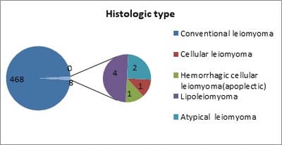
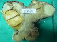
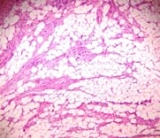
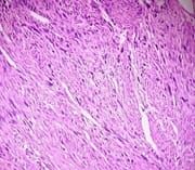
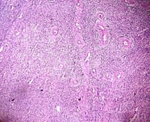
Background: Uterine mesenchymal tumours are a heterogeneous group of neoplasms that can frequently be diagnostically challenging. Most subtypes of leiomyoma are chiefly of interest in that they mimic malignancy in one or more respects. Objective: To evaluate the histomorphological features of uterine leiomyomas and its variants. Materials and methods: Total of 477 cases of uterine leiomyomas and its variants were analysed prospectively in a period of 2years during July 2010 to June 2012 to assess the various pattern of leiomyomas. Cases were studied in detail about complete history, clinical examination and other findings. Results: In the study 468 (98.11%) cases showed features of conventional leiomyoma and 8 cases showed variants of leiomyomas (1.68%). Conclusion: Although their diagnosis is straight forward in most cases, difficulties arise with particular leiomyoma variants, especially highly cellular leiomyoma (often confused with an endometrial stromal tumour) and leiomyoma with bizarre nuclei, mitotically active leiomyoma which may cause concern for leiomyosarcoma.

Keywords: Apoplectic, Cellular, Leiomyoma, Lipoleiomyoma, Uterus, Variants

Corresponding Author How to Cite this Article To Browse
Manasa G. C., Associate Professor, JJM Medical College, Davangere, Karnataka, India.
Email:
Budihal N, Pawar JG, Manasa GC. Histomorphological study of uterine leiomyomas and its variants with brief review of literature. Trop J Pathol Microbiol. 2019;5(7):466-472.
Available From
https://pathology.medresearch.in/index.php/jopm/article/view/289

Manuscript Received Review Round 1 Review Round 2 Review Round 3 Accepted
2019-05-30 2019-05-08 2019-05-16 2019-06-22
Conflict of Interest Funding Ethical Approval Plagiarism X-checker Note
No Nil Yes 8%

© 2019 by Nischita Budihal, Jayashree G. Pawar, Manasa G. C. and Published by Siddharth Health Research and Social Welfare Society. This is an Open Access article licensed under a Creative Commons Attribution 4.0 International License https://creativecommons.org/licenses/by/4.0/ unported [CC BY 4.0].

Introduction

Uterine mesenchymal tumours are a heterogeneous group of neoplasms that can frequently be diagnostically challenging [1]. The most common benign and malignant tumours are leiomyoma and leiomyosarcoma respectively [2]. Leiomyoma is a benign neoplasm composed of smooth muscle cells with a variable amount of fibrous stroma,most commonly affects the body of the uterus [3,4].

It is present in 20-30% of women over 30 years of age rising to more than 40% in those over 40 years old [4,5,6]. Prevalence of fibroids increases to more than 70% with pathological examination of the uterus, indicating that many women who have fibroids are asymptomatic [7].

Most subtypes of leiomyoma are chiefly of interest in that they mimic malignancy in one or more respects [3]. Diverse histological features of uterine leiomyomas are responsible for an erroneous diagnosis of malignancy [8]. Difference between the benign and malignant counterparts of mesenchymal tumours is significant due to the differences in the clinical outcome and the role of the surgical pathologist in making this distinction (especially in difficult cases) cannot be underestimated [1].

Materials and Methods

Setting and type of study: This study was undertaken in the Department of Pathology, JJM Medical College, Davangere. It was prospective study. The study period of two years from July 2010- June 2012.

Sampling method: Material for the study consisted of hysterectomy specimens & myomectomy specimens which were sent for histopathological examination to the Department of Pathology, JJM Medical College from Bapuji Hospital, Chigateri General Hospital and also from private hospitals in and around Davangere. Relevant clinical data was collected from the hospital and laboratory records.

The specimens were received in 10% formalin; after adequate fixation were subjected to thorough gross examination and appropriate sections were taken. After tissue processing, multiple 4-6µ thick paraffin sections were stained with hematoxylin and eosin.

Sample size: Among 12,285 surgical specimens received for histopathological examination in the Department during the study period.

1914 were hysterectomies, 25 were myomectomies, 3 were debulking specimens and one was polypectomy specimen.

Of the total 1943 specimens, 492 cases were mesenchymal tumours (including 8 cases of mixed epithelial and mesenchymal tumours).

Inclusion criteria: Histologically proven mesenchymal tumours and mixed epithelial and mesenchymal tumours like carcinosarcoma, carcinofibroma, adeno-fibroma and adenomyoma of the uterine corpus were included in the present study.

Exclusion criteria: Epithelial tumours and related lesions, gestational trophoblastic disease, lymphoid and hematopoietic tumours and metastatic tumours of the uterine corpus were excluded from the present study.

Ethical considerations and permissions: The study was started after due approval from institutional ethical committee.

Data analysis: The data was entered in MS excel and descriptive statistics were applied

Results

Hysterectomy is the most common surgical operative procedure encountered in gynecological practice.

Among 12,285 surgical specimens received for histopathological examination in the Department during the study period. 1914 were hysterectomies and 25 were myomectomies.

patho_289_01.jpg

Figure 1: Histologic types of leiomyomas

Of the total 1939 specimens, Leiomyoma was diagnosed in 477 cases, of which 24 were myomectomy specimens. Thus, the incidence of leiomyoma was 24.92% of total 1914 hysterectomy specimens. Thus, leiomyoma was the most common tumour 96.95% of all the tumours.


Variants of leiomyoma: Variants of leiomyomas were seen in 8 cases constituting 1.68% of all leiomyomas. Of these, 4 cases were lipoleiomyoma, 2 atypical leiomyoma, one cellular leiomyoma and one apoplectic leiomyoma.

  1. Lipoleiomyoma: 4 cases of lipoleiomyomas were seen between 5th & 6th decades of life. 3 patients presented with mass per abdomen and one with menstrual disturbance (table I). These tumours measured 3-6cm in their greatest dimensions and were intramural and subserosal in location. One of the tumours showed gross evidence of lipoleiomyoma (Figure 2).

Table-1: Details of cases of lipoleiomyoma.

Cases Age Presenting complaint Location of leiomyoma Additional features
Case 1 45 Menstrual disturbance Intramural -
Case 2 47 Mass per abdomen Intramural Cystic change
Case 3 49 Mass per abdomen Intramural -
Case 4 51 Mass per abdomen Subserosal Myxoid change

patho_289_02.jpg

Figure 2: Sectioned surface of uterine lipoleiomyoma showing distinct pale yellowappearance one side and a subserosal leiomyoma on the other side

Microscopically, the tumours were composed of admixture of varying amounts of mature adipose tissue with smooth muscle cells (Figure 3). The adipose tissue was scattered and at places diffusely distributed within leiomyomas.

  1. Atypical leiomyoma: Was diagnosed in two cases, (table II)Cut section of tumour in both the cases was grey white with whorled appearance.

Table 2: Details of cases of atypical leiomyoma.

Feature Case 1 Case 2
Age 32years 36years
Presenting complaint Mass per abdomen Mass per abdomen
Type of specimen received Myomectomy Hysterectomy
Measurement of leiomyoma 13x8x7cm* Subserosal -7x6cm, intramural -2x1cm
Additional features - Mucoid and cystic change

*location could be identified in myomectomy specimen

patho_289_03.jpg

Figure 3: Lipoleiomyomashowing mature adipocytes intermingled with smooth muscle cells

patho_289_04.jpg

Figure 4: Diffusely distributed atypical cells throughout the leiomyoma


Microscopic sections from both the cases showed smooth muscles with moderate to severe cellular atypia arranged in fascicles with areas of smooth muscles with markedly pleomorphic and enlarged nuclei (nuclear atypia appreciated at lower magnification) (Figure 4). Clusters of bizarre hyperchromatic and multinucleated smooth muscle cells were seen. Also noted degenerative cells, smudged chromatin and intranuclear vacuolation. Occasional mitoses (2-3/10 HPFs) were noted. Necrosis was absent.

  1. Haemorrhagic cellular (apoplectic) leiomyoma: A case of haemorrhagic cellular leiomyoma was diagnosed in a 43yr old female who presented with mass per vaginum. On gross examination, tumour showed a polypoidal mass in the endometrial cavity measuring 5x3cm with foci of hemorrhage. Microscopically, the tumour was cellular, with closely packed round to spindle cells with scanty cytoplasm. There was no cytologic atypia. Blood vessels were large with thick muscular wall and cleft like spaces seen. Patchy areas of haemorrhage and oedema were made out. Also noted scattered lymphocytes and hemosiderin macrophages. Occasional MFs (<2/10 HPFs) were seen. The tumour appeared to merge with the surrounding myometrium.

Cellular leiomyoma: A case of cellular leiomyoma was diagnosed in a 40yr old female who presented as mass per abdomen. Two intramural leiomyomas were made out on gross examination, larger measuring 4cm in diameter which was soft in consistency and showed areas of mucoid change on cut section and the smaller one measured 3cm in diameter which showed regular whorled appearance on cut section.

patho_289_05.jpg

Figure 5: Microphotograph showing markedly cellular tumour and the cells are small and round to spindle shaped giving a basoplhilic hue with more blood vessels

Microscopy of both the tumours showed densely cellular benign looking smooth muscle cells arranged in whorled pattern at places. In few areas these smooth muscle cells were small and rounded. Palisading of nuclei was present in some areas. Many large blood vessels with thick muscular wall and cleft like spaces were noted within these tumourcells (Figure5). 5-6 MFs/10 HPFs were noted but there was no atypia. Hydropic, fatty, hyaline and myxoid changes were noted within the tumour.

Discussion

The most common pelvic tumour in the reproductive age group is leiomyoma, occurring in 20–40% of females. Leiomyomas may have a diverse clinical, radiological and morphological pattern and are hormone responsive. Clinically they may be sporadic or syndromic and may present with abdominal pain, menorrhagia and pelvic mass[9].

The World Health Organization has recently (2014) updated its criteria for mesenchymaltumors of the female reproductive tract [10], and variants of benign smooth muscle tumorsare diagnosed according to their unusual histologicfeatures. Mitotically active leiomyomais defined by the presence of 10–15 mitoses/10 high-power fields (hpf) [11,12] and may be seen in patients with enhanced hormonalstates (pregnancy or taking exogenoushormones).

Increased cellularity, higher thanthat of nearby myometrium, is seen in a cellularleiomyoma, and at times, a cellular leiomyomamay have short spindle cells resembling an endometrial stromal tumor. Leiomyomawith bizarre nuclei (also called atypical, symplastic and pleomorphic) show the presenceof scattered large atypical cells.

These atypicalsmooth muscle cells have abundant eosinophiliccytoplasm, irregular nuclear shapes, and multinucleation. The nuclei are hyperchromatic, often with intranuclear inclusions. Occasionally, cells have further chromatin-condensation and fragmentation, resemblingatypical mitotic figures.

A proper pathological study of mesenchymal tumours of the uterus is predicated, on careful gross examination and adequate sectioning.


The tumour should be examined thoroughly, and one block of tissue should be taken for each centimeter of tumour diameter, except from grossly typical leiomyomas; even the latter may have to be examined extensively if the microscopic appearance is unusual [5]. Variants of leiomyomas were seen in eight cases constituting 1.68% of all leiomyomas. Of these, four cases were lipoleiomyomas, two atypical leiomyoma, one cellular leiomyoma and one apoplectic leiomyoma.

  1. Lipoleiomyoma: Lipoleiomyoma is a rare entity which should be differentiated from liposarcoma. Four cases of lipoleiomyoma were diagnosed constituting to 0.84% of all leiomyomas (table III). On gross examination inaddition to white whorled appearance yellowish discoloration was seen. On microscopy interlacingbundles of smooth muscles were mixed with matureadipocytes.

Table-III: Comparison of incidence of lipoleiomyoma in various studies.

Author Year Incidence
Ueda H et al [16] 1999 0.8%
Avritscher [17] 2001 0.03-0.2%
Wang X et al[11] 2006 2.1%
Present study 2012 0.84%

Table-IV: Comparison of age incidence of lipoleiomyoma in various studies.

Author Year Average age in years
Bajaj P [13] 2000 43
Avritscher [11] 2001 48
Wang X et al[17] 2006 53.9
Manjunath HK[12] 2010 50
Present study 2012 47

The mean age of occurrence of lipoleiomyoma in the present study was found to be in 5th decade (table IV)

  1. Atypical leiomyoma: Important entity as it can be misdiagnosed asleiomyosarcoma (Figure-3). It is characterized byspindle cells along with scattered multinucleatedbizarre cells with prominent nucleoli that may mimicatypical cells[13]. A study by Philip PC et al stated that the term atypical leiomyoma is considered if the tumour showed focal or multifocal moderate to severe atypia, and no tumour cell necrosis [14]. Similar features were seen in the present study.

Average rate of mitoses of 2-3/10HPFs noted in the present study also correlated with the study by Oliva E et al [15].

Final diagnosis of atypical leiomyoma with low risk of recurrence was offered according to the study by Philip PC et al[14].

  1. Cellular leiomyoma: A case of cellular leiomyoma was diagnosed in a 40year old female. In the study done by Oliva E et al in 33 cases of cellular leiomyoma the age ranged from 29-65 years [15]. By definition cellular leiomyoma is a smooth muscle tumour with increased cellularity when compared to surrounding myometrium.

The histological features of dense cellularity of cellular leiomyoma in the present study were similar to the features mentioned by various authors [5, 15,16, 17, 18].

The most important differential of cellular leiomyoma is endometrial stromal sarcoma. In most of the cases routine histopathology is sufficient to differentiate these lesions. However, in few cases with extremely cellular leiomyomas immunohistochemistry is used to resolve this dilemma [19].

Conclusion

Smooth muscle tumours and endometrial stromal tumours represent the two main categories of mesenchymal tumours of the uterus. Although their diagnosis is straight forward in most cases, difficulties arise with particular leiomyoma variants, especially highly cellular leiomyoma (often confused with an endometrial stromal tumour) and leiomyoma with bizarre nuclei, mitotically active leiomyoma which may cause concern for leiomyosarcoma.

Differentiation between the benign and malignant counterparts of mesenchymal tumours is through the use of multivariate criteria; that is, criteria that involves several microscopic features such as differentiated cell type, presence and type of tumor necrosis, the degree of cytologic atypia, the mitotic index, and the relationship to surrounding normal structures, including extra uterine sites.

What this study adds to the existing knowledge?

This study contributes to the data regarding histological variants of leiomyoma. Much remains to be learned clinically, particularly regarding those histologic entities that lie between, called leiomyoma variants.


Nonetheless, the bottom line is that, at the present time, the diagnosis of a leiomyoma variant cannot be made with certainty until a pathologist, guided by recently updated 2014 World Health Organization criteria [10], thoroughly examines the specimen.

Author’s Contribution

Nischita Budihal: Data collection, analysis Conceptualization, literature search &writeup. Jayashree G Pawar: Designing the study and supervised the study. Manasa G C: Manuscript preparation and proof reading.

Reference

01. Sangle NA, Lele SM. Uterine mesenchymal tumors. Indian J Pathol Microbiol. 2011;54(2)243-53. doi: 10.4103/0377-4929.81582 [Crossref][PubMed][Google Scholar]

02. Robboy SJ, McCluggage WG. Mesenchymal Uterine tumours, other than pure smooth muscle neoplasms, and adenomyosis, In- Robboy SJ, Mutter GM, Prat J, Bentley R, Russel P, Anderson MC editors. Textbook of Pathology of Female Reproductive Tract, 2ndedn, Elsevier, Churchill Livingstone. 2009;427-55. [Crossref][PubMed][Google Scholar]

03. Hendrickson MR, Tavassoli FA, Kempson RL, McCluggage WG, Haller U, Kubik- Hutch RA. Mesenchymal tumors and related lesions, In- Tavassoli FA, Deville P, editors, World Health Organization Classification of tumours. Pathology and genetics of Tumors of the Breast and Female Genital Organs, Lyon- IARC Press. 2003;233-44. [Crossref][PubMed][Google Scholar]

04. Robboy SJ, Bentley RC, Butnor K, Anderson MC. Pathology and pathophysiology of uterine smooth-muscle tumors. Environ Health Perspect. 2000; 108(5)779-84. [Crossref][PubMed][Google Scholar]

05. Zaloudek CJ, Hendrickson MR, Saslow RA. Mesenchymal tumours of the Uterus, In- Kurman RJ, Elleson LH, Ronett BM, Blaustein`s Pathology of the Female Genital Tract. 6th ed, Springer. 2011;453-527. [Crossref][PubMed][Google Scholar]

06. Blom R, Guerrieri C, Stâl O, Malmström H, Simonsen E. Leiomyosarcoma of the uterus- A clinicopathologic, DNA flow cytometric, p53, and mdm-2 analysis of 49 cases. Gynecol Oncol. 1998;68(1)54-61. doi: 10.1006/gyno.1997.4889 [Crossref][PubMed][Google Scholar]

07. Gross KL, Morton CC. Genetics and the development of fibroids. Clin Obstet Gynecol. 2001; 44(2)335-49. [Crossref][PubMed][Google Scholar]

08. Wantabe K, Suzukki T. Uterine leiomyoma versus leiomyosarcoma- a new attempt at differential diagnosis based on their cellular characteristics. Histopathol. 2006;48(5)563-8. doi: 10.1111/j.1365-2559.2006.02368.x [Crossref][PubMed][Google Scholar]

09. Han SC, Kim MD, Jung DC, Lee M, Lee MS, Park SI, et al. Degeneration of leiomyoma in patients referred for uterine fibroid embolization- incidence, imaging features and clinical characteristics. Yonsei Med J. 2013;54(1)215-9. doi: 10.3349/ymj. 2013.54.1.215 [Crossref][PubMed][Google Scholar]

10. Kurman RJ, Carcangiu ML, Herrington CS, Young RH. WHO classification of tumours of female reproductiveorgans. 4th ed, Lyon, France- International Agency for Research on Cancer. 2014. [Crossref][PubMed][Google Scholar]

11. Bell SW, Kempson RL, Hendrickson MR. Problematic uterine smooth muscle neoplasms- A clinicopathologic study of 213 cases. Am J Surg Pathol. 1994;18(6)535-58. [Crossref][PubMed][Google Scholar]

12. Boyd C, McCluggage WG. Unusual morphological features of uterine leiomyomas treated with progestogens. J Clin Pathol. 2011;64(6)485-9. doi: 10. 1136/jcp. 2011.089664. Epub 2011 Mar 11 [Crossref][PubMed][Google Scholar]

13. Croce S, Young RH, Oliva E. Uterine leiomyomas with bizarre nuclei- a clinicopathologic study of 59 cases. Am J Surg Pathol. 2014;38(10)1330-9. doi: 10.1097/PAS.0000000000000249 [Crossref][PubMed][Google Scholar]

14. Ip PP, Cheung AN. Pathology of uterine leiomyosarcomas and smooth muscle tumours of uncertain malignant potential. Best Pract Res Clin Obstet Gynaecol. 2011;25(6)691-704. doi: 10.1016/j.bpobgyn.2011.07.003 [Crossref][PubMed][Google Scholar]

15. Oliva E, Young RH, Clement PB, Bhan AK, Scully RE. Cellular benign mesenchymal tumors of the uterus- A comparative morphologic and immunohistochemical analysis of 33 highly cellular leiomyomas and six endometrial stromal nodules, two frequently confused tumors. Am J Surg Pathol. 1995;19(7)757-68. [Crossref][PubMed][Google Scholar]


16. Anderson MC. Female genital tract, In- Symmer SW ed, Systemic Pathology. 3rd ed, Edinburg, Churchill Livingstone. 1991. [Crossref][PubMed][Google Scholar]

17. Hendrickson MR and Kempson RL. Pure mesen-chymal tumours of the uterine corpus, In- Fox H Editors Obstetrical & Gynecological Pathology. 4th Ed, New York; Churchill Livingstone. 1995;511-86. [Crossref][PubMed][Google Scholar]

18. Jaiswal CJ. Vaginal management of uterocervical-myomas. J Obstet GynecolIndia. 1996;46;260-3. DOI: 10.18203/2320-1770.ijrcog20173500 [Crossref][PubMed][Google Scholar]

19. Pujani M, Jairajpuri ZS, Rana S, Jetley S, Hassan MJ, Jain R. Cellular leiomyoma versus endometrial stromal tumor- A pathologists' dilemma. J Midlife Health. 2015;6(1)31-4. doi: 10.4103/0976-7800.153619 [Crossref][PubMed][Google Scholar]