E-ISSN:2456-1487
P-ISSN:2456-9887
RNI:MPENG/2017/70771

Case Report

Twin Tales of Rarity

Tropical Journal of Pathology and Microbiology

2025 Volume 11 Number 2 Jul-Dec
Publisherwww.medresearch.in

Twin Tales of Rarity: Sarcina Ventriculi and Colonic Mucormycosis with Unusual Gastrointestinal Presentations

Srivastava R1*, Gowthami N2
DOI:https://doi.org/10.17511/jopm.2025.i02.02

1* Richa Srivastava, Postgraduate, Department of Pathology, MS Ramaiah Medical College, Bengaluru, Karnataka, India.

2 Gowthami N, Assistant Professor, Department of Pathology, MS Ramaiah Medical College, Bengaluru, Karnataka, India.

Sarcina ventriculi and mucormycosis are rare but clinically significant infections that can present as unusual gastrointestinal pathologies. Sarcina ventriculi is a gram-positive, anaerobic coccus associated with delayed gastric emptying, gastric ulcers, emphysematous gastritis, and perforation. Due to its nonspecific symptoms and difficulty in culturing, diagnosis often depends on histopathological identification of characteristic tetrad-forming cocci. Mucormycosis, caused by fungi of the order Mucorales, is an aggressive, angioinvasive infection affecting immunocompromised individuals. Gastrointestinal involvement is rare and frequently misdiagnosed due to overlapping clinical features with malignancy. Histopathology remains the gold standard, revealing broad, aseptate hyphae with right-angle branching and vascular invasion. We present two rare cases: a 39-year-old male with duodenal and antral ulcers harbouring Sarcina ventriculi, and a 43-year-old male with colonic mucormycosis showing extensive necrosis and fungal angioinvasion. These cases emphasize the pivotal role of histopathological examination in diagnosing uncommon gastrointestinal infections and highlight the need for clinical vigilance in atypical presentations.

Keywords: Sarcina ventriculi, mucormycosis, gastrointestinal infections, colonic mucormycosis

Corresponding Author How to Cite this Article To Browse
Richa Srivastava, Postgraduate, Department of Pathology, MS Ramaiah Medical College, Bengaluru, Karnataka, India.
Email:
Srivastava R, Gowthami N, Twin Tales of Rarity: Sarcina Ventriculi and Colonic Mucormycosis with Unusual Gastrointestinal Presentations. Trop J Pathol Microbiol. 2025;11(2):21-25.
Available From
https://pathology.medresearch.in/index.php/jopm/article/view/683

Manuscript Received Review Round 1 Review Round 2 Review Round 3 Accepted
2025-07-21 2025-07-29 2025-08-06 2025-08-14 2025-08-22
Conflict of Interest Funding Ethical Approval Plagiarism X-checker Note
None Nil Yes 11.54

© 2025 by Srivastava R, Gowthami N and Published by Siddharth Health Research and Social Welfare Society. This is an Open Access article licensed under a Creative Commons Attribution 4.0 International License https://creativecommons.org/licenses/by/4.0/ unported [CC BY 4.0].

Download PDFBack To ArticleIntroductionCase Report: 1Case Report: 2DiscussionConclusionReferences

Introduction

Gastrointestinal infections caused by uncommon organisms can present with subtle or misleading clinical findings, often mimicking more common diseases such as malignancies or inflammatory conditions.

Among these, Sarcina ventriculi and mucormycosis represent two rare but clinically significant pathogens. Sarcina ventriculi, a gram-positive, obligate anaerobic coccus, is typically identified histologically by its distinctive tetrad or octet morphology. Though it may be a commensal in some individuals, it has been increasingly recognized in association with delayed gastric emptying, gastric ulcers, emphysematous gastritis, and perforation.[1], [2]

Mucormycosis, a life-threatening fungal infection caused by Mucorales species, is most frequently observed in immunocompromised hosts.[6],[7] Gastrointestinal involvement is particularly rare, often presenting with nonspecific symptoms and overlapping radiological features that simulate malignancy. This report presents two cases: Gastric Sarcina infection and Colonic Mucormycosis, highlighting the pivotal role of histopathology in identifying these rare entities and guiding optimal therapeutic intervention.

Case Report: 1

A 39-year-old male presented with persistent abdominal pain and discomfort of one year's duration. Esophagogastroduodenoscopy revealed duodenal and antral ulcers. Multiple grey-white mucosal biopsies were obtained and submitted for histopathological examination.

Microscopy revealed areas of ulceration with necrotic debris and dense neutrophilic infiltrates. The granulation tissue beneath showed reactive nuclear changes in stromal cells, accompanied by lymphocytic and neutrophilic infiltration. Within the inflammatory exudate, small, basophilic, spherical microorganisms arranged in tetrads were identified, morphologically consistent with Sarcina ventriculi. The organisms exhibited the characteristic refractile appearance with a cuboid structure and size approximating that of red blood cells. Coexistent colonization by Helicobacter pylori was noted. No features of malignancy were identified.

Case Report: 2

A 43-year-old male presented with complaints of abdominal pain, melena, and intermittent constipation over three months. Colonoscopy revealed a polypoidal mass in the colon, and multiple soft tissue fragments were submitted for histological evaluation.

Microscopic examination showed extensive mucosal necrosis with dense chronic inflammatory infiltrates composed of lymphocytes, plasma cells, and histiocytes. Interspersed within necrotic zones were broad, aseptate, ribbon-like fungal hyphae exhibiting right-angle branching, features diagnostic of mucormycosis.

These fungal elements showed clear angioinvasion. Periodic acid–Schiff (PAS) stain confirmed the presence of fungal filaments. The biopsy did not demonstrate evidence of epithelial dysplasia or malignancy.

patho-683-01..JPG

patho-683-02..JPG


patho-683-03..JPG

patho-683-04..JPG
Figure 1: (a) & (b): Esophagogastroduodenoscopy of gastric body & antrum shows multiple 0.25-0.5cm slough-covered ulcers. (c) Basophilic-stained and spherical-shaped sarcina organisms arranged in tetrad packs adjacent to ulcer bed[ H&E stain, 10x magnification] (d) Small spherical microorganisms arranged in tetrads within inflammatory exudate, with flattened thick cell walls at points of contact with adjacent cells [ H&E stain, 40x magnification]

patho-683-05..JPG

patho-683-06..JPG

patho-683-07..JPG
patho-683-08..JPG
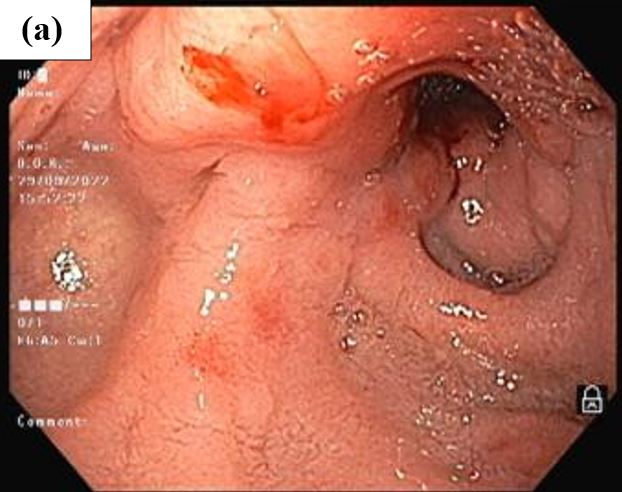
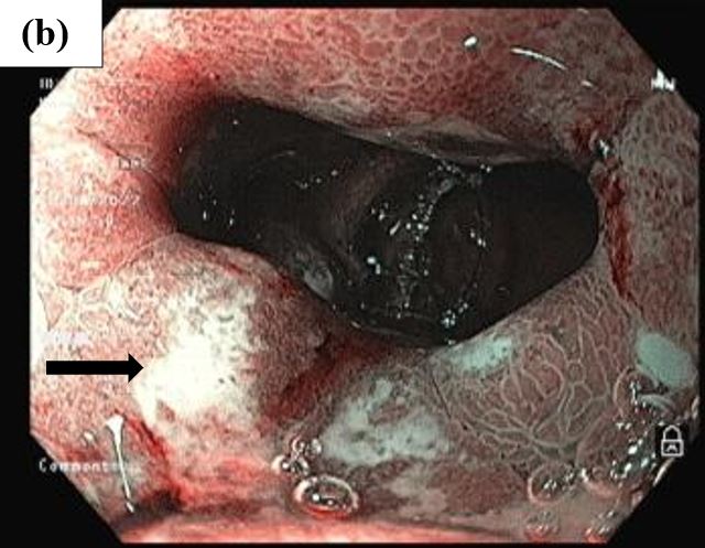
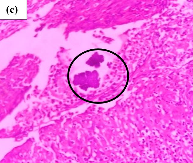
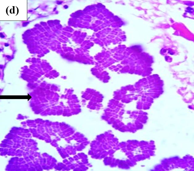
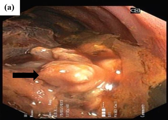
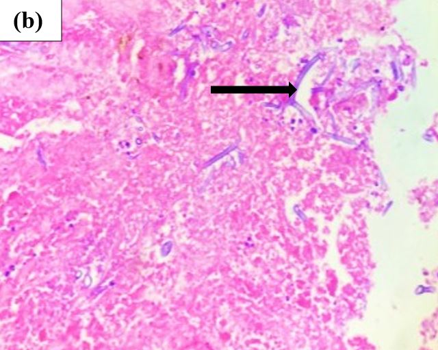
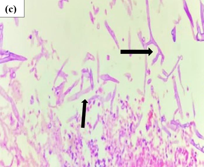
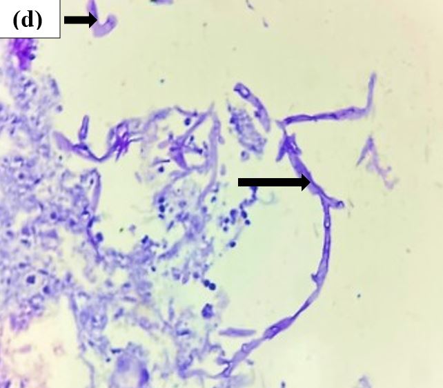
Figure 2: (a) Colonoscopy of the Descending colon showing semi-circumferential polypoidal growth, which was friable and causing luminal narrowing- Suspicion of malignancy. (b) & (c) show fungal hyphae, ribbon-like, broad-based, aseptate exhibiting right-angle branching.  [ H&E stain, 40x magnification] (d) shows fungal hyphae in the areas of necrosis. [ PAS stain, 40x magnification]


Discussion

Sarcina ventriculi was first described in 1842 by Goodsir and can be identified on hematoxylin and eosin (H&E) stained sections as cuboid basophilic cells in tetrad or octet formations. It ferments carbohydrates to produce acetaldehyde, ethanol, hydrogen, and carbon dioxide.[2], [3]

The organism thrives in acidic, anaerobic environments and is often found in patients with delayed gastric emptying due to diabetic gastroparesis, gastric surgery, or pyloric stenosis. It is typically located on the mucosal surface, embedded within gastric mucin or necrotic debris, without invading underlying glands.[2], [3]

Although Sarcina is occasionally found incidentally, its association with severe gastric complications such as emphysematous gastritis, food bezoars, and perforation necessitates its reporting when observed histologically.[2],[4],[5] Historically, the presence of sarcinous vomiting, persistent, frothy emesis was associated with Sarcina overgrowth.[4], [5]

In the present case, the tetrad arrangement, flattened cell walls, and basophilic staining supported the diagnosis of Sarcina ventriculi. Co-infection with Helicobacter pylori may exacerbate mucosal damage.[4] Despite its distinct morphology, Sarcina is often confused with Micrococcus species; however, the latter are smaller, catalase-positive, and aerobic. Sarcina maxima lacks the thick extracellular cellulose layer found in S. ventriculi.[4] Molecular identification through 16S rRNA and pyruvate decarboxylase gene sequencing offers definitive confirmation but remains largely unavailable in routine settings.[1], [3], [5]

Mucormycosis, caused by Mucorales such as Rhizopus, Mucor, Lichtheimia, and Rhizomucor, is an angioinvasive fungal infection with a high mortality rate.[6], [7], [9]While rhinocerebral and pulmonary forms are more prevalent, gastrointestinal mucormycosis constitutes only about 7% of cases.[7,8]Among GI cases, the stomach is the most frequently affected site (57.5%), followed by the colon (32.3%) and ileum (6.9%).[7] The disease is most often seen in immunocompromised individuals, particularly those with uncontrolled diabetes, hematologic mali-gnancies, or post-transplant immunosuppression.[6], [7],[8], [9]

Histopathology remains the cornerstone of diagnosis. The hallmark is vascular invasion by broad, aseptate hyphae with right-angle branching, causing thrombosis, infarction, and hemorrhagic necrosis. Over 94% of mucormycosis cases demonstrate infarction and angioinvasion.[10] Imaging often mimics malignancy, making histology and special stains such as PAS essential for confirmation.[9], [10]

The diagnosis in our case was established through colonoscopic biopsy, with PAS confirming fungal hyphae, sparing the need for culture or molecular diagnostics.

Conclusion

These two cases underscore the importance of recognizing rare gastrointestinal pathogens such as Sarcina ventriculi and mucormycosis, which often mimic more common pathologies both clinically and endoscopically. Timely histopathological examination remains critical for definitive diagnosis, especially in resource-limited settings where advanced molecular tools are not readily available. Early identification and appropriate management of these infections can markedly improve clinical outcomes and reduce morbidity in affected patients.

References

1. Tartaglia D, Coccolini F, Mazzoni A, Strambi S, Cicuttin E, Cremonini C, Taddei G, Puglisi AG, Ugolini C, Di Stefano I, Basolo F, Chiarugi M. Sarcina Ventriculi infection: a rare but fearsome event. A Systematic Review of the Literature. Int J Infect Dis. 2022 Feb;115:48-61 [Crossref][PubMed][Google Scholar]

2. Noor R, Ahsan M, Poombal F, Zaman S. Sarcina ventriculi associated gastritis. Pathologica. 2023 Dec;115(6):341-343. [Crossref][PubMed][Google Scholar]

3. Al Rasheed MR, Senseng CG. Sarcina ventriculi : Review of the Literature. Arch Pathol Lab Med. 2016 Dec;140(12):1441-1445. [Crossref][PubMed][Google Scholar]

4. Attri N, Pareek R, Dhanetwal M, Khan FM, Patel S. Sarcina ventriculi associated gastritis: Mimicking lymphoma on endoscopy. Indian J Pathol Microbiol. 2023 Jan-Mar;66(1):165-167. [Crossref][PubMed][Google Scholar]


5. Ene A, McCoy MH, Qasem S. Sarcina organism of the stomach: Report of a case. Hum Pathol (N Y). 2021;25(200541):200541. [Crossref][PubMed][Google Scholar]

6. Busbait S, AlMusa Z, Al Duhileb M, Algarni AA, Balhareth A. A Cecal Mucormycosis Mass Mimicking Colon Cancer in a Patient with Renal Transplant: A Case Report and Literature Review. Am J Case Rep. 2020 Oct 19;21:e926325. [Crossref][PubMed][Google Scholar]

7. Kumar Debata P, Keshari Panda S, Dash A, Mohanty R, Narayan Mallick B, Tadu D, et al. An unusual presentation of colonic mucormycosis mimicking carcinoma colon- a surgeon’s perspective. Int J Surg Case Rep 2015;10:248–51. . [Crossref][PubMed][Google Scholar]

8. Zhong B, Amundsen T, Farmer C. Invasive gastrointestinal mucormycosis. ACG Case Rep J 2023;10(9):e01161. . [Crossref][PubMed][Google Scholar]

9. Addasi Y, Nguyen AH, Sabri A, Ahmad F, Rangray R, Velagapudi M. Gastrointestinal Mucormycosis: A Clinical Review. Gastroenterology Res. 2023 Oct;16(5):249-253. [Crossref][PubMed][Google Scholar]

10. Parmar M, Bhadreshwara KA, Kanajariya T. A rare case of invasive intestinal mucormycosis: a case report. Int Surg J 2024;11(6):1019–22. . [Crossref][PubMed][Google Scholar]

Disclaimer / Publisher's Note The statements, opinions and data contained in all publications are solely those of the individual author(s) and contributor(s) and not of Journals and/or the editor(s). Journals and/or the editor(s) disclaim responsibility for any injury to people or property resulting from any ideas, methods, instructions or products referred to in the content.