E-ISSN:2456-1487
P-ISSN:2456-9887
RNI:MPENG/2017/70771

Case Report

Enterobius Vermicularis

Tropical Journal of Pathology and Microbiology

2025 Volume 11 Number 2 Jul-Dec
Publisherwww.medresearch.in

A Rare Case Of Acute Appendicitis Due To Enterobius Vermicularis In A Young Man

Verma AK1*, Sheikh F2, Krishnani C3, Simon K Ankita4
DOI:https://doi.org/10.17511/jopm.2025.i02.01

1* Anil Kumar Verma, Assistant Professor, Department of Pathology and Lab Medicine, All India Institute of Medical Sciences, Raipur, Chhattisgarh, India.

2 Firoz Sheikh, Assistant Professor, Department of Pathology, Raipur Institute of Medical Sciences, Raipur, Chhattisgarh, India.

3 Chandni Krishnani, Associate Professor, Department of Pathology, Raipur Institute of Medical Sciences, Raipur, Chhattisgarh, India.

4 K Ankita Simon, Assistant Professor, Department of Pathology and Lab Medicine, All India Institute of Medical Sciences, Raipur, Chhattisgarh, India.

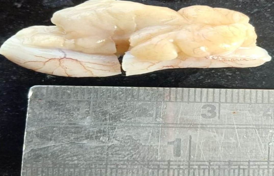
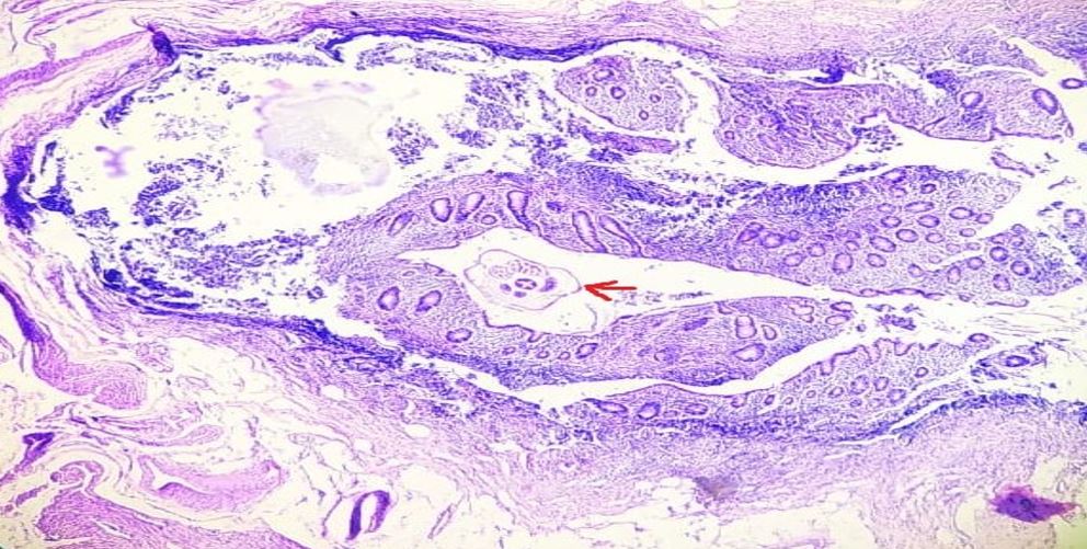
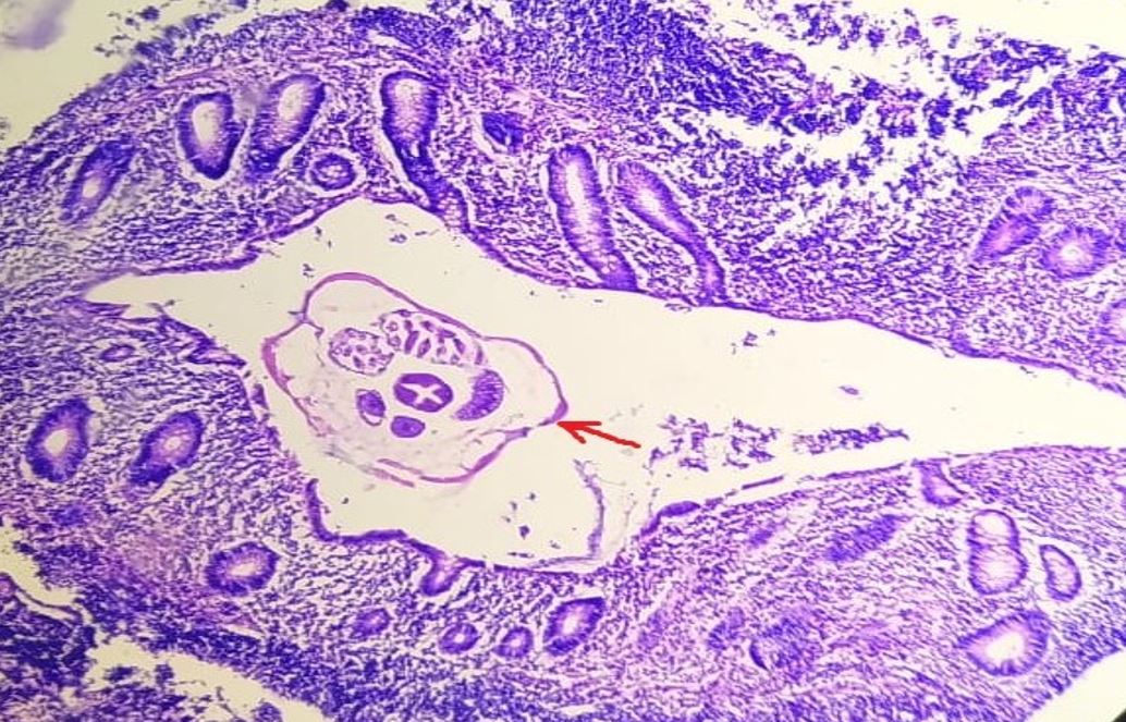
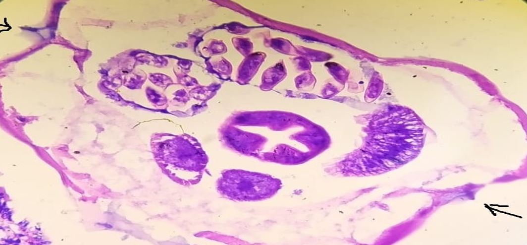
Acute appendicitis is the acute inflammation of the vermiform appendix. Several etiologic factors, including a parasite, may cause it. Many parasites,including Ascaris lumbricoides, taenia, and Enterobius vermicularis, Entamoeba histolytica, etc, can cause acute appendicitis. E. vermicularis is one of the most common parasitic infections around the world, and acute appendicitis, on the other hand, is also a commonly encountered condition in general surgery. However, the association between these two conditions remains rare. We present a case of a young adult male with histopathologically confirmed Enterobius vermicularis infestation in the appendix, highlighting the importance of considering parasitic causes in appendicitis.

Keywords: Parasitological diseases; Appendectomy; Appendicitis; Enterobius vermicularis; Enterobiasis; helminths; Pinworms

Corresponding Author How to Cite this Article To Browse
Anil Kumar Verma, Assistant Professor, Department of Pathology and Lab Medicine, All India Institute of Medical Sciences, Raipur, Chhattisgarh, India.
Email:
Verma AK, Sheikh F, Krishnani C, Simon K Ankita, A Rare Case Of Acute Appendicitis Due To Enterobius Vermicularis In A Young Man. Trop J Pathol Microbiol. 2025;11(2):17-20.
Available From
https://pathology.medresearch.in/index.php/jopm/article/view/681

Manuscript Received Review Round 1 Review Round 2 Review Round 3 Accepted
2025-07-08 2025-07-16 2025-07-24 2025-08-01 2025-08-08
Conflict of Interest Funding Ethical Approval Plagiarism X-checker Note
None Nil Yes 10.55

© 2025by Verma AK, Sheikh F, Krishnani C, Simon K Ankitaand Published by Siddharth Health Research and Social Welfare Society. This is an Open Access article licensed under a Creative Commons Attribution 4.0 International License https://creativecommons.org/licenses/by/4.0/ unported [CC BY 4.0].

Download PDFBack To ArticleIntroductionCase presentationDiscussionConclusionReferences

Introduction

Enterobius vermicularis, also known as pinworm or threadworm, is a common intestinal parasite, particularly in children.Approximately 12.9% of children around the world have been infected withE. vermicularis [1]. The World Health Organization reported that the prevalence of enterobiasis in children is between 4% and 28%. [2].Some patients with enterobiasis are asymptomatic, while others, especially children, may show symptoms such as perianal pruritus, restlessness, loss of appetite, malnutrition, anemia, insomnia, and irritability. Sometimes,enterobiasis can affect the kidneys and fallopian tubes, leading to severe health disorders and even death. [3] [4].

There are multiple ways to transmit enterobiasis, including the faecal-oral route, inhalation, auto-infection, and retrograde infection [5]. The main route of transmission forE. vermicularisis direct contact between infected and uninfected individuals. Therefore, children in crowded environments such as kindergartens, schools, orphanages, and mental institutions are most susceptible to this infection [6].

Case presentation

A 23-year-old male with no significant past medical or surgical history presented to the emergency department at Raipur Institute of Medical Sciences, complaining of right iliac fossa abdominal pain of a one-week duration. The pain was progressive with associated anorexia. He denied any history of fever or change in his bowel habits. On examination, his vital signs were within normal limits. His abdominal examination revealed tenderness in the right iliac fossa with a positive rebound sign. Urine analysis showed that no infection was present. Laboratory investigations showed a hemoglobin level of 13.3 g/dL, a white cellcount of 4100, and a C-reactive protein level of >4.0 mg/L (raised). The coagulation profile, Liver function test, and renal function tests were normal.

The patient’s pelvic ultrasonography showed a blind ended non peristaltic tubular structure (measuring 11.2 mm in diameter) with a thickened wall. A mild patchy increase in mural vascularity and adjoining patchy minimal free fluid is noted in the RIF region. A computed tomography (CT) scan was performed, given the low probability of acute appendicitis,

which showed a retrocecal appendix with a short segmentwall thickening of 6 mm and no significant fat stranding suggestive of borderline tip appendicitis.When a reevaluation was performed, abdominal tenderness was persistent despite adequate analgesia and hydration; therefore, a laparoscopic appendectomy under general anesthesia was performed. Intraoperative findings revealeda mildly hyperemic appendicular tip, which was adherent to the lateral abdominal wall and cecum with minimal hemorrhagic fluid in the pelvis. The appendectomy specimen was sent for histological examination. On gross examination specimen received measured 1.5 x 1.0 x 0.5 cm. The outer surface was smooth. Cut surface shows an obstructed lumen.The Microscopic examination revealed a mild denuded epithelium with an inflammatory infiltrate. E. vermicularis eggs were also seen within the lumen of the appendix. The postoperative course was uneventful, and the patient was discharged after 48 hours. The patient and his family members received 400 mg of albendazole once weekly for three weeksAt his follow-up appointment, he was well with no fresh complaints.

patho-681-01..JPG
Figure 1: Gross specimen of appendix received measuring: 1.5 x 1x 0.5 cm. Outer surface sm-ooth. Cut surface shows an obstructed lumen.

patho-681-02..JPG
Figure 2:
H&E stain, 4×: appendix wall with intraluminal E. vermicularis eggs.


patho-681-03..JPG
Figure 3:
H&E stain, appendix mucosa on a 10 X view showing ulcerated epithelium with inflammatory infiltrate and intraluminal Enterobius vermicularis eggs.

patho-681-04..JPG
Figure 4: H&E stain , appendiceal wall a 40 X magnification, showing evidence of E. vermicularis infection in the form of E. vermicularis eggs.

Discussion

The pathogenesis of acute appendicitis is mainly due to luminal obstruction, with various causes including lymphoid hyperplasia in children, faecoliths, tumors, and helminths, etc. All of these can increase intraluminal pressure and compromise mucosal blood supply, which can lead to hypoxia or ischaemia of the tissue. This later on promotes several microbial invasions of the appendiceal wall, which lead to acute appendicitis. Diagnosis mainly relies on specific clinical presentation—characterized by right lower abdominal pain, nausea, vomiting, and anorexia—along with laboratory findings such as leukocytosis, as well as imaging techniques like ultrasound. Several parasites have been reported as the cause of acute appendicitis. Altun E, Avci V, and Azatcam M carried out a retrospective analysis of 660 patients and a brief literature review that shows Enterobius and Taenia as the main causes of acute appendicitis. [7]. One study shows that Ascaris lumbricoides can cause appendicitis,

particularly in endemic regions, where it may be transmitted through oral ingestion of eggs. [8]. Management typically involves appendectomy, and anthelmintic treatment is necessary to eliminate the parasite from all potential sites. This study highlights how crucial it is to identify the causative agents in acute appendicitis for effective management.

Conclusion

Although Enterobius vermicularis is among the most prevalent intestinal helminths worldwide, its role in acute appendicitis is uncommon and often overlooked. This case highlights the need to consider parasitic causes of appendicitis, particularly in endemic areas. Routine histopathological evaluation of appendectomy specimens is essential for definitive diagnosis and guiding anthelmintic therapy. Early recognition and appropriate management are crucial to prevent recurrence and limit community transmission.

References

1. Global prevalence of enterobiasis in young children over the past 20 years: a systematic review and meta-analysis. Osong Public Health Res Perspect. 2023;14(6):441–450. doi:10. 24171/j.phrp.2023.0204 [Crossref][PubMed][Google Scholar]

2. World Health Organization. Deworming for health and development: report of the third global meeting of the partners for parasite control. Geneva: World Health Organization; 2005. (WHO/CDS/CPE/PVC/2005. 14) [Crossref][PubMed][Google Scholar]

3. Li HM, Zhou CH, Li ZS, Deng ZH, Ruan CW, Zhang QM, et al. Risk factors for Enterobius vermicularis infection in children in Gaozhou, Guangdong, China. Infect Dis Poverty. 2015;4:28. doi:10.1186/s40249-015-0058-9 [Crossref][PubMed][Google Scholar]

4. Akram HE, Al-Warid HS. Evaluation of hematological factors and micronutrients among children infected with Enterobius vermicularis. Iraqi J Sci. 2023;64(4):1625–1634. [Crossref][PubMed][Google Scholar]


5. Ngegba MP, Ngegba AM, Hinckley ES, Koroma MF, Oladele OI. Implications of prevalence and intensity of soil-transmitted helminths on rural farmers' productivity in selected districts of Sierra Leone. Sci J Warsaw Univ Life Sci – SGGW. 2023;23(1):32–45. doi:10.22630/PRS.2023.23.1.3 [Crossref][PubMed][Google Scholar]

6. Al-Daoody AA, Qadir FM, Tahir AA, Mahmood NH, Majeed AF. Risk factors of Enterobius vermicularis infection with symptoms among children in Erbil Governorate. Pak-Euro J Med Life Sci. 2020;3(2):50–58. [Crossref][PubMed][Google Scholar]

7. Altun E, Avci V, Azatçam M. Parasitic infestation in appendicitis: a retrospective analysis of 660 patients and brief literature review. Saudi Med J. 2017;38(3):314–318. doi:10.15537/smj.2017.3.18061 [Crossref][PubMed][Google Scholar]

8. Tsegaye MA, Degefe AW, Fisiha BA. A rare cause of acute appendicitis due to adult Ascaris lumbricoides in a 10-year-old child from Ethiopia: a case report. Int J Surg Case Rep. 2025;128:111119. [Crossref][PubMed][Google Scholar]

Disclaimer / Publisher's Note The statements, opinions and data contained in all publications are solely those of the individual author(s) and contributor(s) and not of Journals and/or the editor(s). Journals and/or the editor(s) disclaim responsibility for any injury to people or property resulting from any ideas, methods, instructions or products referred to in the content.