E-ISSN:2456-1487
P-ISSN:2456-9887
RNI:MPENG/2017/70771

Case Report

Papillary Carcinoma

Tropical Journal of Pathology and Microbiology

2024 Volume 10 Number 4 Oct-Dec
Publisherwww.medresearch.in

A Rare case of papillary carcinoma in branchial cleft cyst

Jadhav SN1*, Shelly D2, Das AK3
DOI:https://doi.org/10.17511/jopm.2024.i04.03

1* Sujata Narendra Jadhav, Senior Resident, Department of Pathology, INHS ASVINI, Mumbai, Maharashtra, India.

2 Divya Shelly, Surg Captain Associate Professor, Department of Pathology, INHS ASVINI, Mumbai, Maharashtra, India.

3 A K Das, (Colonel), Prof and HOD, Pathology, INHS ASVINI, Mumbai, Maharashtra, India.

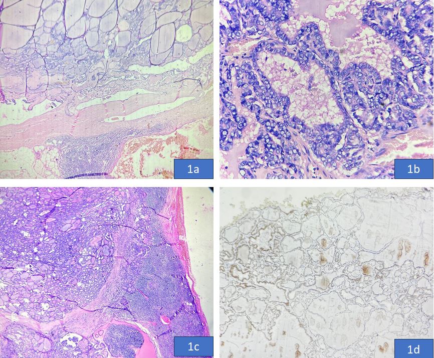
Anomalies of branchial cysts are rare diseases of the head and neck region. Confirmation of definite pathology is always challenging for clinicians. Ectopic thyroid tissue is ubiquitous in the body and a branchial cleft cyst a congenital abnormality can rarely harbor thyroid tissue in the cyst. Primary papillary thyroid carcinoma (PTC) inside ectopic thyroid tissue is extremely rare. We report a very rare case of PTC incidentally arising in a branchial cyst cleft.

Keywords: Branchial cleft cyst, Papillary carcinoma, Thyroid

Corresponding Author How to Cite this Article To Browse
Sujata Narendra Jadhav, Senior Resident, Department of Pathology, INHS ASVINI, Mumbai, Maharashtra, India.
Email:
Jadhav SN, Shelly D, Das AK, A Rare case of papillary carcinoma in branchial cleft cyst. Trop J Pathol Microbiol. 2024;10(4):68-71.
Available From
https://pathology.medresearch.in/index.php/jopm/article/view/671

Manuscript Received Review Round 1 Review Round 2 Review Round 3 Accepted
2024-11-01 2024-11-06 2024-11-14 2024-11-22 2024-11-30
Conflict of Interest Funding Ethical Approval Plagiarism X-checker Note
None Nil Yes 13.64

© 2024by Jadhav SN, Shelly D, Das AKand Published by Siddharth Health Research and Social Welfare Society. This is an Open Access article licensed under a Creative Commons Attribution 4.0 International License https://creativecommons.org/licenses/by/4.0/ unported [CC BY 4.0].

Download PDFBack To ArticleIntroductionCase ReportDiscussionConclusionReferences

Introduction

Brachial cleft cysts are commonly encountered in lateral neck swelling in clinical practice, however, the presence of ectopic thyroid tissue with papillary thyroid carcinoma (PTC) is extremely rare [1]. PTC primarily arising in brachial cleft cyst is indistinguishable from metastatic papillary carcinoma in the cyst and the diagnosis can only be confirmed after total thyroidectomy to exclude occult primary [2]. The first case of PTC in ectopic thyroid of the branchial cyst was reported in 1992 by Balasubramanniam et al [3]. We hereby report a very rare case of papillary thyroid carcinoma incidentally arising in a branchial cleft cyst. To the best of our knowledge, only seventeen cases have reported PTC arising in a branchial cyst without a primary in the thyroid [3], [4], [5], [6].

Case Report

A 70-year-old male presented with a painless right-sided lateral neck mass. Physical examination revealed a 4 cm mass on right side of neck. No significant cervical lymphadenopathy or thyroid mass was noted. Contrast-enhanced computed tomography (CECT) of neck showed a well-demarcated walled, solid cystic-enhancing mass lesion in right carotid space of neck. The solid component was measuring 2.6 mm in size and showed peripheral foci of calcifications. No cervical lymphadenopathy was detected. The differential diagnosis offered in radiology were vagal schwannoma, nerve sheath tumor and paraganglioma. The patient underwent surgical excision of mass and histopathological examination revealed a 5 cm cystic mass with keratin debris confirming a branchial cleft cyst. A solid area measuring 2 x 1.5 cm was identified. Microscopic examination revealed a cyst wall lined by cuboidal epithelium and stratified squamous epithelium with a fibrotic wall showing dense lymphoid infiltrate. The wall also showed few foci of normal thyroid tissue and classical type papillary carcinoma comprising of tumour arranged in form of papillae lined by cells showing nuclear overcrowding, grooving and intranuclear inclusions. Psammoma bodies were also identified. No areas of necrosis were identified. On immunohistochemistry, tumour cells were positive for TTF-1, PAX-8, p-16 and were immunonegative for p63. Two out of four lymph nodes in wall showed metastatic deposits of papillary carcinoma.

To rule out primary thyroid carcinoma and any residual malignancy, a Tc-99m Sestamibi scan was performed, which did not reveal any increased uptake in the thyroid gland. The patient has been under regular follow-up for 6 months, there is no evidence of recurrence.

patho-671-01..JPG
Figure: 1 H&E-stained sections, x40, showing brachial cyst wall and tumor. H & E stained section, 400x, showing papillary architecture of tumor cells H & E stained section, 40x, showing lymph node metastasis IHC, TTF1 positivity

Discussion

The thyroid is the first endocrine organ to develop during fetal embryology and is derived from endoderm. Failure of the thyroid to descend from the thyroid anlage region to its final location in front of the trachea can give rise to ectopic thyroid tissue. The most common location of ectopic thyroid is in the midline-lingual, high cervical, trachea, superior mediastinal and even pericardiac. Very rarely, it can be located in the lateral cervical region. One of the most common cystic lesions of the lateral head-neck region is a branchial cyst. These are located anteriorly to the sternocleidomastoid, although some rare sites have also been reported. The etiology of branchial cysts is not clear, various theories suggest congenital malformation, and cystic degeneration of lymph nodes due to epithelial migration [7]. The proximity of branchial arches during development can explain the presence of ectopic thyroid tissue. The literature studies reveal that ectopic thyroid tissue may clinically present as primary thyroid carcinoma especially papillary carcinoma,


however, most of these reports are from thyroglossal duct cysts, lingual thyroid and struma ovarii. Primary papillary carcinoma of the thyroid arising in branchial cysts is rare. Cochrane search shows only 17 cases have been reported in the literature as PTC arising from branchial cysts without a primary in the thyroid [6]. Two of these cases presented with metastatic lymph node disease at diagnosis. The usual diagnostic dilemma in such situations is that carcinoma in the cyst is a primary or metastatic disease with missed/occult primary in the thyroid. Sidhu et al. proposed the following criteria for diagnosing papillary thyroid carcinoma in branchial cysts [8].

1. an epithelial lining layer with subepithelial lymphoid tissue collection,
2. normal thyroid tissue adjacent to the focus of papillary carcinoma within the wall,
3. and no evidence of papillary carcinoma in the thyroid or other areas.

In our case, tumor showed morphological features of papillary thyroid carcinoma and fulfilled all the above criteria. Metastases in neck nodes are detected in about 20% of cases as per the available evidence and distant metastasis has not been reported [9], [10]. Immunohistochemical positive staining for TTF-1, PAX-8 and thyroglobulin can help in the identification of a PTC, however, it cannot distinguish between primary and metastatic tumors. CT, MRI and scintigraphy imaging can help in detecting ectopic thyroid tissue, however histopathological examination is the gold standard.

Conclusion

In conclusion, we have reported a very rare case of primary PTC located in the branchial cyst. Clinicians should always keep the possibility of ectopic thyroid tissue while dealing with the management of lateral neck mass. The diagnosis of this rare entity requires a multidisciplinary team comprising of radiologists, surgeons and pathologists.

References

1. Matsumoto K, Watanabe Y, Asano G. Thyroid papillary carcinoma arising in ectopic thyroid tissue within a branchial cleft cyst. Pathol Int. 1999 May;49(5):444-6. doi: 10.1046/j.1440-1827.1999.00894.x. PMID: 10417688 [Crossref][PubMed][Google Scholar]

2. Ruhl DS, Sheridan MF, Sniezek JC. Papillary thyroid carcinoma in a branchial cleft cyst without a thyroid primary. navigating a diagnostic dilemma. Case Rep Otolaryngol 2013; 2013: 405342. [Crossref][PubMed][Google Scholar]

3. Balasubramaniam GS, Stillwell RG, Kennedy JT. Papillary carcinoma arising in ectopic thyroid tissue within a branchial cyst. Pathology. 1992 Jul;24(3):214-6. Doi: 10.3109/00313029209063177. PMID: 1437298 [Crossref][PubMed][Google Scholar]

4. Jadusingh W, Shah DJ, Shaw H, Lyn C. Thyroid papillary carcinoma arising in a branchial cleft cyst. West Indian Med J. 1996 Dec;45(4):122-4. PMID: 9033233 [Crossref][PubMed][Google Scholar]

5. Matsumoto K, Watanabe Y, Asano G. Thyroid papillary carcinoma arising in ectopic thyroid tissue within a branchial cleft cyst. Pathol Intern. 1999;49:444–446. Doi: 10.1046/j.1440-1827.1999.00894.x.PMID: 10417688 [Crossref][PubMed][Google Scholar]

6. Cappellani A, Di Vita M, Zanghì A, Di Stefano B, La Porta D, De Luca A, Lanzafame S, D’Angelo A. A case of branchial cyst with an ectopic thyroid papillary carcinoma. Ann Ital Chir. 2004 May-Jun;75(3):349-51; discussion 352. PMID: 15605525 [Crossref][PubMed][Google Scholar]

7. Cho JS, Shin SH, Kim HK, Lee JS, Park MH, Yoon JH, Jegal YJ. Primary papillary carcinoma originated from a branchial cleft cyst. J Korean Surg Soc. 2011 dec;81 Suppl 1(Suppl 1):S12-6. Doi: 10.4174/jkss.2011.81.Suppl1.S12. Epub 2011 Nov 25. PMID: 22319730; PMCID: PMC3267057 [Crossref][PubMed][Google Scholar]

8. Sidhu S, Lioe TF, Clements B. Thyroid papillary carcinoma in lateral neck cyst: missed primary tumour or ectopic thyroid carcinoma within a branchial cyst? J Laryngol Otol. 2000 Sep;114(9):716-8. Doi: 10. 1258/0022215001906598. PMID: 11091840 [Crossref][PubMed][Google Scholar]

9. Mremi A, Nkya G, Amsi P, Sadiq A, Lodhia J, Pallangyo A. Papillary thyroid carcinoma arising from ectopic thyroid tissue in a neck branchial cyst. SAGE Open Med Case Rep. 2023 Apr 20;11:2050313X231168555. Doi: 10.1177/2050313X231168555. PMID: 37122428; PMCID: PMC10134172 [Crossref][PubMed][Google Scholar]


10. Rigas G, Skalias A, Filentas A, Tsantila I, Blioskas S. An Unusual Case of a Papillary Thyroid Carcinoma Arising in a Branchial Cyst. Ear Nose Throat J. 2023 Oct 23:1455613231207819. Doi: 10.1177/01455613231207819. Epub ahead of print. PMID: 37872779 [Crossref][PubMed][Google Scholar]

Disclaimer / Publisher's NoteThe statements, opinions and data contained in all publications are solely those of the individual author(s) and contributor(s) and not of Journals and/or the editor(s). Journals and/or the editor(s) disclaim responsibility for any injury to people or property resulting from any ideas, methods, instructions or products referred to in the content.