E-ISSN:2456-1487
P-ISSN:2456-9887
RNI:MPENG/2017/70771

Research Article

Endometrial Hyperplastic

Tropical Journal of Pathology and Microbiology

2024 Volume 10 Number 3 Jul-Sep
Publisherwww.medresearch.in

BCL2 and Ki 67 expression in endometrial hyperplastic disorders

Somani A1*, Nimbargi R2, Patil A3, Patil A4, Bharadwaj R5
DOI:https://doi.org/10.17511/jopm.2024.i03.02

1* Ashwini Somani, Resident, Department of Pathology, Bharati Hospital and Research Center, Pune, Maharashtra, India.

2 Ravindra Nimbargi, Professor, Department of Pathology, Bharati Hospital and Research Center, Pune, Maharashtra, India.

3 Anuja Patil, Assistant Professor, Department of Pathology, Bharati Hospital and Research Center, Pune, Maharashtra, India.

4 Amrutraj Patil, Senior Resident, Department of Pathology, Bharati Hospital and Research Center, Pune, Maharashtra, India.

5 Reena Bharadwaj, Head and Professor, Department of Pathology, Bharati Hospital and Research Center, Pune, Maharashtra, India.

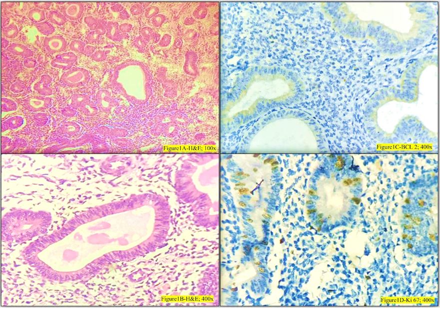
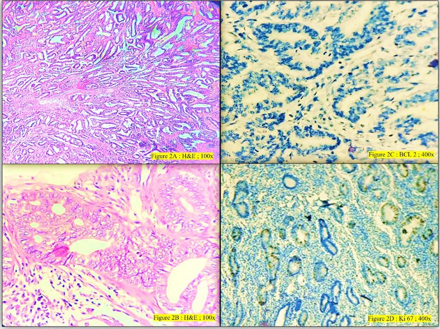
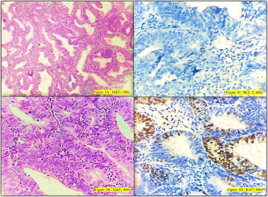
Introduction: The pattern of expression in hyperplastic and premalignant states of endometrium helps us to study the progression of these conditions to frank malignancy. The diagnosis of endometrial hyperplasia with/without atypia is a subjective diagnosis which can affect the treatment line of the patient. This study is done to test the IHC markers BCL-2 and Ki67 on hyperplastic and malignant lesions in an attempt to make the diagnosis of the type of hyperplasia more accurate.

Purpose: The present study attempts to determine the relationship between proliferation and the inhibition of apoptosis in endometrial hyperplastic disorders, using monoclonal antibodies against the proliferation marker, Ki-67 and the anti-apoptotic protein, Bcl-2.

Materials and Methods: Histopathological test requisition forms and paraffin blocks of endometrial biopsy and endometrial curettage reported as endometrial hyperplasia or endometrial carcinoma from February 2022 to May 2023 in the Department of Pathology, Bharati Medical College and Research Center, Pune.were collected. The H & E sections and IHC – BCL-2 and Ki 67 were studied on all 60 cases and the observations noted down to be further statistically analyzed.

Results: Analysis revealed significant p value for both BCL 2 and Ki 67. BCL-2 is an antiapoptotic marker showing positivity in typical hyperplasia cases as compared to atypical or malignant lesions. Ki 67 is a proliferative marker showing an increase in its expression in typical to atypical endometrial hyperplastic lesions to malignant lesions.

Conclusion: BCL-2 and Ki 67 can be used as a marker to assess the endometrial hyperplastic progression towards typia/malignancy.

Keywords: Endometrium, Hyperplasia, Antiapoptotic marker, Proliferative marker

Corresponding Author How to Cite this Article To Browse
Ashwini Somani, Resident, Department of Pathology, Bharati Hospital and Research Center, Pune, Maharashtra, India.
Email:
Somani A, Nimbargi R, Patil A, Patil A, Bharadwaj R, BCL2 and Ki 67 expression in endometrial hyperplastic disorders. Trop J Pathol Microbiol. 2024;10(3):38-44.
Available From
https://pathology.medresearch.in/index.php/jopm/article/view/654

Manuscript Received Review Round 1 Review Round 2 Review Round 3 Accepted
2024-06-13 2024-06-19 2024-06-25 2024-07-01 2024-07-07
Conflict of Interest Funding Ethical Approval Plagiarism X-checker Note
None Nil Yes 13.33

© 2024by Somani A, Nimbargi R, Patil A, Patil A, Bharadwaj Rand Published by Siddharth Health Research and Social Welfare Society. This is an Open Access article licensed under a Creative Commons Attribution 4.0 International License https://creativecommons.org/licenses/by/4.0/ unported [CC BY 4.0].

Introduction

Endometrial cancer is the most common gynaecological cancer in high-income countries and its incidence is rising globally [1]. This cancer is the fourth most common malignancy in women. It mainly affects postmenopausal women, where patients’ mean age at diagnosis is 61 years, and most cases are diagnosed in women aged 45 to 74 years[2]. Ca endometrium has a very good prognosis if diagnosed early with a complete cure possible in the early stages [3].

Most of these cancers present at an early stage and are associated with a good prognosis. The treatment comprises surgical staging and adjuvant radiotherapy and/or chemotherapy depending on the final surgico-pathological stage [4].

The incidence of endometrial hyperplasia (EH) is roughly three times higher than endometrial carcinoma (EC) and certain atypical forms of EH are considered to represent direct precursor lesions to endometrioid EC. There are several lines of evidence that a diagnosis of EH may precede the development of EC and that the two, share common predisposing risk factors [5].

The proliferative endometrium is characterized by decreased apoptotic and increased mitotic activity in response to estrogen. This proves the association of hyperestrogenic states in causing increased proliferation leading to neoplasm [6].

The key markers for endometrial malignancy include estrogen receptor (ER), progesterone receptor (PR), p53, B-Cell Lymphoma 2 (BCL 2), E-cadherin, Cyclin D1, Ki 67, etc. [7, 8]. Proliferation as measured by the expression of the proliferation marker, Ki-67, is distinctly related to estrogen receptor content in normally cycling endometrium6. Apoptosis, or programmed cell death, is a complex process, in which Bcl-2 has an important role.

Bcl-2 is a protooncogene preventing apoptosis and thereby prolonging cell survival. Bcl-2 expression is hormone-regulated in the endometrium, because it closely follows oestrogen receptor (ER) levels, with peak activity in a proliferative phase when maximal ER expression occurs. Analysing the expression of these markers in cyclical endometrium and hyperplasia and with carcinoma will enable determining the malignant potential [9].

Materials And Methods

  • Setting: Department of Pathology, Bharati Vidyapeeth (Deemed to be) University Medical College and Hospital, Pune (Tertiary Care Center)
  • Duration: 1.5 years (February 2022 to May 2023)
  • Type of Study: Prospective observational study
  • Sampling methods: Endometrial curettage
  • Sample size calculation: Minimum of 54 cases (60 cases studied)
  • Inclusion criteria: Endometrial samples obtained from endometrial biopsy or dilatation and curettage and diagnosed as endometrial hyperplasia/ endometrial carcinoma.
  • Exclusion criteria: Inadequate, non-representative or non-fixed samples
  • Data collection procedure: Retrospective and prospective
  • Ethical consideration & permission: The study was conducted using all ethical precautions and with the ethical approval of the institute in which the study was conducted. Funding was provided by the institute.
  • Statistical Analysis: The collected data was coded and entered into a Microsoft Excel sheet. The data will be analyzed using SPSS (Statistical Package for Social Sciences) version 20.0 software. The observations were statistically analysed using the ANOVA test and test of proportions.

Fresh slides were made from paraffin-embedded blocks and stained for H & E using standard protocol. IHC markers BCL 2 and Ki 67 were performed on all the blocks using the standard protocol.

IHC assessment: BCL 2 positivity is indicated by cytoplasmic positivity in glandular and stromal cells. In this study, only the positive cells in the glandular epithelium were considered. Control of Lymph node (HPE- 1802/22/1) was used.

The positive expression of Ki-67 was defined as the presence of brown-yellow granules in cell nuclei or both cell nuclei and cytomembrane.


The manual scoring system was done using the percentage of positively stained nuclei in ten high-power fields selected across the tumour ensuring at least 1000 nuclei were counted. Control of small cell neuroendocrine carcinoma was used (HPE-5236/22/2).

Results

Based on the histopathological features (WHO 5th edition) the endometrial samples were classified as endometrial hyperplasia without atypia, hyperplasia with atypia and endometrial carcinoma.

  • Endometrial hyperplasia without atypia: It is the proliferation of glands of irregular size and shape without significant cytologic atypia.
  • Increased gland to stroma ratio
  • Branching and/or cystically dilated glands
  • Uniform distribution of nuclear features

patho_654_01.JPG
Figure 1:
Endometrial hyperplasia without atypia, 1A-H&E 100x (low power), 1B- H&E 400x (high power), 1C- BCL 2 -positive, 1D- Ki67- 5 to 10% expression

  • Endometrial hyperplasia with atypia/ Endometrial intra epithelial neoplasm: It is the simultaneous change of epithelial cytology and an increased number of endometrial glands in comparison with the stroma within a morphologically defined region.
  • Glands exceed the stroma, reduction in stromal volume.
  • Extensive cytoplasmic clearing and irregularly distributed nuclei
  • Nuclear appearance varied- smaller/elongated/pseudostratified nuclei

patho_654_02.JPG
Figure 2:
Endometrial Hyperplasia with atypia, 2A-H&E 100x (low power), 2B- H&E 400x (high power),

2C- BCL 2 -negative, 2D- Ki67- 11-15% expression

  • Endometrioid carcinoma is a malignant epithelial neoplasm displaying a varying proportion of glandular, papillary and solid architecture with tumour cells showing endometroid differentiation.
  • Serous carcinoma is diffuse with marked nuclear pleomorphism showing typically papillary or glandular patterns.
  • Clear cell carcinoma demonstrates papillary, tubo-cystic patterns with variable pleomorphic polygonal, cuboidal, flat or hobnail cells with clear cytoplasm.

patho_654_03.JPG
Figure 3:
Endometroid Carcinoma, 3A-H&E 100x (low power), 3B- H&E 400x (high power), 3C- BCL 2 -negative, 3D- Ki67- 20% expression

A study of 60 females from 21 to 60 years of age was done. The majority of the women were in the 41 to 50 years age group (40%), followed by 51 to 60 years and 31 to 40 years age group.


The majority of the females had complaints of menorrhagia (33.34%) or post-menopausal bleeding (33.34%). Histopathological diagnosis of endometrial hyperplasia without atypia was made in 34 cases (56.67%), hyperplasia with atypia in 14 cases (23.34%) and endometrial carcinoma in 12 cases (20%).

Table 1: Correlation of clinical symptoms and histopathological diagnosis

Clinical symptomsEndometrial ca IEndometrial ca IIHigh-grade serous caEndometrial hyperplasia with atypiaEndometrial hyperplasia without atypiaTotal
Abdominal pain---235
Abnormal Uterine bleeding-1-3812
Difficulty in procuring a child1---23
Menorrhagia2---21620
Post-menopausal bleed6117520
Total921143460

Out of the 34 cases of endometrial hyperplasia without atypia, BCL 2 was positive in 30 cases (88.24%), and negative in 4 cases (11.76%) with a significant p-value of <0.001. Ki 67 was less than 10% in 31 cases (91.11%) 11 to 20 % in 2 cases (5.88%) and 1 case showed more than 20% (2.94%).

Table 2: BCL 2 expression in various endometrial lesions

BCL 2 expression
Histopathology diagnosisNumber of cases (n)No. of Bcl 2 positive cases No. of Bcl 2 negative casesp-value
EH without atypia34304<0.001
EH with atypia14311<0.002
Endometrial carcinoma11011<0.001
High-grade serous carcinoma110-
Total603426

Table 3: Ki 67 expression in various endometrial lesions

Ki 67 expression grading (overall p-value <0.001)
Histopathology diagnosisNumber of cases (n)<10%11-20%>20%Mean %
EH without atypia3431218.53
EH with atypia14013114.21
Endometrial carcinoma1103822.00
High-grade serous carcinoma100121.00
Total60311811

Out of the 14 cases of endometrial hyperplasia with atypia, BCL 2 was positive in 3 cases (21.43%), and negative in 11 cases (78.57%) giving a significant p-value of <0.002. Ki 67 was expressed in 11 to 20% of the cells in 13 cases (92.85%) and was more than 20 % in 1 case (7.15%).

There were 12 cases of carcinoma of 11 cases of endometrial carcinoma including 9 cases of Grade I and 2 cases of Grade II carcinoma. All 11 cases were negative for BCL 2 (100%) with a significant p-value of <0.001 and showed Ki 67 expression of 11-20 % in 3 cases (27.27%) and more than 20% in 8 cases (72.73%). One case was of high-grade serous carcinoma which showed BCL 2 positivity and Ki 67 expression of more than 20%.

Discussion

In a study by Abd El-Mohsen Hassan & et al at Sohag University, non-malignant endometrial lesions tend to express significantly higher levels of Bcl-2 compared to malignant endometrial lesions (p = 0.014). In malignant neoplastic conditions, the glandular expression of Bcl-2 was significantly higher in endometrial hyperplasia compared to endometrial carcinoma in the same gland (p = 0.006) [10].

Niemann TH et al. researched to investigate the expression of bcl-2 in both endometrial hyperplasia and cancer. The research used immunohistochemical examination of 49 instances, including proliferative and secretory endometrium, complicated and atypical hyperplasia, as well as different stages and forms of endometrial cancer. The presence of Bcl-2 protein was detected in all instances of complex hyperplasia, one instance of complex atypical hyperplasia, and 10 out of 29 instances of cancer.


The expression of complicated atypical hyperplasia and carcinoma was localised and exhibited lower intensity compared to the normal proliferative endometrium. The research found that bcl-2 is seldom overexpressed in complicated atypical hyperplasia or carcinoma, indicating a possible reduction in bcl-2 expression in endometrial cancer relative to hyperplasia. These findings suggest that Bcl-2 likely does not have a substantial impact on the transition from hyperplasia to carcinoma, in contrast to earlier beliefs [11].

Shalini P et al. researched to investigate correlation between cell proliferation and prevention of programmed cell death (apoptosis) in both normal endometrium and hyperplasia. The expression of Bcl-2 dropped, while expression of Ki-67 rose as hyperplasia progressed from a non-atypical state to atypical hyperplasia. This suggests a reduction in apoptosis and an increase in proliferation as hyperplasia advances towards atypia. Therefore, Bcl-2 and Ki-67 might potentially indicate advancement of endometrial hyperplasia and formation of endometrial cancer [9].

Bhati S et al. conducted a hospital-based observational cross-sectional study to evaluate and compare the expression of Bcl-2 and Ki-67 in various endometrial lesions and cyclical endometrium. The study included 57 patients, with tissue blocks processed for haematoxylin and eosin stain and immunohistochemical staining for Bcl-2 and Ki-67. Among the 12 hyperplasia cases, 9 were simple hyperplasia with mean scores of 6.2 for Bcl-2 and 4.8 for Ki-67, and 3 were atypical hyperplasia with mean scores of 4 for Bcl-2 and 2.6 for Ki-67. The study concluded that Bcl-2 and Ki-67 are reliable markers for indicating disease progression and may serve as novel indicators for treatment and follow-up [6].

Ghalib Farhood R et al. did research to assess the expression of the proliferation marker Ki-67 in different types of endometrial lesions. The research included 60 endometrial samples obtained from either endometrial curetting or hysterectomy specimens. These samples were diagnosed as simple hyperplasia (n=10), complicated hyperplasia (n=20), atypical hyperplasia (n=6), and endometrial cancer (n=24). The findings revealed a higher level of Ki-67 expression in endometrial cancer when compared to proliferative endometrium and simple hyperplasia (P=0.0001) [12].

Swami RM et al.68 conducted a retrospective study to examine the immunohistochemistry (IHC) patterns of estrogen receptor (ER), progesterone receptor (PR), p53, and Ki-67 in patients with endometrial adenocarcinoma (EC) and endometrial hyperplasia. Among the 78 cases, 20 were diagnosed with EC (17 endometrioid and three papillary serous adenocarcinomas) and 58 with endometrial hyperplasia. EC was most common in the sixth to seventh decades, while hyperplasia was prevalent in the fourth to fifth decades. Ki-67 expression varied, being <10% in 11 cases, 10-20% in 5 cases, and >20% in 4 cases. Follow-up indicated that 14 out of 15 patients had progression-free survival [13].

In a study conducted by Arjunan A et al Bcl-2 expression was consistently higher in proliferative endometrium than endometrial hyperplasia and diminished significantly in the secretory phase, suggesting estrogen-induced anti-apoptotic activity. As hyperplasia advanced towards atypia, apoptosis decreased while proliferative activity increased. Ki-67 expression was elevated in hyperplastic states, indicating increased mitotic activity and a premalignant status. Bcl-2 and Ki-67 comparison showed Bcl-2 levels were threefold higher in proliferative endometrium and twofold higher in hyperplastic states, reflecting higher anti-apoptotic activity under estrogen influence. The study suggests that Bcl-2 could be a potential therapeutic target in Bcl-2-positive endometrial hyperplasia patients. Larger studies and clinical trials are needed to understand the roles of Bcl-2 and Ki-67 further and explore genomic therapies targeting these genes to treat abnormal uterine bleeding and prevent progression to carcinoma [14].

Riseberg B et al. sought to examine the expression of the proliferation marker Ki-67 and the anti-apoptotic protein Bcl-2 in different types of endometrial lesions. The research included a total of 194 samples, which consisted of endometrial hyperplasia, polyps, carcinomas, and cyclic endometrium. The findings revealed that secretory endometrium, polyps, and atypical hyperplasia had the lowest levels of glandular Ki-67 expression. In contrast, endometrial carcinomas displayed a substantially higher and less varied Ki-67 score (p<0.001). The expression of Bcl-2 was greatest in the proliferative endometrium and polyps, but significantly lower in adenocarcinomas compared to hyperplastic lesions (p=0.002).


Additionally, there was a strong correlation between Ki-67 and Bcl-2 in the proliferative endometrium (p=0.003). The results indicate that the disparity between cell proliferation and programmed cell death (apoptosis) may play a critical role in the formation of several types of endometrial abnormalities, including both non-cancerous and cancerous growths. The variability in Ki-67 expression might assist in distinguishing between various types of endometrial hyperplasia [15].

Laban M et al. conducted a study consisting of 20 normal samples, 40 hyperplastic samples (including simple, complicated, and atypical cases), and 40 samples of endometrioid adenocarcinoma. The results demonstrated a notable rise in Bcl-2 staining, progressing from normal tissue to complicated and atypical hyperplasia, and finally to well-differentiated adenocarcinoma.

The statistical analysis revealed substantial differences in staining intensity, with p-values of 0.002, 0.0008, and 0.0001, respectively. These data indicate that Bcl-2 plays a significant role in the initiation of endometrial carcinogenesis, but it does not contribute to the advancement of the tumour beyond that point [16].

Shevra CR et al. conducted a retrospective case-control study at a tertiary referral centre to examine the expression profile of cell cycle regulatory proteins in normal proliferative endometrium, various types of hyperplasia (simple, complex, and atypical), and endometrial carcinoma.

The study included 61 endometrial samples, comprising 11 cases of simple hyperplasia, 13 of complex hyperplasia, 7 of atypical hyperplasia, and 20 of endometrial carcinoma, obtained from either endometrial curetting or hysterectomy specimens.

Cyclin D1 expression demonstrated a positive correlation with Ki-67 expression. These findings suggest that Cyclin D1 and Ki-67 may be markers for endometrial carcinogenesis and tumour cell proliferation [17].

Conclusion

In this study of 60 females with endometrial hyperplastic disorders, maximum presented with post-menopausal bleeding or menorrhagia. Maximum cases were of endometrial hyperplasia without atypia.

BCL 2 was positive in hyperplasia without atypia but negative in carcinoma cases, whereas Ki67 showed an ascending trend as a proliferative marker in non-atypical to atypical and malignant lesions.

What does this study add to existing knowledge?

Immunohistochemical markers can help make a clearer histopathological diagnosis of endometrial hyperplastic disorders and help remove the individual subjective bias in diagnosis. Studies undertaken on a larger scale can help in better understanding of the same and also eventually help with better patient care in cases of hyperplasia progressing towards carcinoma.

Permission from Institutional Research Board (IRB): Yes

Funding: Nil

Conflict of interest: None

References

1. Crosbie EJ, Kitson SJ, McAlpine JN, Mukhopadhyay A, Powell ME, Singh N. Endometrial cancer. The Lancet. 2022 Apr 9;399(10333):1412-28. [Crossref][PubMed][Google Scholar]

2. Paleari L. New Strategies for Endometrial Cancer Detection and Management. International Journal of Molecular Sciences. 2023 Mar 30;24(7):6462. [Crossref][PubMed][Google Scholar]

3. Endometrial Cancer [Internet]. Kauvery Hospital | Patient Newsletters | A Monthly Newsletter from your Partners in Good Health. 2022 [cited 2024 Aug 3]. Available from: https://www. kauveryhospital.com/ima-journal/ima-journal-april-2022/endometrial-cancer/ [Crossref][PubMed][Google Scholar]

4. Maheshwari A, Kumar N, Mahantshetty U. Gynecological cancers: a summary of published Indian data. South Asian journal of cancer. 2016 Jul;5(03):112-20. [Crossref][PubMed][Google Scholar]

5. Byun JM, Jeong DH, Kim YN, Cho EB, Cha JE, Sung MS, Lee KB, Kim KT. Endometrial cancer arising from atypical complex hyperplasia: The significance in an endometrial biopsy and a diagnostic challenge. Obstetrics & gynecology science. 2015 Nov 16;58(6):468-74. [Crossref][PubMed][Google Scholar]


6. Bhati, S. , & Negi, S. R. Manuscript type: original research article STUDY OF EXPRESSION OF BCL-2 AND Ki-67 IN BIOPSY SPECIMENS OF NORMAL ENDOMETRIUM AND OTHER ENDOMETRIAL LESIONS-AN OBSERVATIONAL STUDY. [Crossref][PubMed][Google Scholar]

7. Hutt, S. , Tailor, A. , Ellis, P. , Michael, A. , Butler-Manuel, S., & Chatterjee, J. (2019). The role of biomarkers in endometrial cancer and hyperplasia: a literature review. Acta oncologica, 58(3), 342-352 [Crossref][PubMed][Google Scholar]

8. Steinbakk, A. , Gudlaugsson, E. , Aasprong, O. G. , Skaland, I., Malpica, A., Feng, W., ... & Baak, J. P. (2011). Molecular biomarkers in endometrial hyperplasias predict cancer progression. American journal of obstetrics and gynecology, 204(4), 357-e1 [Crossref][PubMed][Google Scholar]

9. Shalini, P. , Suresh, N. , & Lilly, S. M. (2021). Expression of BCL-2 and KI-67 in Cyclical Endometrium and in Endometrial Hyperplasia. Journal of Pharmaceutical Research International, 33(21A), 1-11 [Crossref][PubMed][Google Scholar]

10. Hassan, W. A. E. M. , Adly, M. A., & Ahmed, A. (2019). Expression of Bc-l2 in precancerous endometrial lesion and endometrial carcinoma. Sohag Medical Journal, 23(1), 44-49 [Crossref][PubMed][Google Scholar]

11. Niemann, T. H. , Trgovac, T. L. , Mcgaughy, V. R., & Vaccarello, L. (1996). bcl-2 expression in endometrial hyperplasia and carcinoma. Gynecologic oncology, 63(3), 318-322 [Crossref][PubMed][Google Scholar]

12. Abd Ali Al-Humairi, I. (2022). Immunohistochemical study of Ki-67 in hyperplastic and endometrium carcinoma: a comparative study. Archives of Razi Institute, 77(1), 229. [Crossref][PubMed][Google Scholar]

13. Swami, R. M. , Lakhe, R. , Doshi, P. , Karandikar, M. N., Nimbargi, R., & Mani, N. S. Immunohistochemical study of ER, PR, p53 and Ki67 expression in patients with endometrial adenocarcinoma and atypical endometrial hyperplasia [Crossref][PubMed][Google Scholar]

14. Arjunan, A. , Nilavu, J. , ThiriveniBalajji, G. S. , & Praba, V. (2016). Expression of Bcl-2 and Ki-67 in cyclical endometrium and in endometrial hyperplasia–An analysis. IOSR J Dent Med Sci, 15, 43-9 [Crossref][PubMed][Google Scholar]

15. Risberg, B. , Karlsson, K. , Abeler, V. , Lagrelius, A. , Davidson, B., & Karlsson, M. G. (2002). Dissociated expression of Bcl-2 and Ki-67 in endometrial lesions: diagnostic and histogenetic implications. International journal of gynecological pathology, 21(2), 155-160 [Crossref][PubMed][Google Scholar]

16. Laban, M. , Ibrahim, E. A. S. , Agur, W., & Ahmed, A. M. B. E. (2015). Bcl-2 may play a role in the progression of endometrial hyperplasia and early carcinogenesis, but not linked to further tumorigenesis. Journal of microscopy and ultrastructure, 3(1), 19-24 [Crossref][PubMed][Google Scholar]

17. Shevra, C. R. , Ghosh, A. , & Kumar, M. (2015). Cyclin D1 and Ki-67 expression in normal, hyperplastic and neoplastic endometrium. Journal of postgraduate medicine, 61(1), 15-20 [Crossref][PubMed][Google Scholar]