E-ISSN:2456-1487
P-ISSN:2456-9887
RNI:MPENG/2017/70771

Case Report

Pathologist’s Perspective

Tropical Journal of Pathology and Microbiology

2024 Volume 10 Number 2 Apr-Jun
Publisherwww.medresearch.in

Dress Syndrome Demystified: Insights From a Pathologist’s Perspective

Agrawal S1*, Dravid N2, Mahajan D3, Shinde R4, Gadre A5, Patil P6
DOI:https://doi.org/10.17511/jopm.2024.i02.02

1* Sakshi Agrawal, Assistant Professor, Department of Pathology, Acpm Medical College, Dhule, Maharashtra, India.

2 NV Dravid, Emeritus Professor, Department of Pathology, ACPM Medical College, Dhule, Maharashtra, India.

3 DY Mahajan, Professor and HOD, Department of dermatology, ACPM Medical College, Dhule, Maharashtra, India.

4 Rahul Shinde, Senior resident, Department of dermatology, ACPM Medical College, Dhule, Maharashtra, India.

5 AS Gadre, Senior resident, Department of Pathology, ACPM Medical College, Dhule, Maharashtra, India.

6 Priyanka Patil, Junior resident, Department of Pathology, ACPM Medical College, Dhule, Maharashtra, India.

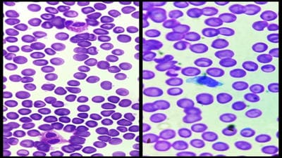
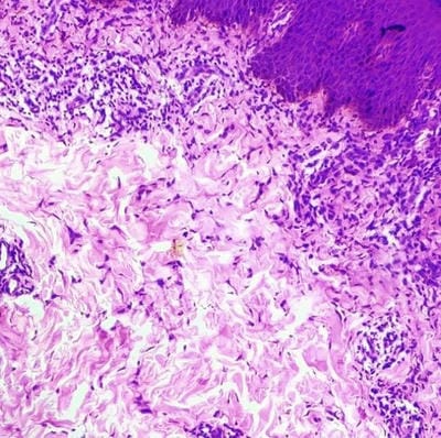
Background: Drug reaction with eosinophilia and systemic symptoms (DRESS) syndrome is a constellation of symptoms that manifest because of certain medications. Several antipsychotics, antibiotics, anti-convulsants, and sulfa-containing drugs are known to be implicated in the etiology of DRESS syndrome. It is a severe idiosyncratic drug reaction with a long latency period and due to the asymptomatic beginning and non-specific nature of symptoms, it is difficult to identify. This disease is diagnosed using the RegisCAR (European Registry of Severe Cutaneous Adverse Reaction) scoring system. The clinical presentation of this disorder consists of a diffuse rash, lymphadenopathy, and systemic organ damage. The diagnostic workup comprises monitoring inflammatory markers on laboratory work, and a skin biopsy (to assess the etiology of the rash).

Case presentation: A 24-year-old male presented with complaints of skin lesions over the whole body for the past 15 days along with erythematous lesions with exfoliation and scaling.

Conclusion: The reported case emphasizes the importance of a thorough medical history including drug reactions in differential diagnosis. The causative drug in this case was found to be antibiotics (cefexime). The treatment for the same is the withdrawal of causative drug and using corticosteroids, in this patient dexamethasone was used.

Keywords: DRESS syndrome, Eosinophilia, Pathology

Corresponding Author How to Cite this Article To Browse
Sakshi Agrawal, Assistant Professor, Department of Pathology, Acpm Medical College, Dhule, Maharashtra, India.
Email:
Agrawal S, Dravid N, Mahajan D, Shinde R, Gadre A, Patil P, Dress Syndrome Demystified: Insights From a Pathologist’s Perspective. Trop J Pathol Microbiol. 2024;10(2):9-13.
Available From
https://pathology.medresearch.in/index.php/jopm/article/view/649

Manuscript Received Review Round 1 Review Round 2 Review Round 3 Accepted
2024-05-02 2024-05-02 2024-05-08 2024-05-14 2024-05-20
Conflict of Interest Funding Ethical Approval Plagiarism X-checker Note
Nil None Yes 13.63

© 2024by Agrawal S, Dravid N, Mahajan D, Shinde R, Gadre A, Patil Pand Published by Siddharth Health Research and Social Welfare Society. This is an Open Access article licensed under a Creative Commons Attribution 4.0 International License https://creativecommons.org/licenses/by/4.0/ unported [CC BY 4.0].

Case report

A 24-year-old male patient came to the dermatology outpatient department with the chief complaints of skin lesions over the whole body for the past 15 days. The patient was relatively asymptomatic before 15 days after which he developed multiple ill-defined areas of erythema, erythematous papules, exfoliation and scaling over lower limbs, chest and abdomen, back and buttocks, upper limbs and palm and sole. This gradually increased in size and number to involve the whole body. Fissures were present over lips and splinter hemorrhage was seen over one fingernail. Patient complaints were associated with occasional itching (mild in intensity), burning sensation, fever (high grade) with chills and rigour, body ache, and backache which were relieved by medications. The patient was also having burning micturition and cough.

patho_649_01.JPG

Figure 1: showing multiple ill-defined areas of erythema, erythematous papules, exfoliation and scaling.

The patient was conscious, cooperative and well-oriented to time, place and person. The patient was examined in natural light. Systemic examination was normal. He was a known case of WIDAL-positive typhoid and was on treatment for 15 days. He was on broad-spectrum antibiotics, cephalosporins, antifungals and antacids. (azithromycin, amoxicillin, ofloxacin, cefpodoxime, domperidone, paracetamol and fluconazole).

There was no history of diabetes, hypertension or any drug allergy. There was no history of similar complaints in the past. Personal history revealed decreased appetite due to a burning sensation in the oral cavity for 15 days. The patient was an alcoholic and tobacco chewer for two years. Family history was insignificant. Temperature was raised with other vitals being within normal range. Ultrasonography of the abdomen and pelvis showed mild hepatomegaly.

Laboratory parameters-

Complete blood count was done on admission and it showed normocytic normochromic red cells. The total white cell count was raised (21000 cells/cumm). Differential count revealed eosinophilia (40%), with relative neutropenia (42%) and lymphopenia (17%), few atypical lymphocytes were seen. Platelet count and morphology were normal.

Table 1: shows CBC parameters during his stay in the hospital

CBC PARAMETER2/4/20244/4/20247/4/202410/4/2024
Hemoglobin (%)12.011.411.112.1
RBC (million/mm3)4.104.194.044.49
WBC (cells/uL)18540176902250017220
Neutrophil (%)42748065
Lymphocyte (%)17184026
Eosinophil (%)40030205
Monocyte (%)01050404
Platelet (lakh/uL)2.32.752.162.16

patho_649_02.JPG
Figure 2: showing eosinophilia and atypical lymphocyte on a stained peripheral smear (100x)

1. The coagulation profile (PT-INR) revealed a test time of 14.9 sec, control time of 14 sec, prothrombin ratio of 1.06 and INR 1.06

2. The urine examination done on admission and on discharge (on 10/4/2024) was normal.


3. Serology tests (HBsAg, HIV and HCV) were non-reactive.

4. The results of random blood sugar levels were normal throughout.

5. Results of renal function tests (creatinine and blood urea nitrogen) were also normal throughout.

6. The results of liver function tests are as follows-

Table 2: showing deranged liver function tests during his stay in the hospital

2/4/20244/4/20247/4/20249/4/202410/4/2024
Total bilirubin (mg/dL)1.31.71.21.51.3
Conjugated bilirubin (mg/dL)0.70.61.00.90.7
Unconjugated bilirubin (mg/dL)0.61.10.20.60.6
SGOT (units/L)170154119141170
SGPT (units/L)310265249294310

7. The serum electrolyte levels on admission were as follows-

Sodium (mEq/L) -135, potassium (mEq/L) - 4.5, chloride (mEq/L) - 104, total protein (g/dL)- 5.3, albumin (g/dL)- 2.8 and globulin (g/dL)- 2.5. Total protein and albumin levels were slightly less than the normal values.

Histopathology report- Microscopy from the skin lesions revealed superficial perivascular lymphocytic infiltrate, spongiosis and interface dermatitis.

patho_649_03.JPG
Figure 3:
showing interface dermatitis and spongiosis Treatment given.

Inj. pantoprazole, Inj. ondensetron, Inj. Paracetamol 100ml BD, Inj. Dexamethasone 4mg, Inj, hydrocort 1mg, tab allegra, tab atarax 25mg 0.5 BD, tab welzine 5mg OD (anti histaminic), tab nitrofurantoin 100mg OD (antibacterial), tab omnacortil 20mg OD (corticosteroid), tab udiliv 300 mg BD (ursodeoxycholic acid), tab mucinac 600mg BD (acetylcysteine) and fudic cream (antibiotic).

Advice given

Avoid following class of drugs-

Non steroidal anti-inflammatory drugs (NSAIDs), penicillins, cephalosporins, fluoroquinolones, sulphonamides and macrolides.

Discussion

Drug-induced hypersensitivity syndrome was first described in 1936 during treatment with anticonvulsant drugs. Later, the association with other drugs was established and the name ‘DRESS syndrome’ was suggested to describe this entity.[1] It is considered to affect 0.9 out of every 1000,000 people. [2]

The pathogenesis of DRESS syndrome is partially understood. Different mechanisms have been implicated in its development, including detoxification defects leading to reactive metabolite formation and subsequent immunological reactions, slow acetylation, and reactivation of human herpes (HHV) and Epstein-Barr virus. The detection of HHV-6 reactivation has even been recently proposed as a diagnostic marker for DRESS syndrome. [3,4]

DRESS syndrome is induced by Th2-lymphocytes and CD8+ cells. Th2 cells probably induce type IV hypersensitivity responses affecting the skin, while CD8 + T cells cause damage to internal organs. Genetic polymorphisms of these elimination mechanisms have been implicated in several skin drug reactions, like DRESS syndrome. [5]

DRESS syndrome manifests as a very wide spectrum of clinical symptoms, including fever >38°C; maculopapular rash, pruritus, erythroderma and exfoliation, occurring mainly on the skin of the face, upper torso and the extremities; painful peripheral lymphadenopathy,


abdominal pain and involvement of internal organs – liver, kidneys, lungs and heart; and changes in peripheral blood profile (eosinophilia, neutrophilia, neutropenia, thrombocytopenia, haemolytic anaemia and presence of atypical lymphocytes in peripheral blood). [6]

These findings guide the diagnosis of DRESS syndrome, however, can sometimes be difficult to distinguish from viral infections such as infection by Epstein-Barr virus or hematologic diseases. Lymphopenia, leukopenia or leukocytosis usually precede, although they often are not detected because they occur several days before installation of the clinical syndrome. Leukocytosis may be high, up until 50,000 leukocytes/mm3, and eosinophilia reaches values higher than 20,000/mm3. [7]

DRESS syndrome usually begins several weeks after exposure to the offending drug of which the most frequently implicated have been anticonvulsants (phenytoin, phenobarbital, and carbamazepine), antibiotics (sulfonamides, dapsone, minocycline, vancomycin, zalcitatbine, nevirapine, efavirenz, and abacavir), and miscellaneous agents including spironolactone, allopurinol, and gold salts. [8]

The most common differential diagnoses for DRESS syndrome are Stevens-Johnson syndrome/toxic epidermal necrolysis, hypereosinophilic syndrome and Kawasaki disease. [9]

Since DRESS syndrome can have variable clinical presentation and at times, differentiation from the benign maculopapular drug reaction can be challenging, histopathology can help differentiate the two up to some extent with resultant better management. Significant findings favouring DRESS syndrome include severe spongiosis, lymphocyte exocytosis, interface vacuolization, papillary dermal edema, moderate to the severe density of perivascular dermal infiltrate, interface dermatitis, dense dermal infiltrate and atypical lymphocytes. [10] On some occasions, there is a band-like infiltrate with atypical lymphocytes simulating epidermotropism like mycosis fungoides. [7] Our finding of the rarity of tissue eosinophilia, despite the common observation of peripheral blood eosinophilia in the study group, was in concordance with previous reports. Tissue eosinophilia rather than eosinophilia in the peripheral blood was found to be a prognostic indicator.

A poor prognosis for patients featuring atypical lymphocytes in peripheral smear has been suggested earlier. [11]

DRESS syndrome must be recognized promptly, and the causative drug is withdrawn. Indeed, it has been reported that the earlier the drug withdrawal, the better the prognosis.

Treatment is largely supportive and symptomatic; corticosteroids are often used. Other immunosuppressants, such as cyclosporin, may also be required. [12]

Conclusion

DRESS syndrome is a severe hypersensitivity reaction caused by a reaction from a drug.

In this case report, the culprit drug was antibiotic cefixime. The diagnosis of DRESS syndrome should be highly suspected with the presence of skin rash, liver involvement, fever, hypereosinophilia and lymphadenopathy. Diagnosis is complicated by the non-specificity of the clinical picture and the initial asymptomatic period.

Histology of DRESS syndrome remains variable with changes involving both epidermis and dermis. The presence of multiple histopathological patterns in a single biopsy helps diagnose DRESS syndrome. Prominent basal vacuolization, spongiosis, presence of lymphocyte exocytosis (>10 lymphocytes/40x in minimum three fields), and superficial and deep dermal infiltrate that is moderate to severe in density help predict DRESS syndrome.

It is extremely important to diagnose this potentially fatal syndrome quickly and implement appropriate treatment.

We need more prospective histological studies with a large sample size to test our observations.

Acknowledgement

We are grateful to all the faculty and postgraduates in the Department of Pathology and Dermatology for their invaluable help.

Financial support and sponsorship: Nil.

Conflicts of interest: There are no conflicts of interest.


References

Cardoso CS, Vieira AM, Oliveira AP. DRESS syndrome: a case report and literature review. BMJ Case Reports 2011; 10. 1136/bcr. 02.2011.3898 [Crossref][PubMed][Google Scholar]

Dhwani Patel , Kinjal V Patel , Archi Desai , S P Srinivas Nayak and G S Chakraborthy. Drug rash with eosinophilia and systemic symptoms (DRESS) syndrome. GSC Biological and Pharmaceutical Sciences. 2023, 22(01), 336–341. [Crossref][PubMed][Google Scholar]

Cacoub et al. The DRESS syndrome: a literature review. The American Journal of Medicine (2011). 124, 588-597. [Crossref][PubMed][Google Scholar]

Suman Rao, Akhila Sunkara, Nimisha Srivastava, Parth Sampat, Christopher Ramos et al. An Uncommon Presentation of DRESS Syndrome Secondary to Leflunomide Use: A Case Report. Journal of Investigative Medicine High Impact Case Reports. 2021, 9: 1–4. [Crossref][PubMed][Google Scholar]

Fatima M, Azimi SS, Ashwini S, Radhakrishna MH. Case Series on DRESS: An Unpredictable Adverse Drug Reaction. Mediterr J Rheumatol 2023;34(2):245-51. . [Crossref][PubMed][Google Scholar]

Monika Prylińska, Marta Dworakowska-Kicińska , Aneta Krogulska. Dress syndrome in 7-year-old male child – case report. Journal of mother and child. 2020;24(3): 45-48. [Crossref][PubMed][Google Scholar]

Criado, Roberta, Avancini, Santi. Drug Reaction with Eosinophilia and Systemic Symptoms (DRESS) / Drug-Induced Hypersensitivity Syndrome (DIHS): a review of current concepts. An Bras Dermatol. 2012;87(3):435-49. [Crossref][PubMed][Google Scholar]

Fareha nawaz, Barry Wall. Drug Rash with Eosinophilia and Systemic Symptoms (DRESS) Syndrome: Suspected Association with Titanium Bioprosthesis. Am J Med Sci 2007; 334(3):215–218. . [Crossref][PubMed][Google Scholar]

Gupta A, Srivastava VK, Rizvi I, Aziz A. DRESS syndrome. BMJ Case Reports 2012;10. 1136/bcr-03-2012-6128. [Crossref][PubMed][Google Scholar]

Jindal R, Chugh R,Chauhan P, Shirazi N, Bisht YS. Histopathological characterization of drug rash with eosinophilia and systemic symptoms (DRESS) and comparison with maculopapular drug rash (MPDR). Indian Dermatol Online J 2022;13:32-9. . [Crossref][PubMed][Google Scholar]

Sasidharanpillai S, Govindan A, Riyaz N, Binitha MP, Muhammed K, Khader A, et al. Drug reaction with eosinophilia and systemic symptoms (DRESS): A histopathology based analysis. Indian J Dermatol Venereol Leprol 2016;82:28-36. . [Crossref][PubMed][Google Scholar]

Sonal Choudhary, Michael Mcleod, Daniele Torchia, Paolo Romanelli. Drug reaction with eosinophilia and systemic symptoms (DRESS) syndrome. J Clin Aesthet Dermatol. 2013;6(6):31–37. [Crossref][PubMed][Google Scholar]