E-ISSN:2456-1487
P-ISSN:2456-9887
RNI:MPENG/2017/70771

Case Report

Submental Lymph Node

Tropical Journal of Pathology and Microbiology

2023 Volume 9 Number 4 October-December
Publisherwww.medresearch.in

Incidental Finding of Microfilaria in Submental Lymph Node Cytology: A Rare Case

Nagar V1*, Patel V2, Gor S3
DOI:https://doi.org/10.17511/jopm.2023.i04.02

1* Vaidehi Nagar, Senior Resident, Department of Pathology, GMERS Medical College and Hospital, Valsad, Gujarat, India.

2 Vibha Patel, Assistant Professor, Department of Pathology, GMERS Medical College and Hospital, Valsad, Gujarat, India.

3 Sharad Gor, Associate Professor, Department of Pathology, GMERS Medical College and Hospital, Valsad, Gujarat, Gujarat India.

Filariasis is a worldwide public health concern. It is frequently examined in peripheral smears obtained from overnight sample collection. In cytology smears, microfilariae are rarely detected, despite their considerable incidence. It is rare for a lymph node to appear as a filarial nodule. Furthermore, it is unusual for lymph node fine needle aspiration cytology (FNAC) to reveal microfilariae. We would like to share the instance of a patient who had submental lymph node swelling and incidentally found microfilariae. For 10–15 days, the patient had a 2 x 2 cm enlargement of the submental lymph node along with a complaint of weight loss. Enlarged lymph nodes in the submental area were seen in the USG-neck region. His condition was tentatively identified as lymph node swelling that was still being assessed. However, microfilariae were unintentionally discovered during the FNAC of the lymph node. When filariasis does not exhibit any clinical symptoms, FNAC can help identify microfilariae in the lymph node.

Keywords: Cytology, Filariasis, Granulomatous Inflammation, Incidental, Submental Lymph Node

Corresponding Author How to Cite this Article To Browse
Vaidehi Nagar, Senior Resident, Department of Pathology, GMERS Medical College and Hospital, Valsad, Gujarat, India.
Email:
Nagar V, Patel V, Gor S. Incidental Finding of Microfilaria in Submental Lymph Node Cytology: A Rare Case. Trop J Pathol Microbiol. 2023;9(4):40-43.
Available From
https://pathology.medresearch.in/index.php/jopm/article/view/633

Manuscript Received Review Round 1 Review Round 2 Review Round 3 Accepted
2023-11-09 2023-11-11 2023-11-18 2023-11-25 2023-12-02
Conflict of Interest Funding Ethical Approval Plagiarism X-checker Note
Nil Nil Yes 16%

© 2023by Nagar V, Patel V, Gor Sand Published by Siddharth Health Research and Social Welfare Society. This is an Open Access article licensed under a Creative Commons Attribution 4.0 International License https://creativecommons.org/licenses/by/4.0/ unported [CC BY 4.0].

Introduction

India is home to an endemic case of filariasis, a serious worldwide public health concern [1]. Wuchereria bancrofti and Brugia malayi are the nematode worms that are the cause of the problem. The infection is spread by mosquitoes, depending on the geographic area, but mostly by Culex and Anopheles mosquitoes [2]. The presence of microfilaria in three consecutive nighttime peripheral smears is often used to make the diagnosis [3]. The presence of filarial nodules and their detection by lymph node fine needle aspiration cytology (FNAC) is rare, despite the high frequency in India [4]. Without any clinical signs of filariasis or microfilaremia, the FNAC of the submental lymph node in our example incidentally detected microfilaria.

Case Report

A 40-year-old male presented with chief complaint of swelling in the submental region of the neck for 10-15 days. It was gradual in onset. The swelling progressively increased in size. He also had a history of right-sided submandibular lymph node swelling five months back. He gave a history of weight loss in the last two to three months. On USG, it was reported as abscess formation. During the local examination, there was a palpable swelling of size 2 cm × 2 cm present over the submental region. It was firm, non-tender, and slightly mobile. Swelling does not move with protrusion of the tongue. There was no local rise in temperature (Figure 1).

patho_633_01.JPG
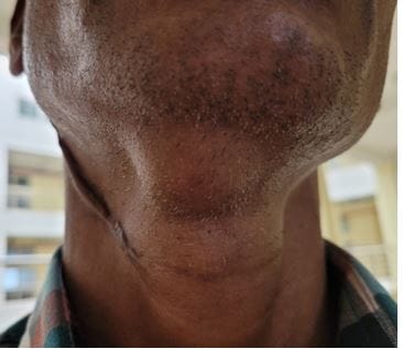
Figure 1: Submental swelling of 2 x 2 cm.

The USG neck revealed a few sub-centimetric and enlarged lymph nodes in the submental region, the largest of which measured 1.7 mm × 9 mm. Few of them showed loss of fatty hilum. The complete blood count was normal. The peripheral smear examination revealed a normal count and morphology of cells with no abnormal findings. ESR was 22 mm/hr. The patient was given a provisional diagnosis of a case of lymphadenopathy under evaluation. The patient was then recommended to undergo FNAC. The smears underwent microscopic examination.

patho_633_02.JPG
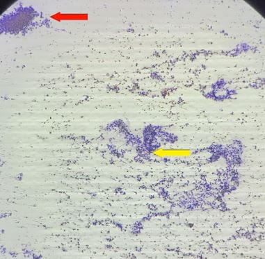
Figure 2: Granuloma formation (red arrow) and microfilaria surrounded by lymphoid cells (yellow arrow)(Pap stain, 100X). 

patho_633_03.JPG
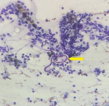
Figure 3: An ensheathed, coiled, and slightly curved microfilaria surrounded by lymphoid cells (yellow arrow) (Pap stain, 400X).


A diagnosis of filariasis with granulomatous lymphadenopathy was given based on cytology of lymph node swelling. In the patient, FNAC lymph node smears were cellular. They revealed granuloma formation which is composed of epithelioid cells with mixed cell populations of small and large lymphoid cells, predominantly mature lymphocytes, many histiocytes, tingible body macrophages, and occasional eosinophils (Figure 2). An ensheathed, coiled, and slightly curved microfilaria was observed (Figure 3). The background contained red blood cells (RBCs).

Discussion

Millions of people are thought to carry microfilaria, and a notably large proportion of those individuals experience filaria-related illnesses [5]. Cases of filariasis are found across the tropics and subtropics of Southeast Asia, India, the Pacific Islands, and sub-Saharan Africa. India recorded the largest number of instances. Although the term "filariasis" refers to any infection brought on by a filarial worm, in everyday usage, it refers only to lymphatic filariasis brought on by Wuchereria or Brugia species. The Culex mosquito served as the transmission vector. Microfilaria in peripheral blood shows nocturnal periodicity. In the latter stages of the illness, it manifests as elephantiasis. In blood samples taken at night between 10 p.m. and 4 a.m., peripheral examination reveals the presence of motile microfilariae parasites [6]. These days, the gold standard for demonstrating micro filarial organisms is the identification of circulating filarial antigens. The significance of FNAC as a diagnostic tool for filariasis is not well documented in the literature [7].

The diagnosis of clinically suspected filariasis is not usually made by FNAC. Nonetheless, there have been reports of unintentional fine-needle aspiration cytology smear detections [8, 9]. Peripheral blood film testing may not reveal microfilariae in instances of elephantiasis, occult filariasis, lymphangitis, and early allergy presentations [2, 10].

Rare case reports exist of microfilariae accidentally discovered in cytology smears of breast lesions, axillary lymph nodes, thyroid nodules, and bronchial aspirates [3]. In the absence of any filariasis-related clinical symptoms, our patient exhibited enlargement in the submental lymph nodes.

The detection of microfilaria in the lymph node FNAC in our instance was accidental, which highlights the significance of extensive screening for individuals who have no clinical signs of filariasis or microfilaremia in their blood circulation [11].

Conclusion

Microfilariasis should be taken into consideration in endemic regions like India. For the sensitive, economical, and essential diagnosis of helminthic etiology in unexplained lymph node swellings, fine needle aspiration cytology can be a valuable help. This highlights how crucial it is to carefully screen cytology smears for microfilaria in individuals who do not exhibit microfilaremia or clinical signs of the parasite. This will make it easier to treat patients accurately and promptly.

References

1. Pantola C, Kala S, Agarwal A, Khan L: Microfilaria in cytological smears at rare sites coexisting with unusual pathology: A series of seven cases. Trop Parasitol. 2012, 2:61-3. doi:10. 4103/2229-5070.97244 [Crossref][PubMed][Google Scholar]

2. Sharma J, Chaudhary N, Bordia S: Microfilariae in lymph node aspirate: a case report. J Med Sci Clin Res. 2017, 5:18694-18697. doi:10. 18535/jmscr/v5i3.69 [Crossref][PubMed][Google Scholar]

3. Sharma A, Sudhamani S, Pandit A, Kiri VM: Microfilariae in lymph node aspirate. Bombay Hospital J. 2012, 54:302. . [Crossref][PubMed][Google Scholar]

4. Chauhan K, Agarwal S: Incidental detection of microfilaria in lymph node aspirate: a case report. IJSR. 2012, 2:424-425. doi:10. 15373/22778179/DEC2013/129 [Crossref][PubMed][Google Scholar]

5. Agrawal VK, Sashindran VK: Lymphatic filariasis in India: problems, challenges and new initiatives. Med J Armed Forces India. 2006, 62:359-62. doi:10. 1016/S0377-1237(06)80109-7 [Crossref][PubMed][Google Scholar]

6. Pathak B, Maimoon S (November 08, 2022) Incidental Finding of Microfilaria in Lymph Node Cytology: A Case Report. Cureus 14(11): e31275. doi:10. 7759/cureus. 31275 [Crossref][PubMed][Google Scholar]


7. Suthar K, Kashyap R, Mewada B, Suthar H: Filariasis presenting as generalized lymphadenopathy: a rare case report. Int J Med Sci Public Health. 2013, 2:755. doi:10. 5455/ijmsph.2013.290420131 [Crossref][PubMed][Google Scholar]

8. Gite V, Dhakane M: Incidental microfilaria in cervical lymph node cytology. Apollo Med. 2018, 15:24. doi:10. 4103/am.am_34_17 [Crossref][PubMed][Google Scholar]

9. Chakrabarti N, Konar K, Mala, Bandyopadhyay A: Incidental detection of microfilaria in cytology: report of three cases. Annals of Clinical Pathology. 2014, 1033. doi:10. 47739/2373-9282/1033 [Crossref][PubMed][Google Scholar]

10. Gupta D, Gupta P, Jain S, Rahar S: Cytological diagnosis of microfilariae in clinically unsuspected cases: a retrospective review of 12 cases. Cytopathology. 2021, 32:807-12. doi:10. 1111/cyt.13035 [Crossref][PubMed][Google Scholar]

11. Bharti S, Agarwal NK, Katiyar R, Dwivedi S, Shri M: Cytological diagnosis of microfilaria at unusual sites in clinically unsuspected cases: some rare presentations of filariasis. Trop Doct. 2018, 48:373-5. doi:10. 1177/0049475518793630 [Crossref][PubMed][Google Scholar]