E-ISSN:2456-1487
P-ISSN:2456-9887
RNI:MPENG/2017/70771

Case Report

Osteoclast-Like Giant Cells

Tropical Journal of Pathology and Microbiology

2023 Volume 9 Number 1 January-March
Publisherwww.medresearch.in

Invasive ductal carcinoma of the breast with stromal osteoclast-like giant cells

Fatima S.1*, Ahmed Sadek S.2
DOI: https://doi.org/10.17511/jopm.2023.i01.01

1* Sohaila Fatima, Assistant Professor, Department of Pathology, King Khalid University, Abha, Kingdom of Saudi Arabia.

2 Shaymaa Ahmed Sadek, Specialist, Department of Laboratory Medicine, Aseer Central Hospital, Abha, Kingdom of Saudi Arabia.

Carcinomas containing multinucleated osteoclast-like giant cells (OGCs) arise in many organs. It is extremely rare, occurring in less than 2% of breast cancer patients. It is associated with ductal, lobular, mucinous, cribriform, papillary, and metaplastic histological subtypes of invasive breast carcinomas. OGCs are thought to be histiocytic in origin, developing in response to breast cancer-induced hypervascular microenvironments. Their significance and role as prognostic markers are not clear. Here we present a female with a mass in her left breast which was diagnosed as invasive ductal carcinoma with stromal osteoclast-like giant cells.

Keywords: Invasive ductal carcinoma, breast, osteoclast-like giant cells

Corresponding Author How to Cite this Article To Browse
Sohaila Fatima, Assistant Professor, Department of Pathology, King Khalid University, Abha, , Kingdom of Saudi Arabia.
Email:
Sohaila Fatima, Shaymaa Ahmed Sadek, Invasive ductal carcinoma of the breast with stromal osteoclast-like giant cells. Trop J Pathol Microbiol. 2023;9(1):1-4.
Available From
https://pathology.medresearch.in/index.php/jopm/article/view/620

Manuscript Received Review Round 1 Review Round 2 Review Round 3 Accepted
2023-02-04 2023-02-07 2023-02-14 2023-02-21 2023-02-28
Conflict of Interest Funding Ethical Approval Plagiarism X-checker Note
Nil Nil Yes 19%

© 2023by Sohaila Fatima, Shaymaa Ahmed Sadekand Published by Siddharth Health Research and Social Welfare Society. This is an Open Access article licensed under a Creative Commons Attribution 4.0 International License https://creativecommons.org/licenses/by/4.0/ unported [CC BY 4.0].

Introduction

Carcinomas containing multinucleated osteoclast-like giant cells (OGCs) arise in many organs including the breast, lung, pancreas, small intestine, and thyroid gland [1]. Carcinoma of the breast containing OGCs is uncommon and present in less than 2% of breast cancer patients [2,3]. OGCs have been seen in different histological subtypes of breast carcinoma. Invasive ductal carcinoma represents the prototypic expression of breast carcinoma [4]. Invasive breast carcinoma of no special type (NST) comprises the largest group of invasive breast cancers. It is not an easily defined entity as it represents the heterogeneous group of tumors that fail to exhibit sufficient characteristics to achieve classification as a specific histological type [5]. OGCs are histiocytic and their prognostic significance in breast carcinoma is uncertain. Here we present a 36-year-old female with a mass in her left breast which was reported as breast imaging reporting and data system (BI-RADS) VI and on biopsy was diagnosed as invasive ductal carcinoma with stromal osteoclast-like giant cells.

Case Report

A 36-year-old female presented with a mass in her left breast. A bilateral breast mammogram was performed and the left breast revealed a well-defined dense lesion with spiculated border situated at the outer deep retro-areolar region. No malignant features of micro-calcification, nipple retraction or skin thickening were seen. The axillary lymph node was enlarged. Ultrasound revealed a well-defined hypoechoic soft tissue lesion measuring 2x 1x 2 cm with spiculated border 4 cm from the nipple and a well-defined round shape anechoic simple cyst measuring 0.4 x 0.5 cm located 10 cm from the nipple. Multiple axillary lymph-adenopathy with loss of fatty hilum and cortical thickness were seen. Impression: Left breast malignant lesion with axillary lymphadenopathy and BI-RADS VI. Tru-cut biopsy from the mass showed infiltration of the breast parenchyma by a malignant neoplasm, composed of nests and some tubules of pleomorphic cells, exhibiting a moderate amount of eosinophilic cytoplasm and large hyperchromatic nuclei with some prominent nucleoli. The intervening stroma showed dense lymphoplasmacytic infiltrate and numerous, widely scattered osteoclast-like giant cells (OGC). Foci of ductal carcinoma in-situ,

solid and cribriform patterns with areas of cancerization of lobules were also noted. Immunohistochemistry showed estrogen receptor (ER) positivity in more than 90% of tumour cells, progesterone receptor positivity (PR) in more than 90% of tumour cells, Her2/neu: negativity, CD68: positivity in the stromal OGC. The diagnosis was invasive ductal carcinoma (IDC) with stromal OGC.

patho_620_01.JPG

Figure 1: a. Mammogram of left breast showing well-defined dense lesion with spiculated border situated at the outer deep retro-areolar region with an enlarged axillary lymph node. b. Ultrasound left breast showed well-defined hypoechoic soft tissue lesion with spiculated border.

patho_620_02.JPG

Figure 2: a, b. Sections show infiltration of the breast parenchyma by a malignant neoplasm, composed of nests and some tubules of pleomorphic cells, exhibiting a moderate amount of eosinophilic cytoplasm and large hyperchromatic nuclei with some prominent nucleoli. The intervening stroma shows dense lymphoplasmacytic infiltrate and numerous widely scattered osteoclast-like giant cells (Hematoxylin and Eosin: a,b x20X).


patho_620_03.JPG
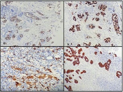
Figure 3: Immunohistochemical study a. Showing estrogen receptor positivity in malignant cells (ER x20X). b. Showing progesterone receptor positivity in malignant cells (PR x20X). c. Showing CD68 positivity in osteoclast-like giant cells (CD68 x40X). d. Showing AE1/AE3 positivity in malignant cells (AE1/AE3 x20X)

Discussion

OGCs are seen admixed with different histological subtypes of invasive breast carcinomas- ductal, lobular, mucinous, cribriform, papillary, and metaplastic. Factor et al reported two similar cases in 1977[6] followed by a series of eight cases by Agnantis and Rosen in 1979 [7]. Most of these lesions are moderately or poorly differentiated IDC with cribriform growth patterns [1]. The gross appearance of carcinomas with OGCs tends to be brown. The giant cells are generally associated with an inflammatory, fibroblastic, hypervascular stroma, with extravasated erythrocytes, lymphocytes and monocytes along with mononucleated and binucleated histiocytes, some containing hemosiderin. The giant cells vary in size and have a variable number of nuclei [5]. The OGCs range from 20 to 180 um in diameter. They contain abundant cytoplasm and many evenly distributed and usually centrally located oval nuclei, some of which contain small nucleoli. They tend to cluster close to the edges of carcinomatous glands or in intervening stroma and they may be found in the glandular lumens. The stroma typically contains mononuclear histiocytes whose cytological features resemble those of the OGCs [1]. The osteoclast-like elements are of a non-neoplastic histiocytic nature and they form from the fusion of mononuclear precursors [4]. The majority of the tumours are ER, PR positive, and Her2 negative with

luminal phenotype especially luminal A subtype [8]. In our case also it was IDC with ER, PR positive and HER2 negative.

The mechanism for the formation of OGCs is unknown. A recent study showed that secretion of specific cytokines, such as vascular endothelial growth factor (VEGF) and matrix metalloproteinases (MMP)12, lead to a charac­teristic inflammatory and hypervascular stro­ma, which is commonly observed in breast car­cinoma with OGCs, regardless of histology of tumoral cells. Therefore, the appearance of OGCs may not be antitumoral immunological reac­tion, but rather pro-tumoral differentiation of macrophage responding to hypervascular microenvironments induced by breast cancer [9]. OGCs were found to express markers associated with osseous osteoclasts (MMP-9, tartrate-resistant acid phosphatase, and cathepsin K), as well as the histiocytic marker CD68. Peripheral blood monocytes' transformation into osteoclast-like giant cells in breast carcinoma could be virally induced [8]. A study found that OGCs isolated from invasive breast cancer were able to digest bone directly in vitro. Unlike osteoclasts, which require the presence of osteoblasts to be stimulated, these OGCs were directly activated by the presence of the parathyroid hormone. Additionally, the cells were not inhibited by calcitonin, demonstrating another key distinction between OGCs and osteoclasts [10].

Different histological subtypes of breast carcinomas can be associated with giant cells. When giant cells appear in conjunction with mesenchymal elements tumours should be regarded as a variant of metaplastic carcinoma. When OGCs are seen in the stroma of a typical carcinoma lacking mesenchymal differentiation, the tumor is classified as IDC with OGCs as in our case. Fibroblastic reaction, collagenization, angiogenesis, and lymphocytic infiltration are variably present in the stroma. Pleomorphic giant cell carcinoma contains multinucleated giant cells which are large, and hyperchromatic with irregular nuclei, and prominent nucleoli. They also show positivity for epithelial markers and are not immunoreactive for CD68 as OGCs [4]. The differential diagnosis of OGCs includes megakaryocytes in myeloid metaplasia and granulomatous foci in inflammatory conditions such as sarcoidosis or coexistent with carcinoma or the stroma of fibroepithelial tumors. They lack the relatively abundant cytoplasm seen in OGCs.


The prognosis is related to the characteristics of the associated breast carcinoma and does not appear to be influenced by the presence of stromal giant cells [5]. These tumours were found to have similar clinical courses, prognosis and outcomes when compared to typical invasive carcinomas [2,7]. Cai reviewed cases of breast carcinoma with OGCs and found the majority had a relationship to marked angiogenesis which may signal a poorer prognosis [11].

In conclusion, carcinomas in many organs contain multinucleated OGCs but their presence in breast carcinomas is rare. They resemble osteoclasts immunohistochemically however they can be activated in absence of osteoblasts being functionally different from them. Further studies are required to study their role in carcinomas and whether they can be targeted for therapeutic purposes.

Reference

1. Rosen PP: Mammary carcinoma with osteoclast-like giant cells. Rosen’s Breast Pathology, 4 ed. Philadelphia (PA): Lippincott Williams & Wilkins; 2014.

2. Holland R, van Haelst UJ: Mammary carcinoma with osteoclast-like giant cells: additional observations on six cases. Cancer 1984, 53:1963-1973.

3. Cai N, Koizumi J, Vazquez M: Mammary carcinoma with osteoclast-like giant cells: a study of four cases and a review of literature. Diagn Cytopathol 2005, 33:246-251.

4. Collins LC. Breast. In: Goldblum, JR; Lamps, LW; McKenney, JK; Myers, JL, editors. Rosai and Ackermans surgical pathology. Mosby St. Louis, Missouri; 11, 2017, 1434-1527.

5. Ellis I O, Collins L, Ichihara S, MacGrogan G. Invasive carcinoma of no special type. In: Lakhani SR, Ellis IO, Schnitt SJ, Tan PH, van de Vijver MJ, editors. WHO Classification of Tumours of the Breast. IARC press, 2012; p.36-38.

6. Factor SM, Biempica L, Ratner I, Ahuja KK, Biempica S. Carcinoma of the breast with multinucleated reactive stromal giant cells. A light and electron microscopic study of two cases. Virchows Arch A Pathol Anat Histol. 1977 May 13;374(1):1-12. doi: 10.1007/BF00430566. PMID: 141778.

7. Agnantis NT, Rosen PP: Mammary carcinoma with osteoclast-like giant cells: a study of eight cases with follow up. Am J Clin Pathol 1979, 72: 383-9.

8. Zhou S, Yu L, Zhou R, Li X, Yang W. Invasive breast carcinomas of no special type with osteoclast-like cells frequently have a luminal phenotype. Virchows Arch 2014; 464: 681-8.

9. Shishido-Hara Y, Kurata A, Fujiwara M, Itoh H, Imoto S and Kamma H. Two cases of breast carcinoma with osteoclastic giant cells: are the osteoclastic giant cells pro-tumoural differen¬tiation of macrophages? Diagn Pathol 2010; 5: 55.

10. Athanasou NA, et al. The origin and nature of stromal osteoclast-like multinucleated giant cells in breast carcinoma: implications for tumour osteolysis and macrophage biology. Br J Cancer. 1989;59(4):491–8.

11. Cai G, Simsir A, Cangiarella J. Invasive mammary carcinoma with osteoclastlike giant cells diagnosed by fine-needle aspiration biopsy: review of the cytologic literature and distinction from other mammary lesions containing giant cells. Diagn Cytopathol. 2004; 30(6):396–400.