E-ISSN:2456-1487
P-ISSN:2456-9887
RNI:MPENG/2017/70771

Research Article

Nasal Cavity

Tropical Journal of Pathology and Microbiology

2022 Volume 8 Number 5 September October
Publisherwww.medresearch.in

The Histopathological study of Lesions of the Nasal Cavity, Paranasal Sinuses and Nasopharynx in Eastern UP, India

Bundela A.1, Bundela A.2*, Mitra S.3
DOI: https://doi.org/10.17511/jopm.2022.i05.02

1 Archana Bundela, Associate professor, Department of Pathology, B.R.D. Medical College, Gorakhpur, Uttar Pradesh, India.

2* Alpana Bundela, Assistant professor, Department of Pathology, B.R.D. Medical College, Gorakhpur, Uttar Pradesh, India.

3 Shaila Mitra, Professor, Department of Pathology, B.R.D. Medical College, Gorakhpur, Uttar Pradesh, India.

Background: Masses in the nasal cavity, paranasal sinuses and nasopharynx are a heterogeneous group of lesions with a broad spectrum of histopathology features, So careful histological workup is essential for a correct diagnosis and to determine the extent of involvement and prompt treatment. The present study has been conducted on 158 cases in the department of pathology, BRD Medical College, Gorakhpur. Aims and Objective: (1) To establish the histopathological diagnosis of Inflammatory, benign and malignant lesions of the nasal cavity, paranasal sinuses and nasopharynx (2) To categorize these lesions into non-neoplastic and neoplastic lesions and to study their histopathological patterns. (3) To compare the findings of our study with other studies. Material and Method: This was a prospective study of sinonasal lesions that were biopsied or surgically excised and received in the pathology Department. Result: A total of 158 cases of nasal, and paranasal lesions were reported during the study period. Out of a total of 158 cases, 135 cases were inflammatory and non-neoplastic lesions,20 cases were benign and 03 cases were malignant lesions. Age ranged from 08 yrs to 78 yrs with male predominance and the majority of cases belonged to the Hindu community. Out of 135 inflammatory or non-neoplastic cases, the most common lesion was nasal polyp and among 20 benign cases, Inverted papilloma was the commonest lesion. Out of 03 malignant lesions, Squamous cell carcinoma was the predominant histological type of lesion. Conclusion: Nasal, Paranasal and Nasopharyngeal lesions comprise a wide spectrum of lesions but their clinical manifestations are very limited. Hence a careful histopathological examination is mandatory for a proper diagnosis so that a correct and timely intervention can be made. In this study, we concluded that in Eastern UP, malignant lesions of nasal, paranasal and nasopharyngeal regions are very less in comparison to inflammatory and benign cases.

Keywords: Nasal, Paranasal, Nasopharyngeal Masses

Corresponding Author How to Cite this Article To Browse
Alpana Bundela, Assistant professor, Department of Pathology, B.R.D. Medical College, Gorakhpur, Uttar Pradesh, India.
Email:
Archana Bundela, Alpana Bundela, Shaila Mitra, The Histopathological study of Lesions of the Nasal Cavity, Paranasal Sinuses and Nasopharynx in Eastern UP, India. Trop J Pathol Microbiol. 2022;8(5):65-70.
Available From
https://pathology.medresearch.in/index.php/jopm/article/view/610

Manuscript Received Review Round 1 Review Round 2 Review Round 3 Accepted
2022-06-07 2022-06-09 2022-06-16 2022-06-23 2022-06-30
Conflict of Interest Funding Ethical Approval Plagiarism X-checker Note
Nil Nil Yes 17%

© 2022by Archana Bundela, Alpana Bundela, Shaila Mitraand Published by Siddharth Health Research and Social Welfare Society. This is an Open Access article licensed under a Creative Commons Attribution 4.0 International License https://creativecommons.org/licenses/by/4.0/ unported [CC BY 4.0].

Introduction

The nasal cavity, Paranasal sinuses and nasopharynx through the continuity form a complex system of the upper respiratory tract; this region is endowed with a variety of elements such as epithelial, glandular, lymphoid, cartilage and bone and is exposed to a variety of diseases like infections and tumours.[1]. Masses can be congenital or acquired and their presenting features are diverse and depend upon the type, spread and extent of the primary disease. According to, the patients may have nasal features, (obstruction, discharge, nasal mass, epistaxis, smell abnormalities) and sometimes metastatic lymph nodes.[2]. Nasal polyps are the most common cause of nasal obstruction but it is quite impossible to distinguish clinically between simple nasal polyps, and polypoidal lesions which are caused by specific granulomatous diseases, hence it becomes important that all masses of the nose should be submitted for histopathological examination.[3,4,5]. However studies on nasal masses are lacking in medical literature in Eastern Uttar Pradesh. With this background the present study was conducted to categorize various lesions of the nasal cavity, PNS and Nasopharynx into non-neoplastic and neoplastic and to study their histopathological patterns along with their incidences in Eastern Uttar Pradesh.

Material and Method

The study was a prospective study conducted in the department of Pathology at B.R.D medical college Gorakhpur, on the patients attending the OPD and on admitted patients in the ward of the ENT department, Nehru Chikitsalaya, Gorakhpur during a period from August 2019 to September 2021, on the patients suffering from lesions of the nasal cavity, Paranasal sinuses and nasopharynx, during a period of 2 years.

Histopathological examination was done to categorize the nature of various lesions into non-neoplastic and neoplastic lesions and analyze their histopathological pattern and relative distribution of various lesions with regard to age and sex in our setting of OPD. Freshly biopsied specimens were fixed in 10% formalin and processed as a routine procedure. Thin sections of 4-5 µm have been cut after dewaxing and then stained with hematoxylin and eosin stain. Histopathological diagnoses were made. All slides were evaluated.

We have taken ethical clearance from the ethical committee of our institution.

Result

The present study was conducted on 158 cases of the nasal cavity, Paranasal sinuses and nasopharyngeal lesions including 135 cases of inflammatory and non – neoplastic, 20 cases of benign neoplastic and 03 cases of malignant lesions.

Nasal cavity, paranasal sinuses and parapharyngeal lesions were diagnosed based on histomorphological features.

The age of total patients ranged from 08 to 78 years with the mean age of presentation being 34.5 years. (table 2)

The majority of cases were seen in the second (46% ), third ( 28%)and fourth( 11%) decade of life. .(table 2)

Out of the total 158 cases, 68.62% were non-neoplastic and 31.38% were neoplastic lesions. .(table 1)

The majority of cases of Nasal cavity, PNS and nasopharyngeal lesions belong to rural areas (62.69%) and less

to urban areas (37.31%). The rural-to-urban ratio was 1.68:1 and the male-to-female ratio was 1.52:1 revealing male predominance. .(table 3)

In the present study, the majority of cases (72.57%) belong to the Hindu community followed by the Muslim community (27.13%).

The most common site was the nasal cavity (72.57%) followed by paranasal sinuses (20.70%) and nasopharynx (6.73%)..(table 4)

On histopathological examination, Out of 135 cases of non-neoplastic lesions, the nasal polyp was the commonest lesion(78.48%)observed in this region. .(table 5)

Out of 20 benign neoplastic lesions, inverted papilloma (40%) was the commonest benign neoplastic lesion followed by angiofibroma (25%) and capillary hemangioma (20%)..(table 6)

Among 03% of malignant cases squamous cell carcinoma was the predominant histological type constituting 66.66% of all cases, followed by nasopharyngeal carcinoma(33.33%). .(table 7)


Table 1: Distribution of total non-neoplastic and neoplastic lesions of the nasal cavity, paranasal sinuses and nasopharynx studied.

Nature of lesions No of cases percentage
Inflammatory and Non-neoplastic lesions 135 85.44%
Benign lesions 20 12.65%
Malignant lesions 03 1.89%
Total 158 100%

Table 2: Age-wise Distribution of non-neoplastic and neoplastic lesions of the nasal cavity, paranasal sinuses and nasopharynx.

Age Group (In Years) Non Neoplastic Cases Benign Cases Malignant Cases
0-10 04(2.96%) 01(5.0%)  
11-20 38(28.14%) 07(35%)  
21-30 46(34.07%) 04(20.0%)  
31-40 28(20.7%) 03(15%)  
41-50 11(8.14%) 02(10%)  
51-60 02(1.48%) 01(05%)  
61-70 06(4.44%) 02(10%) 02(66.34%)
71-80 - - 01(33.64%)
Total Cases 135(100%) 20(100%) 03(100%)

Table 3: Gender Wise Distribution of Various Lesions of the Nasal Cavity, Paranasal Sinuses and Nasopharynx. 

Gender Inflammatory And Non Neoplastic Lesions   (Number and percentage) Benign Neoplastic Lesions   (Number and percentage) Malignant Lesions     (Number and percentage) Total
Male 78(57.78%) 12(60%) 03(100%)  
Female 57(42.22%) 08(40%) -  
M: F Ratio 1.36:1 1.5:1    
Total 135 20 03  

Table 4: Distribution of non-neoplastic and neoplastic lesions of the nasal cavity, Paranasal sinuses and nasopharynx based on site. 

Site of origin Inflammatory and non-neoplastic lesions Benign neoplasm Malignant neoplasm
Nasal Cavity 118 17 02
Paranasal Sinuses 17 02 00
Maxillary 16 02  
Ethmoid 00    
Frontal 01    
Sphenoid 00    
Nasopharynx 00 01 01
Total 135 20 03


Table 5: Distribution of non-neoplastic lesions of Nose and PNS. 

Lesions Number Of Cases Percentage (%)
Nasal Polyp 113 83.70%                    
Allergic polyp 15 11.11%
Rhinosporidiosis 04 2.96%
Rhinoscleroma 02 1.48%
Tuberculosis 01 0.78%
Total 135 100%

Table 6: Distribution of Benign neoplastic lesions of nose and PNS.

Lesions Number of cases Percentage (%)
Inverted papilloma 09 40%
Angiofibroma 05 25%
Capillary hemangioma 05 25%
Basal cell adenoma 01 10%
Total 20 100%

Table 7: Distribution of malignant neoplastic lesions of nose and PNS.

Lesions Number of lesions Percentage (%)
Squamous cell carcinoma 02 66.66%
Nasopharyngeal carcinoma 01 33.33%
Total 03 100%

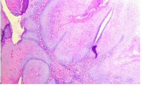
Figure 1: Micophotograph of Inverted sinonasal papilloma Showing inward Growth (H&E 40X)

Figure 2: Microphotograph of Rhinosporodiosis showing globular sporangia(H&E40X)


Figure 3: Microphotograph of tubercular lesion showing granulomas with necrosis(H&E40X)

Figure 4: Microphotograph of Capillary Haemangioma Showing Closely packed spindle cells with space containing little blood (H&E 40X)

Figure 5: Microphotograph Of Rhinoscleroma Showing (Foamy Histiocytes) Mikulicz Cells[H&E 40X]

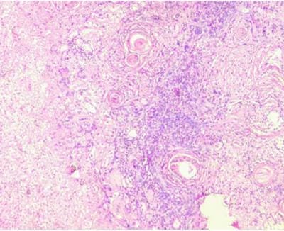
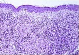
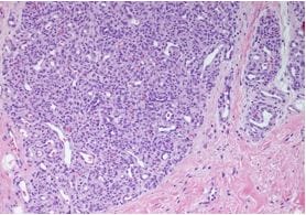
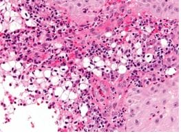
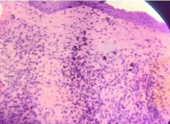
Figure 6: Microphotograph Of Nasal polyp Showing sinonasal epithelium with underlying oedematous stroma and inflammatory cells [H&E 40X]

Discussion

In the present study, Out of 158 cases, inflammatory and non-neoplastic lesions(85.44%) were the commonest lesions followed by Benign(12.65%) and then malignant lesions (1.89%). These findings were compared with other studies.

  Studies  Total cases studied Non-neoplastic lesions Benign Lesions Malignant lesions
Chopra et al, 2010 100 84% 11% 05%
Aparna M et al, 2012 117 86% 11% 2.6%
Bist et al study, 2012 101 56.4% 19.8% 23.76%
Present study 158 85.44% 12.65% 1.89%

These findings were well correlated with the findings of Chopra et al 2010 [7]. and Aparna M et al 2012 [8]. but different from Bist et al 2012[6]. study.


The most affected age group in our study was from 2nd decade to 4th decade. These findings are similar to the findings of Tandon et al (1971) [9]. who reported incidence of non-neoplastic lesions as maximum in 20-29 years, while benign lesions were maximum(35%) in 11-20 years. These findings correlate with those of Satarkar RN 2016[1]. who reported 47.7% of cases in the same age group.

The malignant lesions were predominant in the sixth and seventh decades of our study, similar findings were observed in the study by Frazell & Lewis[10] and Ghosh &Bhattacharya[11]. who reported a maximum number of malignant cases in the fifth to seventh decades. Sinonasal and nasopharyngeal masses had a predilection for males with a male-female ratio of 1.52:1. Lathi et al 2011[12]. showed male to female ratio of 1.5:1, however a Nigerian study conducted by Bakari et al [13]. 2010 revealed female preponderance with an opposite M: F ratio of 1:1.2.

In the present study cases from rural areas predominated (62.69%) over urban areas (37.30%) and the maximum cases (64.87%)were from the Hindu community. Our findings are comparable with the findings of Maheshwari A et al 2017[14] study. In their study, 63.75% of cases were from Rural areas and 60% of cases were from Hindu communities.

In our study, the majority of lesions occurred in the nasal cavity(84.81%)followed by the Paranasal sinuses(12.65%)and nasopharynx(3.44%). Our findings are in concordance with the findings of Shah SN et al 2012.[15].

On analyzing histopathological diagnosis in the present study among inflammatory and non-neoplastic lesions the most common lesion was the nasal polyp (94.20%). This finding was similar(85%) to the observation by Tondon et al 2016[9]. The second most common lesion was Rhinosporidiosis which is endemic in Asian countries. Only 4 cases (2.53%) were reported in our study which is in accordance with the study of Ambreen B et al [16] who observed Rhinosporidiosis in 2.6% of cases.

The incidence of Rhinoscleroma in the present study was lower (1.48%)than that observed by Tondon et al 1971 [9].


Inverted papilloma (45%) in the nose and paranasal sinuses are the most common Benign epithelial neoplasm observed in our study. The second most common benign lesion was angiofibroma (30%), these above findings are comparable with the findings of Garg D et al 2014 [17]. And 25% of cases of capillary hemangioma reported in our study are in concordance with the study of Garg D et al 2014[17]. while Shah H et al 2017[18] reported capillary Hemangioma as the commonest benign lesion in their study.

Sinonasal carcinoma is a common neoplasm .In our study, 66.66% of cases of squamous cell carcinoma were reported and nasopharyngeal carcinoma was reported in 33.33% of cases. Our findings are comparable with the findings of Khan N et al 2006[19] who reported squamous cell carcinoma as the most common lesion (37.5%) followed by Nasopharyngeal carcinoma (25%)

Conclusion

Amongst the non-inflammatory lesions, the nasal polyp is the commonest lesion followed by Rhinosporidiosis and Rhinoscleroma. The common age group is second and third decades with male predominance, These lesions are mainly seen in Hindus and the rural population in Eastern UP.

Inverted papilloma was common among benign neoplastic lesions followed by angiofibroma. The common age group is the second and third decades with male predominance.

Malignant lesions were comparatively less than benign neoplastic lesions. Differences between benign and malignant lesions of the nasal cavity, paranasal sinuses and nasopharynx were found statistically significant (P<0.0001).

What this study adds to existing knowledge

Our data suggest that nasal polyp is commonest in noninflammatory lesions, inverted papilloma is commonest in benign neoplastic lesions and malignant lesions are less common in comparison to benign lesions in EASTERN UP.

Acknowledgement: We wish to thank our co-authors and pathologists Dr Shaila Mitra and Dr Archana Bundela and other members of the pathology department for their valuable contribution to the present study.


Reference

01. Satarkar RN, Srikanth S. Tumors and tumor-like conditions of the nasal cavity, paranasal sinuses, and nasopharynx: A study of 206 cases. Indian J Cancer. 2016 Oct-Dec;53(4):478-482. doi: 10.4103/ijc.IJC_551_16 [Crossref][PubMed][Google Scholar]

02. Somani, S. , P. Kamble, and S. Khandekar. Mischievous presentation of nasal masses in rural areas. Asian J Ear Nose Throat 2 (2004): 9-17 [Crossref][PubMed][Google Scholar]

03. Dasgupta A, Ghosh RN, Mukherjee C. Nasal polyps - histopathologic spectrum. Indian J Otolaryngol Head Neck Surg. 1997 Jan;49(1):32-7. doi: 10.1007/BF02991708 [Crossref][PubMed][Google Scholar]

04. Friedman I. Inflammatory conditions of the nose. In Symmers WSTC,ed. Nose and Ear. 3rd ed.Edinburgh: Churchil Livingstone 1986;19-23 [Crossref][PubMed][Google Scholar]

05. Kamel RH, Ishak EA. Enlarged adenoid and adenoidectomy in adults: endoscopic approach and histopathological study. J Laryngol Otol. 1990 Dec;104(12):965-7. doi: 10.1017/s0022215100114495 [Crossref][PubMed][Google Scholar]

06. Bist SS, Varshney S, Baunthiyal V, Bhagat S, Kusum A. Clinico-pathological profile of sinonasal masses: An experience in tertiary care hospital of Uttarakhand. Natl J Maxillofac Surg. 2012 Jul;3(2):180-6. doi: 10.4103/0975-5950.111375 [Crossref][PubMed][Google Scholar]

07. Dua, Kapil, et al. Histopathology of nasal masses. An International Journal Clinical Rhinology 3. 2 (2010): 81-85. [Crossref][PubMed][Google Scholar]

08. M Kulkarni A, G Mudholkar V, S Acharya A, V Ramteke R. Histopathological study of lesions of nose and paranasal sinuses. Indian J Otolaryngol Head Neck Surg. 2012 Sep;64(3):275-9. doi: 10.1007/s12070-011-0286-7 [Crossref][PubMed][Google Scholar]

09. Tondon, P. L. , J. Gulati, and N. Mehta. Histological study of polypoidal lesions in the nasal cavity. Indian Journal of Otolaryngology 23.1 (1971): 3-11 [Crossref][PubMed][Google Scholar]

10. Evans, Audrey E. , et al. Vincristine in the treatment of acute leukemia in children. Cancer 16. 10 (1963): 1302-1306 [Crossref][PubMed][Google Scholar]

11. Ghosh, A. , and K. Bhattarcharya. Nasal and nasopharyngeal growth-A 10 year survey. J Ind Med Ass 47 (1966): 13 [Crossref][PubMed][Google Scholar]

12. Bakari, Aminu, et al. Clinico-pathological profile of sinonasal masses: an experience in national ear care center Kaduna, Nigeria. " BMC Research Notes 3. 1 (2010): 1-5. [Crossref][PubMed][Google Scholar]

13. Lathi, A. , et al. Clinico-pathological profile of sinonasal masses: a study from a tertiary care hospital of India. ACTA otorhinolaryngologica italica 31. 6 (2011): 372 [Crossref][PubMed][Google Scholar]

14. Ambreen, Beige, et al. Histopathological study of lesions of nose and paranasal sinuses and association of Human Papilloma Virus (HPV) with sinonasal papillomas and squamous cell carcinoma. " International Journal of Medical Research & Health Sciences 5. 6 (2016): 7-16. [Crossref][PubMed][Google Scholar]

15. Shah, Shaila N. , and Yatish Goswami. Study of lesions of nasal cavity, nasopharynx and paranasal sinuses by histopathological examination. " Gujarat Med J 67. 2 (2012): 70-72 [Crossref][PubMed][Google Scholar]

16. Maheshwari A et al International J of Otorhinolaryngology and Head and Neck Surgery. 2017;3(4):1015-1019doi http://dx. doi. org/10. 18203/issn-2454-5929 [Crossref][PubMed][Google Scholar]

17. Panchal, L. , et al. Sino-nasal epithelial tumors: a pathological study of 69 cases. " Journal of postgraduate medicine 51. 1 (2005): 30 [Crossref][PubMed][Google Scholar]