E-ISSN:2456-1487
P-ISSN:2456-9887
RNI:MPENG/2017/70771

Research Article

Histopathology

Tropical Journal of Pathology and Microbiology

2022 Volume 8 Number 1 January February
Publisherwww.medresearch.in

Histopathological spectrum of gastrointestinal lesions in patients undergoing gastrointestinal endoscopic biopsies- A Prospective study

Mishra R.1*, Vahikar S.2, Shrivastava K.3, Kumari Mitra S.4, Mishra V.5
DOI: https://doi.org/10.17511/jopm.2022.i01.04

1* Ritambhara Mishra, Junior Resident, , Baba Raghav Das Medical College, Gorakhpur, U.P, India.

2 Shilpa Vahikar, Professor, Department of Pathology, Baba Raghav Das Medical College, Gorakhpur, U.P, India.

3 Kanchan Shrivastava, Professor, Department. of Pathology, Baba Raghav Das Medical College, Gorakhpur, U.P, India.

4 Shaila Kumari Mitra, Professor and Head, Department of Pathology, Baba Raghav Das Medical College, Gorakhpur, U.P, India.

5 Vivek Mishra, DM, Gastroenterologist, Baba Raghav Das Medical College, Gorakhpur, U.P, India.

Introduction: The disorders of gastrointestinal tract are responsible for a great deal of morbidity and mortality. Endoscopy in combination with biopsy plays an important role in the exact diagnosis of gastrointestinal tract disorders for further management. Objectives: The study was carried out to determine the spectrum of histopathological lesions of GIT and to compare it with its endoscopic findings. Methods: A prospective study was conducted on the GIT endoscopic biopsies and their histopathological assessment was done at the department of pathology, B.R.D. medical college from July 2020 to June 2021. Endoscopies were performed using an endoscope. The biopsy specimens received were fixed in 10.0% formalin and routinely processed in H & E stain. Results: Out of 100 endoscopic biopsies, the majority of cases were male predominance. In oesophagal biopsies, the most common non-neoplastic lesion was chronic non-specific esophagitis and SCC was the most common malignancy in neoplastic lesions. Among stomach biopsies, the most common non-neoplastic lesion was chronic non-specific gastritis and malignancy was adenocarcinoma. In duodenal biopsies, 50% constituted non-neoplastic lesions and 50% had neoplastic pathology. Among colorectal biopsies, the most common non-neoplastic lesion was chronic non-specific colitis and the most common malignancy was adenocarcinoma. Conclusion: The incidence of the non-neoplastic lesion is higher than the neoplastic lesion. The most common non-neoplastic lesion is chronic non-specific gastritis and the most common malignancy is squamous cell carcinoma of the oesophagus. Among 100 cases, endoscopy reported 46% cases as neoplastic and 54% as non-neoplastic, whereas Histopathology revealed 35% as neoplastic and 65% as nonneoplastic.

Keywords: Histopathology, Endoscopy, Squamous cell carcinoma, Adenocarcinoma

Corresponding Author How to Cite this Article To Browse
Ritambhara Mishra, Junior Resident, , Baba Raghav Das Medical College, Gorakhpur, U.P, India.
Email:
Ritambhara Mishra, Shilpa Vahikar, Kanchan Shrivastava, Shaila Kumari Mitra, Vivek Mishra, Histopathological spectrum of gastrointestinal lesions in patients undergoing gastrointestinal endoscopic biopsies- A Prospective study. Trop J Pathol Microbiol. 2022;8(1):22-28.
Available From
https://pathology.medresearch.in/index.php/jopm/article/view/593

Manuscript Received Review Round 1 Review Round 2 Review Round 3 Accepted
2021-12-22 2021-12-24 2021-12-31 2022-01-07 2022-01-14
Conflict of Interest Funding Ethical Approval Plagiarism X-checker Note
Nil Nil Yes 16%

© 2022by Ritambhara Mishra, Shilpa Vahikar, Kanchan Shrivastava, Shaila Kumari Mitra, Vivek Mishraand Published by Siddharth Health Research and Social Welfare Society. This is an Open Access article licensed under a Creative Commons Attribution 4.0 International License https://creativecommons.org/licenses/by/4.0/ unported [CC BY 4.0].

Introduction

Disorders of the gastrointestinal tract (GIT) are the most commonly encountered problems in clinical practice. They cause a high degree of morbidity and mortality. [1]. GIT endoscopy along with biopsy is an established procedure for investigating a wide range of gastrointestinal conditions, especially inflammatory and malignant diseases. It is not only used to diagnose malignant and inflammatory lesions but is also used for monitoring the course, the extent of the disease, response of the therapy and early detection of complications. [2].

A wide variety of infections, inflammatory disorders, vascular disorders, mechanical conditions, and toxic and physical reactions, including radiation injury and neoplasm may occur in the esophagus and stomach [3]. Endoscopic biopsy examination followed by histopathologic assessment is a relatively safe procedure and the current gold standard to assess patients with symptoms of GIT [4].

Our study aims to analyse the various histopathological categories of gastrointestinal tract lesions, with endoscopic and histopathological correlation and evaluation of the usefulness of endoscopic biopsy for efficient diagnosis and management.

Aims and objectives

The study was carried out to determine the spectrum of histopathological lesions of the gastrointestinal tract and to compare it with its endoscopic findings.

Materials and Methods

A prospective study was conducted on the GIT endoscopic biopsies and the histopathological assessment was done at the department of pathology, Baba Raghav Das medical college from July 2020 to June 2021. Endoscopies were performed using OLYMPUS of model CV170 processor Fujifilm EC600WLV2, FujifilmED 580XT model of the endoscope. The biopsy specimen was put in saline and placed on the filter paper with mucosal surface upwards. Then the filter paper was immersed in 10% formalin for fixation. After adequate fixation entire tissue was routinely processed and embedded in paraffin

with mucosal surfaces uppermost. Five micron thick sections were cut perpendicular to this surface and four to five sections were prepared on each slide. Each section was stained with H & E stain and studied microscopically. Adequacy of biopsy was assessed. An attempt was made to diagnose the lesion on gross visualization during endoscopy and to correlate them histopathologically. Special stains were done whenever required. Tumours were diagnosed as per the WHO histological classification of gastrointestinal tumors.

Inclusion criteria: All endoscopic gastrointestinal biopsies (esophageal, gastric duodenal and colorectal biopsies) received in the department of Pathology B.R.D. Medical college Gorakhpur for histopathological examination.

Exclusion criteria: Inadequate specimens and autolyzed specimens.

Statistical method: The data collected for the study were statistically analysed using the chi-square test. P-value < 0.05 was considered significant.

Results

Gender, age group and site distribution: Out of 100 cases, 62 (62%) patients were males and 38 (38%) were females, the male to female ratio was 1.6:1. The sex distributions are shown in figure I. The age of the study population ranges from a minimum of 10 to a maximum of 95 yrs, with the majority of cases in the age group 41-60 yrs.

patho_593_01.JPG
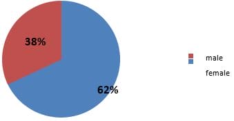
Figure 1: Distribution of cases according to sex (n=100)

Out of 100 biopsies, non-neoplastic cases were 65 (65%) and 35(35%) cases were neoplastic nature. The site-wise distributions of endoscopic biopsies were oesophagus 33 (33%), stomach 38 (38%), duodenum 4 (4%) and colorectum 25(25%).


Esophageal biopsies comprised 33 cases. Chronic regurgitation and heartburn were the chief complaints of the patients along with chest pain, dysphagia, vomiting, pain abdomen and consistent cough. Amongst the various lesions most common lesion was non-specific esophagitis with the least common being Barrett’s esophagus and hyperplastic polyp. (Table -1).All the malignant neoplastic lesions of esophagus were squamous cell carcinoma. The majority of squamous cell carcinoma was in the lower esophagus 10(71.43%) followed by the middle esophagus 3 (21.43%) and 1 case in the upper esophagus (7.14%). The majority of esophageal squamous cell carcinoma were moderately differentiated 10(71.43%) cases followed by 3 cases (21.43%) of well-differentiated squamous cell carcinoma and 1 case (7.14%) was poorly differentiated. The esophagus is the most common site for malignancy in the whole GIT.

Table 1: Distribution of Esophageal lesions

Histopathological Diagnosis No. of cases Percentage
Chronic Non-Specific esophagitis 6 18.18%
Hyperplastic Polyp. 2 6.06%
Barrett’s esophagus 2 6.06%
Sq. Cell Hyperplasia 6 18.18%
Dysplastic Sq.Epithelium 3 9.10%
Sq. Cell Carcinoma 14 42.42%
Total 33 100%

Endoscopic findings of esophageal lesions: Endoscopic features of esophagitis maximally included areas of hemorrhage and erosions/ulceration along with the areas of erythema and friability of the mucosa, hyperplastic polyp showed polyp and Barrett’s esophagus showed pink colon mucosa having tongue shape projections(figure-2). Squamous cell carcinoma of esophagus endoscopically presented with a different pattern of growth as ulcero-proliferative in maximum cases followed by ulcerated, ulcer infiltrative, stenosing/stricture and proliferative lesion

Gastric lesions: It comprised a total of 38 cases. The most common complaints were those of pain in the epigastrium, vomiting and pain abdomen along with dyspepsia, nausea, weight loss, faecal blood loss/ melena, and bloody vomitus. Chronic non-specific gastritis was the most commonly diagnosed lesion in 14 cases (36.84%) followed by 8 cases of chronic active gastritis with H. Pylori (21.06%), 3(7.89%) cases of Hyperplastic

Polyp, 2(5.26%) cases of Adenoma with least common cases seen of Non-Hodgkin’s lymphoma, Heterotopic pancreas and Intestinal metaplasia accounting for one case each. Adenocarcinoma was the most common malignant neoplastic lesion with 8 cases (21.06%). Concerning differentiation, moderately differentiated adenocarcinoma with 4 cases (50%) was slightly more than poorly differentiated lesions with 3 cases (37.50%) followed by well-differentiated adenocarcinoma with 1 case (12.50%). The antrum was the most common site of gastric carcinoma followed by the body.

Endoscopic findings of gastric lesions: On endoscopy; the majority of cases of gastritis showed erosion and hemorrhage in superficial mucosa, along with erythema and mucosal irregularities. Heterotopic pancreas showed multiple mucosal nodular deposits(figure-2)and NHL presented as ulcerated growth at antro pyloric area and stenosis(figure- 4). Adenocarcinoma of the stomach endoscopically presented as ulcerative growth, ulceroproliferative growth, proliferative growth, flattening of mucosa and erythematous appearance.

Duodenal lesions: A total of 4 duodenal biopsies were analyzed. Pain abdomen was the most common complaint of the patients along with other complaints such as loss of appetite, weight loss, diarrhea, vomiting and fever. There was 1 case (25%) each of non-specific duodenitis and celiac sprue. Adenocarcinoma was the most common neoplastic lesion with 2 cases (50%) and both cases were well-differentiated adenocarcinoma of the ampulla of Vater. Endoscopic findings of duodenal lesions specific duodenitis showed erythematous patch and erosion on endoscopically. Celiac sprue presented as scalloping or loss of fold and adenocarcinoma endoscopically presented as ulcerating fungating growth.

Table 2: Distribution of colorectal lesions 

Histopathological Diagnosis No. of cases Percentage
  Chronic non-specific colitis   11   44.00%
Adenocarcinoma 10 40.00%
Juvenile Polyp. 1 4.00%
Ulcerative Colitis 1 4.00%
SRUS (Solitary Rectal Ulcer Syndrome) 1 4.00%
Lymphocytic colitis 1 4.00%
Total 25 100%

This group comprised a total of 25 cases. The most common chief complaints in the pathologies of this region were pain abdomen, rectal bleeding, vomiting, diarrhea, fever, weight loss and faecal urgency. The most common complaint in the patient was pain abdomen. Patients who commonly complained of straining during defecation were diagnosed as the case of solitary rectal ulcer syndrome (SRUS). Children mostly presenting with rectal bleeding were diagnosed with a juvenile rectal polyp. The maximum number of cases were in the category of Chronic non-specific colitis 11(44%). (Table-2 ) . Adenocarcinoma was the most common malignant neoplastic lesion with 8 cases (40%). The majority of colorectal Adenocarcinoma were moderately differentiated by 5(62.50%) cases followed by 2(25%) cases of well-differentiated Adenocarcinoma and 1(12.50%) case was poorly differentiated.

Endoscopic findings of colorectal lesions:Majority of colitis presented as erythematous patch, erosion and ulceration on sigmoidoscopy and colonoscopy. On sigmoidoscopy, ulcerative colitis was characterized by erythematous and friable mucosa with mucosal granularity. Juvenile polyp showed polyp on endoscopy. Adenocarcinoma of the colon presented as ulcerative and ulceroproliferative growth on colonoscopy.

patho_593_02.JPG Figure 2: Barrett’s esophagus (H&E Stain- 40x), pink mucosa with tongue shape projections (Upper GI Endoscopy).

patho_593_03.jpg
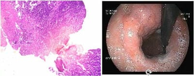
Figure 3: NHL Stomach (H&E Stain- 40x), Enlarged folds and diffuse thickening in fundus ( Upper GI Endoscopy)

patho_593_04.jpg
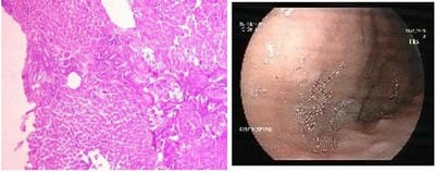
Figure 4: Heterotopic Pancreas in Stomach (H&E Stain- 40x), Multiple mucosal Nodular Deposits in Stomach in(upper GI endoscopy)

Table 3: Endoscopic and histopathological findings 

Histopath Ology Endoscopic Findings Total Chi-Square Value P. Value
NL UL IL Polyp. BO HP
Adeno Carcinoma 20 (100%) 0 (00.00%) 0 (00.00%) 0 (00.00%) 0 (00.00%) 0 (00.00%) 20(100%)                 20.4017                 0.000416 <0.01
SCC 14 (100%) 0 (00.00%) 0 (00.00%) 0 (00.00%) 0 (00.00%) 0 (00.00%) 14(100%)
NHL 1 (100%) 0 (00.00%) 0 (00.00%) 0 (00.0%) 0 (00.00%) 0 (00.00%) 1(100%)
Inflammatory Lesion 7 (15.91%) 11 (25.00%) 26 (59.09%) 0 (00.00%) 0 (00.00%) 0 (00.00%) 44(100%)
Ulcer 0 (00.00%) 1 (100.0%) 0 (00.00%) 0 (00.00%) 0 (00.00%) 0 (00.00%) 1(100%)
Dysplasia with mild hyperplasia 3 (33.33%) 3 (33.33%) 3 (33.34%) 0 (00.00%) 0 (00.00%) 0 (00.00%) 9(100%)
Polyp. 0 (00.00%) 0 (00.00%) 0 (00.00%) 6 (100.00%) 0 (00.00%) 0 (00.00%) 6(100%)
Adenoma 1 (50.00%) 0 (100.0%) 1 (50.00%) 0 (00.00%) 0 (00.00%) 0 (00.00%) 2(100%)
Barrett’s esophagus 0 (00.00%) 0 (00.00%) 0 (00.00%) 0 (00.00%) 2 (100.0%) 0 (00.00%) 2(100%)
Heterotopic Pancreas 0 (00.00%) 0 (00.00%) 0 (00.00%) 0 (00.00%) 0 (00.00%) 1 (100.0%) 1(100%)
Total 46(46.00%) 15(15.00%) 30(30.00%) 6(6.00%) 2(2.00%) 1(1.00%) 100(100%)

NL= Neoplastic Lesion, UL=Ulcerative Lesion, IL=Inflammatory Lesion, BO= Barrett’s esophagus, HP – Heterotopic pancreas.


Out of 100 cases, endoscopy revealed 46 (46%) neoplastic lesions, of which 35 (35%) cases were histologically diagnosed as neoplastic. On endoscopy 30 (30%) cases were inflammatory lesions whereas 44 (44%) cases were diagnosed as inflammatory on histopathology. On the other hand, 15 (15%) cases were revealed as an ulcerative lesion on endoscopy, of which 1case (1%) was a benign ulcer on histopathology. 6 (6%) cases were diagnosed as a polyp on both endoscopy and histopathology. 2 (2%) cases of Barrett’s esophagus on endoscopy, both cases were histologically diagnosed as Barrett’sesophagus. On endoscopy 1 (1%) case was of the heterotopic pancreas, it was diagnosed as heterotopic pancreas histologically. Moreover, 9 (9%) cases were diagnosed as dysplasia with mild hyperplasia on histopathology (Table-3). In the present study, we included dysplastic squamous epithelium of the esophagus and adenoma of the stomach in the non-neoplastic category because no obvious malignancy was found in the histopathology of those lesions. Endoscopy finding inflammatory lesions included erythema, erosion, scalloping or loss of fold, friable mucosa and mucosal granularity.

Among 100 cases, endoscopy reported 46(46%) cases as neoplastic and 54(54%) as non-neoplastic, whereas Histopathology revealed 35(35%) as neoplastic and 65 (65%) as non-neoplastic. All neoplastic lesions are malignant (100%). The correlation of endoscopic and histopathological findings in neoplastic lesions revealed a sensitivity and specificity of 100% and 83.08% respectively with a positive predictive value of 76.09%. The chi-square statics is 20.4017. The p-value =<0.000416, which is statistically significant. The result is significant at p<0.01.

Discussion

Disorders of the Gastrointestinal tract are responsible for a great deal of morbidity and mortality. A wide range of tests is available for the investigation of patients with gastrointestinal symptoms. Upper GIT endoscopes, sigmoidoscopy and colonoscopy had a revelatory impact on the diagnosis of whole GIT lesions and their histopathological interpretation. This study was conducted from July 2020 to June 2021 dealing with 100 gastrointestinal endoscopic biopsies.

Site wise distribution of all cases; In the present

study, the most common biopsies received are gastric biopsies, which constituted 38% of the total 100 gastrointestinal biopsies. This is similar to a study by Krishnappa R et al [2]. andPrasaad PR et al [5]. who also observed that the most common biopsies received were gastric biopsy, accounting for 68% and 56%.of the total gastrointestinal biopsy.

Sex distribution of all cases: Of the 100 patients with gastrointestinal tract endoscopic biopsies, 32% were females and 68% were males. This was also proved by another study done by Krishnappa R et al [2], Shennak MM et al [6]. The male: female ratio was 1.6: 1. A similar study was done by Hussain et al [7]

Age distribution of all cases: In the present study gastrointestinal tract disease was predominant between the age groups of 41-60 yrs which showed a similar trend to the study done by Shennak MM et al [6].


Esophageal lesions: Among the esophageal biopsies received in the present study (33cases) 19 were non-neoplastic and 14 were malignancies. This data is similar to other studies by Shennak MM et al [6]andAbilash SC et al [8]. Among the malignant neoplasms, squamous cell carcinoma was the most common constituting 42.42% of esophageal biopsies. Oesophageal carcinoma was most commonly seen in the lower end (10)71.43 % in our study followed by middle esophagus (3) 21.43% and upper esophagus (1) 7.14%. The finding of our study was similar to the study of Rumana et al[9] Squamous cell carcinoma of esophagus endoscopically presented as ulceroproliferative in 7 cases (50%) ulcerated in 4 (28.57%) cases ulcer infiltrative, stenosing/stricture and proliferative lesion in 3 cases each (21.43%) similar study done by Bhat et al.[10]

Gastric Lesions: Of the total 38 biopsies from the stomach, 29 were non-neoplastic and 9 were neoplastic. The most common lesion in gastric biopsy in this study was chronic gastritis, which is similar to the study done by Hirachand et al [1] and Islam et al. [4]

Eight cases were malignant of which moderately differentiated adenocarcinoma was the most common which is similar to other studies by Hirachand et al [1]. and Sharma


P et al [11]. In contrast, Bilal A sheikh et al [12]. in his study, observed that poorly differentiated adenocarcinoma was more common than moderately differentiated adenocarcinoma. Antrum was the most commonest site of gastric carcinoma followed by the body of the stomach. A study byAbilash SC et al [8]. also showed similar results. On endoscopy; the majority of cases of gastritis showed erosion and hemorrhage in superficial mucosa, along with erythema and mucosal irregularities similar to the study done by Kaur M et al [13]. In our study, adenocarcinoma of the stomach endoscopically presented as ulcerative growth (40%), followed by ulceroproliferative growth (30%) proliferative growth (20%), flattening of mucosa and erythematous appearance (5% each), which is similar to study done by Krishnappa R et al [2].

Duodenal lesions: In the present study, only 4 duodenal biopsies were received, out of which 1 showed chronic non-specific duodenitis and 1 case of celiac sprue. Two cases were reported of adenocarcinoma, both were well-differentiated adenocarcinoma of the ampulla of Vater, which is similar to a study done by Krishnappa R et al [2]. In endoscopy, the loss of scalloping or folds of the mucosa is the commonest finding to diagnose celiac disease similar to a study done by Kaur M et al[13].

Colonic Lesions: Among the lower gastrointestinal biopsies, colonic biopsies were the most common. 25 colorectal biopsies were received. The most common lesion in the colonic biopsies in the present study was chronic non-specific colitis (44%). Among the malignancies of the colon, moderately differentiated adenocarcinoma was the most common constituting 80% of colonic malignancies, which is similar to the study by Durrani A[14].On sigmoidoscopy, the erythematous and granular mucosa is the commonest findings to diagnose ulcerative colitis similar findings were found study done by Kaur M et al [13].

Among 100 cases endoscopy reported 46(46%) cases as neoplastic and 54(54%) as non-neoplastic, whereas Histopathology revealed 35(35%) as neoplastic and 65 (65%) as non-neoplastic. All neoplastic lesions       were malignant(100%), which is higher than the study done by Islam et al (46.36%) [4]. and Nazrin et al (55.56%)[15]. Endoscopy and biopsy have certain advantages and limitations. Advantages: It is a minimally

invasive procedure. It is sensitive for diagnosing mucosal diseases. Limitations: It cannot assess functional diseases. It cannot detect wall thickness and luminal diameter. It is difficult to diagnose if biopsy samples are very small. Complications of endoscopic biopsy are very rare with a well experienced endoscopic surgeon. They include perforation, laceration of major blood vessels and mucosal bleeding. The present study highlights the correlation of endoscopic findings such as erythema, edema, exudates, loss of scalloping and mucosal granularity with the histopathology findings for reaching the correct diagnosis.

Conclusion

In conclusion variety of non-neoplastic and neoplastic lesions are reported in the present study. A common site of GIT endoscopic biopsy in the stomach. The incidence of non-neoplastic lesions is higher than the neoplastic lesion. The most common non-neoplastic lesion is chronic non-specific gastritis and the most common malignancy is squamous cell carcinoma of the esophagus. Among 100 cases, endoscopy reported 46(46%) cases as neoplastic and 54(54%) as non-neoplastic, whereas Histopathology revealed 35(35%) as neoplastic and 65 (65%) as non-neoplastic. All neoplastic lesions are malignant (100%). The correlation of endoscopic and histopathological findings in neoplastic lesions revealed a sensitivity and specificity of100% and 83.08% respectively with a positive predictive value of 76.09%. The chi-square statics is 20.4017. The p-value =<0.000416, which is statistically significant. The result is significant at p<0.01.

Authors' Contribution: Dr Ritambhara Mishra-Manuscript preparation, Data collection, Dr Shilpa Vahikar and Dr Kanchan Shrivastava –Analyzing the Manuscript, Dr Shaila K. Mitra- Reviewing the Manuscript, Dr Vivek Mishra –Endoscopy data collection.

What does the study add to existing knowledge?

There could be a mismatch between endoscopic diagnosis and histopathological diagnosis so endoscopy alone can be less reliable. Therefore histopathological examination of biopsies is considered the gold standard for final diagnosis.


Reference

01. Hirachand, Suspana, et al. Histopathological spectrum of upper gastrointestinal endoscopic biopsies. . [Crossref][PubMed][Google Scholar]

02. 1 (2018): 67-74. . . [Crossref][PubMed][Google Scholar] [Crossref][PubMed][Google Scholar]

03. Krishnappa, Rashmi, et al. A study on histopathological spectrum of upper gastrointestinal tract endoscopic biopsies. International Journal of Medical Research & Health Sciences 2. 3 (2013): 418-424. [Crossref][PubMed][Google Scholar]

04. Rosai, Juan. Special techniques in surgical pathology. Rosai and Ackerman's Surgical Pathology (2004): 37-91. [Crossref][PubMed][Google Scholar]

05. Islam, Sk Md Jaynul, et al. Endoscopic and histologic diagnosis of upper gastrointestinal lesions, experience in a port city of Bangladesh. Chattagram Maa-O-Shishu Hospital Medical College Journal 13. 3 (2014): 11-14. [Crossref][PubMed][Google Scholar]

06. Prasaad, Priavadhana Rajan, and Bheema Rao. Histo-pathological spectrum of gastrointestinal lesions-an experience in a tertiary care centre in South India. Int J Res Med Sci 4. 8 (2016): 3407-3412. [Crossref][PubMed][Google Scholar]

07. Shennak MM, Tarawneh MS, Al-Sheikh TM. Upper gastrointestinal diseases in symptomatic Jordanians: A prospective endoscopic study. Ann Saudi Med. 1997 Jul;17(4):471-4. doi: 10.5144/0256-4947.1997.471 [Crossref][PubMed][Google Scholar]

08. Hussain, Syed Imtiyaz, et al. Clinico histopathological study of upper gastrointestinal tract endoscopic biopsies. International Journal of Current Research and Review 7. 16 (2015): 78. [Crossref][PubMed][Google Scholar]

09. Abilash, S. C. , et al. Histopathologic spectrum of upper gastrointestinal tract mucosal biopsies: A retrospective study. " Sch. J. App. Med. Sci 4.5E (2016): 1807-13 [Crossref][PubMed][Google Scholar]

10. Rumana M, Khan AR, Khurshid N, Seema A, Besina S, et al. (2005) The changing pattern of esophago-gastric cancer in Kashmir. JK Practitioner Int 12: 189-192. [Crossref][PubMed][Google Scholar]

11. Bhat, N. , et al. Histopathological study of upper gastrointestinal endoscopic biopsies-1 year prospective study. Br Biomed Bull 6. 2 (2018): 315 [Crossref][PubMed][Google Scholar]

12. Sharma, Poonam, et al. Histopathological Spectrum of various gastroduodenal lesions in North India and prevalence of Helicobacter pylori infection in these lesions: a prospective study. Int J Res Med Sci 3. 5 (2015): 1236-41. [Crossref][PubMed][Google Scholar]

13. Sheikh, Bilal A. , Shaista M. Hamdani, and Roohi Malik. Histopathological spectrum of lesions of upper gastrointestinal tract: a study of endoscopic biopsies. Global J Med Public Health 4.4 (2015): 1-8 [Crossref][PubMed][Google Scholar]

14. Kaur, Manpreet, et al. Correlation between histopathological and endoscopic findings of non-malignant gastrointestinal lesions: an experience of a tertiary care teaching hospital from Northern India. Journal of Pathology of Nepal 8. 1 (2018): 1289-1296. [Crossref][PubMed][Google Scholar]

15. Durrani, A. A. , et al. Pattern of upper gastrointestinal malignancies in Northern Punjab. Pak J Med Sci 25.2 (2009): 302-7 [Crossref][PubMed][Google Scholar]

16. Nazrin, Mosammat Suchana, et al. Histopathological study of upper gastrointestinal tract endoscopic biopsies. " Journal of Current and Advance Medical Research 6. 1 (2019): 42-46. [Crossref][PubMed][Google Scholar]