E-ISSN:2456-1487
P-ISSN:2456-9887
RNI:MPENG/2017/70771

Research Article

vaginitis

Tropical Journal of Pathology and Microbiology

2021 Volume 7 Number 6 November December
Publisherwww.medresearch.in

Candida and Aerobic Bacterial Isolates with Antibiotic Susceptibility pattern from vaginitis among Reproductive age group women in a Tertiary Care Center, a Retrospective Analysis

Namitha B.1*, Natarajan A.2, Gomathy .3
DOI: https://doi.org/10.17511/jopm.2021.i06.06

1* B N Namitha, Assistant Professor, Department of Microbiology, Sri Devaraj Urs Medical College, Kolar, Karnataka, India.

2 Arvind Natarajan, Professor and HOD, Department of Microbiology, Sri Devaraj Urs Medical College, Kolar, Karnataka, India.

3 Gomathy, Professor, Department of Obstetrics and Gynaecology, Sri Devaraj Urs Medical College, Kolar, Karnataka, India.

Introduction: Vaginitis, a common clinical condition among women of reproductive age, results in considerable morbidity. This study aimed to analyze the frequency of Candida and aerobic bacterial isolates from the vagina with antibiotic susceptibility testing. Methods: A retrospective study was done in a tertiary care center, South India, Kolar. Data was collected from one year. Two high vaginal swabs collected were subjected to Gram staining, culture. The isolate was identified phenotypically, and an antibiotic sensitivity test was performed. Result: Prevalence of aerobic and Candidal vaginitis is 19.43% in the age group of women between 16 to 53years. The majority of the subjects, 50.8%, belongs to the 21-25yrs age group, and the highest positivity was seen among women in the age group 16 to 20 years. Of the 97 isolates, bacterial isolates included 60(61.85%), 37(38.15%) had Candida species. The most common etiological agent in aerobic vaginitis was Escherichia coli (E Coli), followed by Staphylococcus aureus. 35(94.60%) were Candida albicans, 1(2.70%) each was Candida krusei and Candida paropsilosis Gram-negative organisms showed more sensitivity to Carbapenems, least sensitivity was seen among Ampicillin, third-generation Cephalosporin Piperacillin and Ciprofloxacin. Conclusion: Gynecological conditions like vaginitis and vaginosis causes severe morbidity and mortality especially in pregnancy which needs to be addressed appropriately and adequately hence identification of the etiologic agents becomes the necessity to provide adequate and appropriate treatment

Keywords: Aerobic vaginitis, Candida, Escherichia coli, Antibiotic susceptibility testing

Corresponding Author How to Cite this Article To Browse
B N Namitha, Assistant Professor, Department of Microbiology, Sri Devaraj Urs Medical College, Kolar, Karnataka, India.
Email:
B N Namitha, Arvind Natarajan, Gomathy, Candida and Aerobic Bacterial Isolates with Antibiotic Susceptibility pattern from vaginitis among Reproductive age group women in a Tertiary Care Center, a Retrospective Analysis. Trop J Pathol Microbiol. 2021;7(6):305-312.
Available From
https://pathology.medresearch.in/index.php/jopm/article/view/591

Manuscript Received Review Round 1 Review Round 2 Review Round 3 Accepted
2021-12-08 2021-12-10 2021-12-17 2021-12-24 2021-12-31
Conflict of Interest Funding Ethical Approval Plagiarism X-checker Note
Nil Nil Yes 18%

© 2021by B N Namitha, Arvind Natarajan, Gomathyand Published by Siddharth Health Research and Social Welfare Society. This is an Open Access article licensed under a Creative Commons Attribution 4.0 International License https://creativecommons.org/licenses/by/4.0/ unported [CC BY 4.0].

Introduction

The healthy flora of the vagina, made up of diverse microorganisms in different quantities and ratios, is predominated by Lactobacilli responsible for acidic PH, remaining in harmony with the host [1]. Vaginitis (inflammation of the vagina) or vaginosis is a common clinical condition among women of reproductive age group where other diverse pathogenic agents replace normal vaginal flora [2]. The term Aerobic Vaginitis (AV) and Bacterial Vaginitis (BV), though there is a thin silver line to differentiate these two terms where there is a dysbiotic outgrowth of typical commensal flora, though they may coexist, they are different entities having different treatment options and clinical risk [3, 4].  

Etiology among AV differs from BV, including aerobic and facultative anaerobic bacteria. Anaerobic bacteria are mainly associated with BV [3]. Other agents like Candida, Trichomonas vaginitis and Herpes simplex implicates different complications [2]. Commensals become opportunistic pathogens if the environment favors their overgrowth [5]. 3/4th of the women is most likely to experience Vulvovaginal Candidiasis (VVC) in their life time which often leads to recurrences. Treating appropriately and adequately is the need of the hour [6]. Though Candida albicans has been reported to be the commonest isolate in most of the studies, there is some proportion of non-albicans Candida that is responsible for treatment failure with first-line antifungal agents [7].

These conditions, if not diagnosed, can lead to various complications like pelvic inflammatory disease, infertility, ectopic pregnancy, reproductive dysfunction. Vaginal infections in pregnancy lead to serious consequences like miscarriage, chorioamnionitis, premature rupture of membranes, and preterm delivery. Ascending infection during pregnancy may also result in maternal complications such as sepsis, septic arthritis, maternal respiratory distress and even menstrual toxic shock syndrome [8]. Though the etiology differs, there are no pathognomic clinical features to differentiate any entities like bacterial vaginitis, aerobic vaginitis, vaginal candidiasis, which can occur either as a single or mixed infection. The syndromic approach in the diagnosis and treatment may often be unsuccessful,

resulting in complications. Therefore, microbiological evaluation has considerable value in providing appropriate treatment and reducing complications. This study aimed to analyze the frequency of Candida and aerobic bacterial isolates from the vagina with their antibiotic susceptibility testing.

Material and methods

This study was conducted in the Department of Microbiology and Obstetrics and Gynecology at Sri Devaraj Urs Medical College, South India, Kolar. It is a cross-sectional observational, retrospective study. Data for one year (January 2019 to February 2020) was collected from the Laboratory information system in the Central diagnostic laboratory services, Microbiology section and was entered in an Excel sheet and analyzed.

Inclusion criteria: Symptomatic women with complaints of vaginal discharge in the reproductive age group who attended Obstetrics and Gynecologic outpatient and inpatient department were enrolled in the study.

Two high vaginal swabs were collected from each woman. One swab was used for Gram staining, and the other swab was used for inoculation into culture media. 5% sheep Blood agar, Chocolate agar and Mac Conkey agar were used for inoculation incubated at 37OC for 48hrs at atmospheric air. Chocolate agar was incubated in a CO2 incubator. The colonies were identified phenotypically by morphological characteristics, Gram staining reactions and standard biochemical tests [9]. The isolate was considered significant if the Gram staining picture showed a considerable number of pus cells, Para basal epithelial cells, altered microbial flora with scanty or no lactobacilli associated with predominant heavy to moderate growth of one or two organisms and the absence or suppression of normal flora Candida has grown on the culture media were identified by Gram staining and speciated by inoculation onto Hichrom agar which was recognized by the colour of the colony.

Antimicrobial susceptibility test: Antibiotic susceptibility testing of all the bacterial isolates was done on Mueller Hinton agar (MHA) plate by modified Kirby Bauer’s disc diffusion technique [10]. Following antibiotic disc (procured from Hi-Media Mumbai) was used according to CLSI (Clinical and


Laboratory Standards Institute, 2018) guidelines [11].

Amoxycillin/ Clavulanic acid (20/10mcg), Gentamicin (10mcg), Ciprofloxacin (5mcg), Tetracycline (30mcg), Doxycycline (30mcg), Linezolid (30mcg), Erythromycin (15mcg), and Clindamycin (2mcg), Cotrimoxazole(1.25/23.75mcg), Chloramphenicol(30mcg) were put for Gram positive organisms and Gentamicin (10mcg), Amikacin (30mcg), Tobramycin (10mcg), Imipenem (10mcg), Meropenem (10mcg), Ertapenem (10mcg) Piperacillin/Tazobactam (100/10mcg), Cefepime (30mcg), Ampicillin (10mcg), Amoxycillin Clavulanic acid (20/10mcg), Ceftriaxone (30mcg), Cefotaxime (30mcg), Piperacillin (100mcg), Cotrimoxazole (1.25/23.75mcg), Chloramphenicol (30mcg), Ciprofloxacin (5mcg), Levofloxacin (5mcg), Doxycycline (30mcg), Tetracycline (30mcg) were put for Gram negative organisms. For non-fermenters following antibiotics were used, Amikacin(30mcg), Gentamicin (10mcg), Tobramycin (10mcg), Piperacillin (100mcg), Piperacillin/Tazobactam (100/10mcg), Ceftazidime (30mcg), Ceftriaxone (30mcg), Cefotaxime (30mcg), Ciprofloxacin (5mcg), Levofloxacin (5mcg), Imipenem (10mcg), Meropenem (10mcg).

The inhibition zone diameter was measured and interpreted according to the standards recommended by CLSI 2018.

Statistical analysis: Data were entered into a Microsoft Excel datasheet and was analyzed using SPSS 22 version software. Categorical data was represented in the form of Frequencies and proportions.

Graphical representation of data: MS Excel and MS word were used to obtain various types of graphs.

P-value (Probability that the result is true) of <0.05 was considered statistically significant after assuming all the rules of statistical tests.

Statistical software:  MS Excel, SPSS version 22 (IBM SPSS Statistics, Somers NY, USA) was used to analyze data.

Results

The total number of samples received in the microbiology laboratory included 463.

Out of 463 subjects, growth was positive in 90(19.43%), which say the prevalence of vaginitis is 19.43%.  AV included 11.44 % (53/463), and Candidal vulvovaginitis had 7.99 % (37/463).

The age group of the women was between 16 to 53years. The majority of the subjects, 50.8%, belongs to the 21-25yrs age group, and the isolation rate was more in the 16 to 20 years age group (Table 1).

Table 1­: Distribution of subjects studied and positive cases according to age group.

Age group Number of cases studied Number of positive cases
16-20yrs 97(21%) 27(27.8%)
21-25yrs 235(50.8%) 41(17.4%)
26-30yrs 102(22%) 17(16.66%)
31-35yrs 18(3.9%) 1(5.5%)
36-40yrs 8(1.7%) 3(37.5%)
>40yrs 3(0.6%) 1(33.3%)
Total 463 90(19.43%)

Ninety-seven isolates were obtained from the 90 women with positive growth, of which bacterial isolates included 60(61.85%) of which Gram-negative were 43(71.66%), Gram-positive were 17(28.34%), and 37(38.15%) included Candida species (Figure 1 &2).

PATHO_591_01.JPG
Figure 1:
Percentage of aerobic bacterial and Candida isolates.

Total 97isolates among these bacterial isolates were 60 (61.85%), and Candida were 37 (38.15%).

patho_591_02.JPG
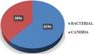
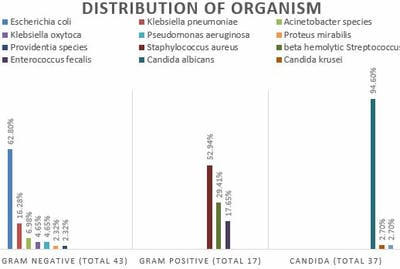
Figure 2: Total isolates.


The most common etiological agent in aerobic vaginitis was Escherichia coli (E Coli), followed by Staphylococcus aureus (Table 2).

Table 2­: Distribution of organisms isolated from the study subjects.

Organism Frequency(n) Percentage (%)
Candida albicans 35 38.89
Escherichia coli 27 30
Staphylococcus aureus 9 10
Klebsiella pneumoniae 7 7.77
β haemolytic Streptococcus 5 5.55
Enterococcus species 3 3.33
Acinetobacter species 3 3.33
Pseudomonas aeruginosa 2 2.22
Klebsiella oxytoca 2 2.22
Proteus mirabilis 1 1.11
Providentia species 1 1.11
Candida krusei 1 1.11
Candida Paropsilosis 1 1.11

Out of 90 subject’s single organism was found in 83(92.33%) subjects, and more than one organism was present in 7(7.77%) subjects (Figure 3& 4).

patho_591_03.JPG
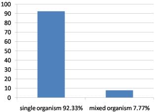
Figure 3: Percentage of single and mixed organisms.

patho_591_04.JPG
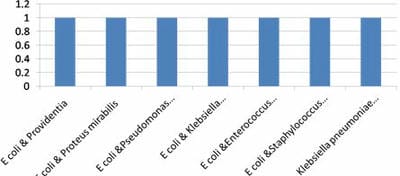
Figure 4: Distribution of mixed organism.

Enterobacteriaceae group of organisms showed higher sensitivity to Carbapenems, Chloramphenicol, Amikacin, and combination drugs

like Ceftazidime & Clavulanic acid, Piperacillin & Tazobactam.

Moderate sensitivity was seen among Gentamicin, Tobramycin, Doxycycline, Cotrimoxazole and Levofloxacin. Least sensitivity was seen among Ampicillin, Ampicillin & Clavulanic acid, third-generation Cephalosporin like Ceftriaxone & Cefotaxime, Ceftazidime, Piperacillin and Ciprofloxacin (Table 3).

Table 3: Antibiotic sensitivity of Enterobacteriaceae group of organisms.

Antibiotic E coli (n=27) % K. pneumoniae (n=7) % K. oxytoca (n=2) %  Provi dentia (n=1) % Proteus mirabilis s(n=1) % Overall sensitivity %
PI (3)11.1 (2)28.5 00 00 00 7.92
PIT (21)77.7 (4)57.1 (2)100 00 (1)100 66.96
AMP (3)11.1 (1)14.2 00 00 00 5.06
AMC (3)11.1 (2)28.5 00 00 00 7.92
CTR (3)11.1 (2)28.5 00 00 00 7.92
CTX (4)14.8 (2)28.5 00 00 00 8.66
CAZ (4)14.8 (3)42.8 00 00 00 11.52
CAC (19)70.3 (4)57.1 (2)100 00 (1)100 65.48
COT (15)62.9 (5)71.4 00 (1)100 00 46.86
AK (22)81.4 (4)57.1 (2)100 (1)100 00 67.7
GEN (17)62.9 (6)85.7 (1)50 (1)100 00 59.72
TOB (15)55.5 (6)85.7 (1)50 (1)100 00 58.24
CIP (11)40.7 (3)42.8 00 (1)100 00 36.7
LE (12)44.4 (4)57.1 00 (1)100 00 40.3
C (23)85.1 (4)57.1 (2)100 (1)100 00 68.44
DO (11)40.7 (3)42.8 00 (1)100 (1)100 56.7
IPM (15)55.5 (4)57.1 (1)50 (1)100 NT 65.65
MRP (27)100 (4)57.1 (2)100 (1)100 (1)100 91.42
ETP (22)81.4 (4)57.1 (2)100 (1)100 00 67.7

PI- Piperacillin, PIT- Piperacillin/Tazobactam, AMP- Ampicillin, AMC- Amoxycillin/ Clavulanic acid, CTR- Ceftriaxone, CTX- Cefotaxime, CAZ- Ceftazidime, CAC- Ceftazidime/ Clavulanic acid/ COT- Cotrimoxazole, AK- Amikacin, GEN- Gentamicin, TOB- Tobramycin, CIP- Ciprofloxacin, LE- Levofloxacin, C- Chloramphenicol, DO- Doxycycline, IPM- Imipenem, MRP- Meropenem, ETP- Ertapenem, NT- Not Tested.

Non-fermenters like Acinetobacter species and Pseudomonas aeruginosa showed 100% sensitivity to Carbapenems, Aminoglycosides, Fluoroquinolones and Cotrimoxazole. Moderate sensitivity was seen among Ceftriaxone, Cefotaxime, Ceftazidime, Piperacillin, and Piperacillin/ Tazobactam (Table 4).


Table 4 Antibiotic sensitivity of non-fermenters.

Antibiotic Acineto bacter (n=3) % Pseudo monas aeruginosa (n=1) % Overall sensitivity       %
PI (1)33.3 (1)100 66.65
PIT (1)33.3 (1)100 66.65
CTR (2)66.6 NT 66.6
CTX (2)66.6 NT 66.6
CAZ (1)33.3 (1)100 66.65
COT (3)100 NT 100
AK (3)100 (1)100 100
GEN (3)100 (1)100 100
TOB (3)100 (1)100 100
CIP NT (1)100 100
LE NT (1)100 100
IPM (3)100 (1)100 100
MRP    (3)100 (1)100 100

PI- Piperacillin, PIT- Piperacillin/ Tazobactam, CTR- Ceftriaxone. CTX- Cefotaxime, CAZ- Ceftazidime, COT- Cotrimoxazole, AK- Amikacin, GEN- Gentamicin, TOB- Tobramycin, CIP- Ciprofloxacin, LE- Levofloxacin, IPM- Imipenem, MRP- Meropenem, NY- Not Tested.

Table 5: Antibiotic sensitivity of Gram-positive organisms. 

Antibiotic  Staphylococcus aureus(n=9) % β haemolytic Streptococcus  (n=5) % Enterococcus faecalis (n=3) % Overall sensitivity %
AMC (2)22.2 NT NT 22.2
P 00 (5)100 NT 50
E (1)11.1 (1)20 00 10.36
CD (4)44.4 (3)60 NT 52.2
TE (7)77.7 (2)40 (1)33.3 50.33
DO (9)100 NT (1)33.3 66.65
COT (7)77.7 NT NT 77.7
C (7)77.7 (4)80 (3)100 85.9
GEN (6)66.6 NT NT 66.6
CIP (1)11.1 NT NT 11.1
LE NT 00 (1)33.3 16.65
LZ (9)100 (5)100 (3)100 100
VA NT (5)100 (3)100 100
HLG NT NT (2)66.6 66.6
AMP NT NT (2)66.6 66.6

AMC- Amoxycillin/ Clavulanic acid, P- Penicillin, E- Erythromycin, CD- Clindamycin, TE- Tetracycline, DO- Doxycycline, COT- Cotrimoxazole, C- Chloramphenicol, GEN- Gentamicin, CIR- Ciprofloxacin, LE- Levofloxacin, LZ-

Linezolid, VA- Vancomycin, HLG- High-level Gentamicin, AMP- Ampicillin, NT- Not Tested.

Discussion

The etiology of vaginitis is primarily due to microbial causes, which can be detected in the laboratory by standard techniques [3]. Misdiagnosis of AV as any other cause of vaginitis leads to incorrect treatment, leading to complications like desquamative inflammatory vaginitis, which can be deleterious in pregnancy [4]. Vulvovaginal candidiasis being the second most common infection of the vulvovaginal area, requires specific treatment with antifungal agents, [5]. the prevalence of which in our study was 7.99% Prevalence of aerobic vaginitis in different studies when compared to this study (11.43%) was higher. It was reported as 26%, 21.25 and 20.8% in the study conducted by Nahar et al. [12] Krishnaswamy et al. [13]. (2019) and Sangeetha et al. [14].  (2015) respectively. Still higher rate of 58.5% was reported by Pal K et al. [15]. (2017). The study by Wang ZL et al. (2016) showed an almost similar frequency of AV as that of our study as 15.40% [16].

Overall vaginal infection in our study due to AV & VVC was 19.43%. The other causes of vaginitis due to Trichomonas BV were not included, as their diagnostic methods mainly rely on microscopy. A retrospective study in Solapur by Shaikh et al. showed the overall prevalence of vaginitis as 37%, and AV and VVC was 66.48% and 21.62%, respectively [17]. In another study AV & VVC reported was 51% & 17% respectively [3]. Looking into this prevalence of vaginitis mandates the isolation and identification of the organism followed by susceptibility testing. Our study had the frequency of infection higher in the age group of 16 to 20 years (27.8%) followed by 21 to 25 years (17.4%). Our institute having access to the rural population, early marriage, and early sexual activity may be the reason for this population group having the highest frequency.

25 to 30 years followed by 31 to 35 years had the highest frequency of infection in the study by Pal et al. [15].  Sangeetha KT et al. [14] and Krishnaswamy L et al. [13]. Mumtaz et al. noted the highest frequency of AV among 31 to 40 years (39.5%) followed by 41 to 50 years (35.8%) [18]. Vulvovaginal candidiasis was reported to be 13.1% and 5.3%, highest in the age group of 30 to 39years and 18 to 25years, respectively [19].


There is a vast difference in the Candida species isolated from the vagina worldwide, which depends on various factors such as geographical area, presence of risk factors like diabetes, use of antibiotics, type of population studied. However, Candida albicans (94.60%) is the commonest species of VVC in our study, and there was an increase in non-albicans Candida (58%) isolated. The main species were Candida glabrata (71%) and Candida krusei (29%) [20].

Gram-positive isolates were lesser compared to Gram-negative isolates. Escherichia coli was the commonest organism, followed by Staphylococcus aureus, similar to the study by Pal et al., Jahic et al., Mulu et al.  In contrast to this, Gram-positive were 83.8%. Gram-negatives were 16.3%, and the predominant species was Streptococcus agalactiae which was detected by amplicon sequencing 5 targeting the 16S rRNA V4 region and also by culture [21]. 9.68% and 1.2% β hemolytic Streptococcus was detected respectively by Sangeetha et al. [14], and Mulu et al., which was 5.5% in our study Staphylococcus aureus were most common by Mumtaz et al. [18]. Enterococcus fecalis was the commonest organism as per Sangeetha KT et al. Most of the microorganisms isolated in AV are the bacteria that colonize the intestinal tract. Escherichia coli being the highest community in the intestinal tract, is most likely to have gained entry into the vulvovaginal region, causing infection due to proximity of anal orifice with the vaginal orifice and also an imbalance in the normal microbiota of the vagina along with other contributing factors like poor hygiene [5, 22].

7.7% of individuals had AV caused by more than one organism, and one incidence of AV combined with VVC was noted in our study.  16% of the combination of AV with VVC was reported by Wang et al. by using the rapid enzyme kit detection method and microscopy.   Polymicrobial growth was also noted to the extent of 19.23% by Sangeetha et al. Enterobacteriaceae group of organisms showed maximum sensitivity to Meropenem (91.42%), almost similar to Krishnamurthy et al., where the sensitivity was 100%. Most minor sensitivities were observed with Ampicillin, Ceftriaxone, Fluoroquinolones similar to study by Sangeetha et al., Mumtaz et al. and Pal K et al. One Pseudomonas aeruginosa isolated was sensitive to all antibiotics, sensitivity to Piperacillin/ Tazobactam and Carbapenems was highest for Pseudomonas

by Sangeetha et al. and Krishnamurthy et al. With this scenario Carbapenems to be considered especially while treating pregnant women with aerobic vaginitis to prevent the risk of transmission of infection to neonates Isolation of Gram positive’s though lesser than Gram-negatives, drug resistance has significant impact such as infertility, toxic shock syndrome, neonatal septicemia. Clindamycin sensitivity 52.2% noted elevates the use of most common antibiotic in the form of pessary Aerobic vaginitis is a newly recognized entity that has a various impact both in pregnant and nonpregnant women which has to be addressed appropriately and adequately to combat the adverse outcome in pregnancy and to prevent infertility, discomfort like dyspareunia [23, 24].

Conclusion

It is necessary to deal with the pathogens of vaginitis after microbiological evaluation. This will differentiate the causes of vaginitis like Vulvovaginal Candidiasis and aerobic vaginitis, included in this study. Error in treatment can be avoided like unnecessary administration of broad-spectrum antibiotics, which carries the risk of depletion of a beneficial organism like Lactobacilli, the emergence of drug resistance and hence to focus on the appropriate treatment

What does this study add to existing knowledge?

Aerobic vaginitis being a common condition, most of the time, no efforts will be made to identify the causative agent. In our study, to our surprise, we found a high incidence of resistance and the organisms being resistant to most of the commonly used antibiotics.

Permission from the Institutional Research Board (IRB): Yes.


Reference

01. Witkin, Steven S. , and William J. Ledger. Complexities of the uniquely human vagina. " Science Translational Medicine 4.132 (2012): 132fs11-132fs11 [Crossref][PubMed][Google Scholar]

02. Mulu W, Yimer M, Zenebe


Y, Abera B. Common causes of vaginal infections and antibiotic susceptibility of aerobic bacterial isolates in women of reproductive age attending at Felegehiwot referral Hospital, Ethiopia: a cross sectional study. " BMC women's health 15. 1 (2015): 1-9. [Crossref][PubMed][Google Scholar]

03. Donders, G. G. , Vereecken, A. , Bosmans, E. , Dekeersmaecker, A., Salembier, G. and Spitz, B. Definition of a type of abnormal vaginal flora that is distinct from bacterial vaginosis: aerobic vaginitis." BJOG: An International Journal of Obstetrics & Gynaecology 109.1 (2002): 34-43 [Crossref][PubMed][Google Scholar]

04. Jahic M, Mulavdic M, Nurkic J, Jahic E, Nurkic M. Clinical characteristics of aerobic vaginitis and its association to vaginal candidiasis, trichomonas vaginitis and bacterial vaginosis. " Medical archives 67. 6 (2013): 428. [Crossref][PubMed][Google Scholar]

05. Kaambo E, Africa C, Chambuso R, Passmore JA. Vaginal microbiomes associated with aerobic vaginitis and bacterial vaginosis. " Frontiers in public health 6 (2018): 78. [Crossref][PubMed][Google Scholar]

06. Bitew, Adane, and Yeshiwork Abebaw. Vulvovaginal candidiasis: species distribution of Candida and their antifungal susceptibility pattern. " BMC women's health 18. 1 (2018): 1-10. [Crossref][PubMed][Google Scholar]

07. Pirotta, Marie V. , and Suzanne M. Garland. Genital Candida species detected in samples from women in Melbourne, Australia, before and after treatment with antibiotics. " Journal of clinical microbiology 44.9 (2006): 3213-3217 [Crossref][PubMed][Google Scholar]

08. Choi S. J. , Park S. D. , Jang I.H., Uh Y., Lee A. The prevalence of vaginal microorganisms in pregnant women with preterm labor and preterm birth." Annals of laboratory medicine 32.3 (2012): 194-200 [Crossref][PubMed][Google Scholar]

09. Collee, John Gerald, Thomas Jones Mackie, and James Elvins McCartney. Mackie & McCartney practical medical microbiology. Harcourt Health Sciences, 1996. [Crossref][PubMed][Google Scholar]

10. Bauer A. W. , Kirby W. M. , Sherris J.C., Truck M. Antibiotic susceptibility testing by a standardized

single disc method." Am J clin pathol 45.4 (1966): 493-496 [Crossref][PubMed][Google Scholar]

11. CLSI. Performance standards for Antimicrobial Susceptibility Testing. 28th ed. CLSI supplements M100. Wayne PA: Clinical and Laboratory Standard Institute; 2018. [Google Scholar] [Crossref][PubMed][Google Scholar]

12. Nahar D, Soni G, Chand AE, Mourya S. Bacterial etiology and their antibiogram in aerobic vaginitis patients at Tertiary Care Hospital, Kota, Rajasthan. " International Journal of Scientific Study 4. 3 (2016): 103-107. [Crossref][PubMed][Google Scholar]

13. Krishnasamy, Lakshmi, Chitralekha Saikumar, and Govindasamy Kumaramanickavel. Aerobic Bacterial Pathogens Causing Vaginitis in Patients Attending A Tertiary Care Hospital and their Antibiotic Susceptibility Pattern. " J Pure Appl Microbiol 13. 2 (2019): 1169-1174. [Crossref][PubMed][Google Scholar]

14. Sangeetha, K. T. , Saroj Golia, and C. L. Vasudha. A study of aerobic bacterial pathogens associated with vaginitis in reproductive age group women (15-45 years) and their sensitivity pattern. (2015) [Crossref][PubMed][Google Scholar]

15. Pal K, Sidhu SK, Devi P, et al; Department of Microbiology, Government Medical College, Amritsar, Punjab, India. Etiology of vaginal infections and antimicrobial resistance pattern of aerobic bacterial isolates in women of reproductive age group attending a tertiary care hospital. Asian Pac. J. Health Sci. 2017;4(4):15. doi:10.21276/apjhs.2017.4.4.5 [CrossRef] [Google Scholar] [Crossref][PubMed][Google Scholar]

16. Wang ZL, Fu LY, Xiong ZA, Qin Q, Yu TH, Wu YT, et al. Diagnosis and microecological characteristics of aerobic vaginitis in outpatients based on preformed enzymes. " Taiwanese Journal of Obstetrics and Gynecology 55. 1 (2016): 40-44. [Crossref][PubMed][Google Scholar]

17. Sadiya Shaikh, Prakash Waghmare, Anu Sharma, Kishor Ingole and Rashmi Bawane. A Retrospective Evaluation of Vaginitis in Women of Reproductive Age Group in a Tertiary Care Hospital in Solapur, India. " Int. J. Curr. Microbiol. App. Sci 7.02 (2018): 762-768 [Crossref][PubMed][Google Scholar]


18. Mumtaz S, Ahmad M, Aftab I, Akhtar N, Ul Hassan M, Hamid A. Aerobic vaginal pathogens and their sensitivity pattern. " J Ayub Med Coll Abbottabad 20. 1 (2008): 113-117. [Crossref][PubMed][Google Scholar]

19. Jacob, L. , John, M. , Kalder, M. , & Kostev, K. Prevalence of vulvovaginal candidiasis in gynecological practices in Germany: A retrospective study of 954,186 patients." Current medical mycology 4.1 (2018): 6 [Crossref][PubMed][Google Scholar]

20. Ghaddar N. , Anastasiadis E. , Halimeh R. , Prevalence and antifungal susceptibility of Candida albicans causing vaginal discharge among pregnant women in Lebanon. " BMC infectious diseases 20.1 (2020): 1-9 [Crossref][PubMed][Google Scholar]

21. Wang C. , Fan A. , Li H. , Yan Y. , Qi W., Wang Y., et al. Vaginal bacterial profiles of aerobic vaginitis: a case–control study." Diagnostic microbiology and infectious disease 96.4 (2020): 114981 [Crossref][PubMed][Google Scholar]

22. Tempera, Gianna, and Pio Maria Furneri. Management of aerobic vaginitis. " Gynecologic and obstetric investigation 70. 4 (2010): 244-249. [Crossref][PubMed][Google Scholar]

23. Mahmoud F. Hassan, Nancy M. A. Rund, Does aerobic vaginitis have adverse pregnancy outcomes? prospective observational study. " Infectious diseases in obstetrics and gynecology 2020 (2020) [Crossref][PubMed][Google Scholar]

24. Donders, Gilbert GG, Gert Bellen, and Kateryna S. Ruban. Abnormal vaginal microbioma is associated with severity of localized provoked vulvodynia. Role of aerobic vaginitis and Candida in the pathogenesis of vulvodynia. " European Journal of Clinical Microbiology & Infectious Diseases 37.9 (2018): 1679-1685 [Crossref][PubMed][Google Scholar]