E-ISSN:2456-1487
P-ISSN:2456-9887
RNI:MPENG/2017/70771

Case Report

Enterobius Vermicularis

Tropical Journal of Pathology and Microbiology

2022 Volume 8 Number 1 January February
Publisherwww.medresearch.in

Appendiceal Enterobius Vermicularis infestation discovered incidentally in a case of sigmoid volvulus: A Rare Co-occurrence

Shetty A.1, Gaffoor N.2, Muralidhar A.3*
DOI: https://doi.org/10.17511/jopm.2022.i01.05

1 Archana Shetty, Associate Professor, Department of Pathology, CDSIMER, Kanakapura, Karnataka, India.

2 Nidha Gaffoor, Senior Resident, Department of Pathology, CDSIMER, Kanakapura, Karnataka, India.

3* Aparna Muralidhar, Assistant Professor, Department of Pathology, CDSIMER, Kanakapura, Karnataka, India.

Enterobius Vermicularis is a gastrointestinal parasite mainly affecting children. It is one of the many causes of acute appendicitis. The probability that every case of infestation leads to an appendiceal inflammatory response is controversial, as cases with normal histology have been reported. Appendix is subjected to examination only when the patient presents, with symptoms primarily related to it or attributed to obstructive causes of acute abdomen like intussusception, mass lesions, strictures or volvulus. The case presented is an adult who was operated for sigmoid volvulus with incidental finding of Enterobius Vermicularis infestation in the appendix. Though incidental, identification of worms is important as antihelminthic therapy can be curative, avoiding undue surgical interventions

Keywords: Appendectomy, Enterobius, Parasites, Inflammation, Microscopy

Corresponding Author How to Cite this Article To Browse
Aparna Muralidhar, Assistant Professor, Department of Pathology, CDSIMER, Kanakapura, Karnataka, India.
Email:
Archana Shetty, Nidha Gaffoor, Aparna Muralidhar, Appendiceal Enterobius Vermicularis infestation discovered incidentally in a case of sigmoid volvulus: A Rare Co-occurrence. Trop J Pathol Microbiol. 2022;8(1):29-32.
Available From
https://pathology.medresearch.in/index.php/jopm/article/view/586

Manuscript Received Review Round 1 Review Round 2 Review Round 3 Accepted
2021-12-14 2021-12-16 2021-12-23 2021-12-30 2022-01-06
Conflict of Interest Funding Ethical Approval Plagiarism X-checker Note
Nil Nil Yes 18%

© 2022by Archana Shetty, Nidha Gaffoor, Aparna Muralidharand Published by Siddharth Health Research and Social Welfare Society. This is an Open Access article licensed under a Creative Commons Attribution 4.0 International License https://creativecommons.org/licenses/by/4.0/ unported [CC BY 4.0].

Introduction

Enterobius vermicularis (EV) infestation is a paediatric helminthic infection, uncommonly affecting adults. The cecum is the major site where the worm resides. It can reach the appendix causing serious morbidity. However, the primary parasitic infestation of the appendix is rare. The role of this worm in the aetiology of acute appendicitis is also controversial and is more often an incidental finding. Pathologists should be aware of the histomorphology of the worm to avoid underreporting of this entity which can be controlled with anti-helminthics. [1]. We report one such incidental finding of a pinworm  in the appendix of a patient who was operated on for sigmoid volvulus.

Case report

A fifty-one-year-old male presented with complaints of pain abdomen and vomiting for two days, relieved on taking medications and passing stools. History revealed that the patient in addition had on and off episodes of constipation for which he had taken over the counter medications. On admission, an Erect X-ray abdomen revealed marked distention of a loop of the large bowel in the left upper quadrant (Figure 1).

patho_586_01.JPG
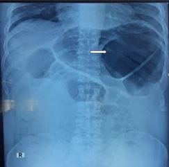
Figure 1: Erect X-Ray abdomen showing distended large bowel loop in the left upper quadrant of the abdomen (arrow).

CT abdomen (plain and contrast) showed gross dilatation of the colon up to the rectosigmoid region with evidence of twisting of sigmoid mesocolon – features suspicious of volvulus. Intraoperatively, grossly dilated sigmoid colon with mobile ascending

& descending colon without evidence of gangrene was noted. The patient was taken up for surgery – sentence to be deleted.  Sigmoid volvulus was de-rotated and decompressed using suction. The sigmoid colon, part of descending colon (suspecting gangrene) and appendix were resected after ligating the mesocolon.

Grossly, appendix with mesoappendix measured 4.5 cm in length, with an intact tip. Cut section showed a thickened wall with a small white thread-like structure protruding from the lumen.The colonic segment measured 30 cm in length with a dilated area of 8 cm seen from one surgical end. Cut section of the dilated segment showed flattening of mucosa with mild brownish discolouration. (Figure 2).

patho_586_02.JPGpatho_586_2.JPG
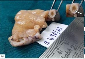
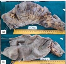
Figure 2a: The gross image of the appendix, inset shows threadlike structure - worm (arrow) Figures 2b and 2c: The gross image of external surface (2b) and cut surface (2c) of the colon showing flattening of mucosa in the dilated segment.

Microscopically, the lumen of the appendix showed a parasite morphologically resembling Enterobius Vermicularis along with faecal matter and features of subacute appendicitis.


The dilated colonic segment showed ischemic changes with unremarkable surgical margins. (Figure 3).

patho_586_03.JPG
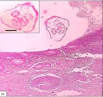
Figure 3 a: Photomicrograph of appendix showing Enterobius Vermicularis in the lumen, H&E, 100x. Inset – Cross-section of Enterobius Vermicularis with prominent ala (arrow), H&E,400x

patho_586_3.JPG
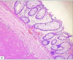
Figure 3b: Photomicrograph of an ischemic segment of the colon showing edematous mucosa and submucosa with few congested blood vessels, H&E, 100x

The patient was started on antihelminthics, had an uneventful postoperative period and is currently doing well on follow up.

Discussion

Sigmoid volvulus (twisting of a loop of intestine around its mesentery) is commonly related to malrotation of the gut at birth. It is usually observed in neonates and children, rarely in adults due to postoperative adhesions or chronic constipation leading to stagnation of the colon, extreme high fibre diet, or rarely

underlying psychiatric illness.[2]. On literature search, reports mentioning the incidental finding of worms in cases of sigmoid volvulus have been documented. N Mourra et al [3]. have reported an unusual presentation of chronic intestinal schistosomiasis, masquerading as recurrent volvulus. In our case, we had a coincidental finding of Enterobius Vermicularis infestation of the appendix in a patient who was operated for sigmoid volvulus, which was diagnosed radiologically. Gastrointestinal infection due to Enterobius Vermicularis occurs worldwide and is considered to be the most common helminthic infection.[4]. A clinical picture resembling acute appendicitis may be caused by the worm obstructing the lumen or causing a hypersensitivity reaction in the tissue. However, it is not clear whether the invading organism causes the inflammation or if the parasite is an incidental finding when inflammation is already present.[5]. Usually, the pathogenicity of Enterobius Vermicularis is mild, ranging from asymptomatic to nocturnal anal pruritus. Radiological and laboratory findings are not helpful in pre-operatively distinguishing acute appendicitis related to parasites.[6]. Humans are the only known natural hosts of this parasite. It can have a wide range of pathogenicity, implying that pathologists should be familiar with its histological aspects for accurate diagnosis. The histologic identification of Enterobius Vermicularis or its eggs on resected specimens is not difficult when the pathologist performs a careful search for this parasite. In our case, we could find parasites in the lumen, even macroscopically. Whether they are the causative agents of acute appendicitis or an incidental finding, remains uncertain.[7]. The common histological findings in resected appendiceal specimens range from normal to various inflammatory patterns such as lymphoid hyperplasia, eosinophilic or neutrophilic infiltrate. Enterobius Vermicularis may occasionally be associated with severe inflammation, ulceration and perforation.[6]. The cross-section shows characteristic double and lateral “thorn-like” extensions (alae) from the thin eosinophilic wall of the parasite. Within the wall of parasites, annular structures are easily seen, corresponding to the intestine. Numerous oval eggs, with flattened edges, can be found in gravid females. In solid organs like ovaries, spleen or liver, the histological diagnosis can be tricky as parasites have no sufficient space to develop. Granulomas and other inflammatory changes around oval structures


(eggs) can be the main features to lead the pathologist to the correct diagnosis. In summary, histological patterns of acute appendicitis associated with E. vermicularis are a very rare finding.[7].

Conclusion

Identification of Enterobius Vermicularis in appendectomy specimens appears to be incidental, then being the aetiology of appendicitis, especially when the appendix is resected for other surgical causes. Pathologists must be aware of its histological features to avoid under-reporting this entity, which if recognized can be cured with anti-helminthics.

Reference

01. Hasan A, Nafie K, El-Sayed S, Nasr M, Abdulmohaymen A, Baheeg M, et al. Enterobius vermicularis in appendectomy specimens; Clinicopathological assessment: Cross-sectional study. Ann Med Surg (Lond). 2020 Oct 28;60:168-172. doi: 10.1016/j.amsu.2020.10.057 [Crossref][PubMed][Google Scholar]

02. Reddy YPS, Senthil Kumaran S, Vanka V, Rab A, Patel V. Abdominal pain - a common presentation with unusual diagnosis: a case report. J Community Hosp Intern Med Perspect. 2020 Oct 29;10(6):604-608. doi: 10.1080/20009666.2020.1821469 [Crossref][PubMed][Google Scholar]

03. Mourra N, Lesurtel M, Paye F, Flejou JF. Chronic schistosomiasis: an incidental finding in sigmoid volvulus. J Clin Pathol. 2006 Jan;59(1):111. [Crossref][PubMed][Google Scholar]

04. Uysal E, Dokur M. The Helminths Causing Surgical or Endoscopic Abdominal Intervention: A Review Article. Iran J Parasitol. 2017 Apr-Jun;12(2):156-168. [Crossref][PubMed][Google Scholar]

05. Panidis S, Paramythiotis D, Panagiotou D, Batsis G, Salonikidis S, Kaloutsi V, et al. Acute appendicitis secondary to Enterobius vermicularis infection in a middle-aged man: a case report. J Med Case Rep. 2011 Nov 30;5:559. doi: 10.1186/1752-1947-5-559 [Crossref][PubMed][Google Scholar]

06. Altun E, Avci V, Azatcam M. Parasitic infestation in appendicitis. A retrospective analysis of 660 patients and brief literature review. Saudi Med J.

2017 Mar;38(3):314-318. doi: 10.15537/smj.2017.3.18061 [Crossref][PubMed][Google Scholar]

07. Efared B, Atsame-Ebang G, Soumana BM, Tahiri L, Hammas N, El Fatemi H, et al. Acute suppurative appendicitis associated with Enterobius vermicularis: an incidental finding or a causative agent? A case report. BMC Res Notes. 2017 Oct 6;10(1):494. doi: 10.1186/s13104-017-2822-3 [Crossref][PubMed][Google Scholar]