E-ISSN:2456-1487
P-ISSN:2456-9887
RNI:MPENG/2017/70771

Research Article

Colorectal Cancer

Tropical Journal of Pathology and Microbiology

2021 Volume 7 Number 6 November December
Publisherwww.medresearch.in

Histological Variants and Tumor Heterogeneity in Colorectal Cancer

Gupta P.1, Gupta R.2*, Sahoo T.3, Sharma A.4
DOI: https://doi.org/10.17511/jopm.2021.i06.05

1 Priyanka Gupta, Assistant Professor, Department of Pathology, Chirayu Medical College and Hospital, Bhopal, M.P, India.

2* Rohan Gupta, Assistant Professor, Department of Pathology, RKDF Medical College and Hospital, Bhopal, M.P, India.

3 T.P Sahoo, DM, Medical Oncology, Silverline Hospital, Bhopal, M.P, India.

4 Abhishek Sharma, Professor, Department of Surgery, Peoples Medical College and Research Centre, Bhopal, M.P, India.

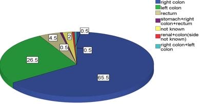
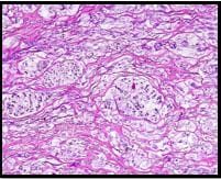
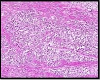
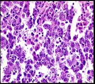
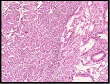
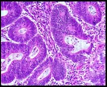
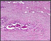
Introduction: Colorectal cancer is not one disease but rather a collection of neoplastic diseases. Due to heterogeneity in the disease definite categorization, TNM staging, and different therapy responses and prognosis, extensive disease stratification is required. Therefore TNM Staging, Microsatellite stability status, Tumor Grade, Lymphovascular emboli, Intraepithelial lymphocytes, Tumor budding and other parameters are assessed in the pathology report to indicate the extent and prognosis of the disease. Objectives and Method: This retrospective study includes 200 resected specimens of Colorectal Cancer cases. The histological parameters of 200 Colorectal Cancer cases without prior radiotherapy and chemotherapy were considered. All radical specimens of Colorectal cancers were fixed in formalin and were subjected to processing and staining by Hematoxylin and Eosin. The sections obtained were microscopically examined by two pathologists individually. The histological parameters were recorded. Results and Conclusion: Out of the 200 cases, 119 (59.5%) were males, and 81 (40.5%) were females with a Male: Female ratio of 1.4:1.The common locations of the tumors as shown in Table no.1 are the right colon (n=131, 65.5%) left colon (n=53, 26.5%), rectum (n=9, 4.5%), synchronous tumors (n=3, 1.5%) and colon with site not specified (n=4,2.0%). Mucinous carcinoma 23 cases, Signet Ring Cell carcinoma 17 cases, Diffuse increase in Intraepithelial Lymphocytes, Undifferentiated Carcinoma 48 cases and Heterogeneous morphologies 35 cases, Tumor Budding in 49 cases, Crohn's like lymphocytic infiltrate in 3 cases, Synchronous tumour 3 cases, Cribriform morphology in 31 cases along with Right-sided tumours in 134 cases.

Keywords: Colorectal Cancer, Heterogeneous Morphology, Intraepithelial Lymphocytes and Microsatellite stability

Corresponding Author How to Cite this Article To Browse
Rohan Gupta, Assistant Professor, Department of Pathology, RKDF Medical College and Hospital, Bhopal, M.P, India.
Email:
Priyanka Gupta, Rohan Gupta, T.P Sahoo, Abhishek Sharma, Histological Variants and Tumor Heterogeneity in Colorectal Cancer. Trop J Pathol Microbiol. 2021;7(6):298-304.
Available From
https://pathology.medresearch.in/index.php/jopm/article/view/583

Manuscript Received Review Round 1 Review Round 2 Review Round 3 Accepted
2021-11-30 2021-12-01 2022-12-08 2021-12-15 2021-12-22
Conflict of Interest Funding Ethical Approval Plagiarism X-checker Note
Nil Nil Yes 19%

© 2021by Priyanka Gupta, Rohan Gupta, T.P Sahoo, Abhishek Sharmaand Published by Siddharth Health Research and Social Welfare Society. This is an Open Access article licensed under a Creative Commons Attribution 4.0 International License https://creativecommons.org/licenses/by/4.0/ unported [CC BY 4.0].

Introduction

Colorectal cancer (CRC) is the second most common cancer in women and the third most common cancer in men [1,2]. CRC incidence rates generally are higher among males than females at all anatomic subsites. [3, 4]. The mean age for CRC is 62 years, and the risk group includes a range from 60-79 years. The incidence rates vary among different world regions, with a tenfold higher incidence in western countries than developing countries. Various well-established risk factors for developing CRC are Environmental factors, age, a family history of CRC, a personal history of cancer (e.g., colon, rectum, ovary, and Endometrium), chronic inflammatory bowel diseases, and a history of polyps in the colon. The TNM classification system, designed by the American Joint Committee on Cancer and the Union for  International Cancer Control, remains the most robust prognostic parameter to stratify patients. The TNM system describes the depth of tumour invasion throughout the wall of the colon (T), the number of affected lymph nodes (N), and the presence of metastasis (M). [5]. Based on the T, N, and M classifiers, tumours are divided into four stages with different prognostic and therapeutic consequences. Stage 1 tumours have a 5-year survival rate of 94%. Stage 2 tumours have a 5-year survival rate of 82%, which drops to 67% for stage 3 patients. Metastatic or stage 4 colon cancers have a 5-year survival rate of only 11%. The therapeutic strategy is highly dependent on the cancer stage at the time of diagnosis. Surgery is considered a first-line treatment for stage 1, 2, and 3 tumours, and it is followed by adjuvant chemotherapy for stage 3 tumors and stage 2 tumors with adverse risk factors. In stage 4 tumors, treatment options for resectable metastases include neoadjuvant chemotherapy, metastectomy, and colectomy. Tumour heterogeneity in Colorectal cancer is divided into Intertumoral and Intratumoral. Tumour heterogeneity has been investigated to different levels, including genomics, histopathologic features and characteristics of inflammatory infiltrate, especially Tumour-infiltrating lymphocytes.

Four kinds of genomic or epigenetic instability have been described in colorectal cancers [6].
1. Chromosomal instability (CIN).
2. Microsatellite instability (MSI).
3. CpG island methylator phenotype (CIMP).
4. Global DNA hypomethylation.

patho_583_01.JPG

Figure 1:adenoma –carcinoma progression sequence: CRC progresses through at least two well-defined pathways. CIN pathway starts with classical tubular adenoma, then the early acquisition of APC gene mutations leads to deranged Wnt signalling, K ras oncogene mutations at early adenoma stage and loss of heterozygosity of 18 chromosomes at advanced adenoma stage and ultimate TP 53 mutations translating it to frank malignancy. On the other hand, the MSI pathway acquires BRAF mutation and is not associated with 18q loss and TP53 mutations.[6].
- polyposis.
- Familial colorectal cancer type X.

Table 1: Important clinicopathological and molecular differences between Lynch syndrome and sporadic colorectal carcinomas:[7, 8]. 

Clinical Parameters Lynch Syndrome Sporadic Crc
Age < 50 years > 70 years
Sex F = M F >> M
Other Lynch associated tumours Yes No
Proximal Location of tumour Yes Yes
Histology Features:    
Mucinous carcinoma Yes Yes
Signet ring cell carcinoma Yes Yes
Medullary carcinoma Yes Yes
Intraepithelial lymphocytes Yes Yes
Crohn's like reaction Yes Yes
Pushing borders Yes Yes
Poor differentiation Yes Yes
Tumour budding Yes No
Tumour heterogeneity Yes Yes
Serration/serrated precursor No Yes
Immunohistochemistry    
Absence of MLH1 and PMS2 Yes, some Yes
Absence of MSH2 and MSH6 Yes, some No
Absence of MSH6 or PMS2 Yes, few No
Molecular Features:    
MSI-H Yes Yes
BRAF mutation No Yes
MLH1 promoter methylation Rare Yes
Germline mutation in mismatch repair gene Yes No

Materials and Methods

Settings: The study was conducted in Gupta Diagnostic Laboratory, Bhopal, in association with Silverline Hospital

Duration of study: Retrospective study and was conducted for five years of duration 2017-2021. A Retrospective Study of 200 cases of sporadic Colorectal adenocarcinoma was done within five years of duration. A sampling of the cases was done consecutively.

Inclusion Criteria: Individuals operated for Colorectal Cancer and given informed consent were selected for the study.

Exclusion Criteria: Individuals with colorectal cancer cases who were subjected to Neo-adjuvant chemotherapy were excluded from the study

Sample Size: All individuals who satisfied the inclusion criteria during the study period were included in the study.

Surgical Procedure: Right and Left Hemicolectomy. Total Colectomy, Abdominal Perineal Resection for Rectal cancer cases and Lower Abdominal Resections. The cases were retrieved from Gupta Diagnostic Laboratory. The specimens were noted for Location, including Right colon, Left Colon, Synchronous tumours, Left and Right Colon and Rectum. Depth of penetration, number of lymph node positivity and TNM staging. The histological parameters were reviewed by two observers subsequently.

patho_583_02.JPG
Figure 2: Location of Tumors.

They were: Mucinous carcinoma, Signet ring cell adenocarcinoma, Undifferentiated carcinoma, and Crohn's like lymphocytic infiltration. We also

tested Cribriform patterns, heterogeneous morphologic patterns (combination of more than one of the above patterns and tumour budding as additional histological parameters. Documentation of other clinic-pathological parameters was done: Age of the patient, Synchronous/ Metachronous tumours, T3N0 tumours, and Right-sided tumour.

Results

We reviewed 200 colorectal carcinomas in the form of 200 resected specimens over five years. The study cohort of 200 was received from Silverline Hospital.  The patients' age ranged from 14 to 88 years, with a mean age of 51.06 years and a median of 51.00 years, respectively. Ninety patients were less than 50 years of age, and 110 patients were more than 50 years of age. Out of the 200 cases, 119 (59.5%) were males, and 81 (40.5%) were females with a Male: Female ratio of 1.4:1.The common locations of the tumors as shown in Table no.1 are the right colon (n=131, 65.5%) left colon (n=53, 26.5%), rectum (n=9, 4.5%), synchronous tumors (n=3, 1.5%) and colon with site not specified (n=4,2.0%). Various morphologies and Histological parameters that we came across were Mucinous carcinoma 23 cases FIG 3, Signet Ring Cell carcinoma 17 cases FIG 4, Diffuse increase in Intraepithelial Lymphocytes (FIG5)  Undifferentiated Carcinoma 48 cases and Heterogeneous morphologies 35 cases FIG 6, Tumor Budding in 49 cases FIG 8,  Crohn's like lymphocytic infiltrate in 3 cases, Synchronous tumour 3 cases, Cribriform morphology in 31 cases FIG 7 along with Right-sided tumours in 134 cases.

Discussion

According to WHO, 2010 "Mucinous adenocarcinoma is defined by the presence of pools of extracellular mucin covering more than 50% of the lesion. Tumours in which extracellular mucin covers <50% of the lesion are designated as adenocarcinoma with a mucinous component. Mucinous carcinoma, which accounts for 10% of overall colon cancer, are predominantly located on the right side of the colon and have high rates of MSI, BRAF, KRAS and PIK3CA mutations.

In our study, we found 23 cases of mucinous carcinoma with a mucinous component of more than 50% and 10 points showing Mucinous differentiation ( mucinous


area less than 50%). Most of the mucinous carcinoma in our studies are right-sided, as mentioned in other studies. [9, 10]. The prognostic outcome of mucinous tumours is somewhat controversial. Mucinous morphology was initially considered a negative prognostic marker; however, when corrected for the tumour stage, no difference in survival rates between glandular and mucinous tumours was observed. [11].

Signet ring cell adenocarcinoma is a rare histological subtype and accounts for less than 1% of cases of CRC. [12]. It is characterized by the proliferation of signet ring cells with intracellular mucin pools that push the nucleus to the periphery FIG 3, Mucin-rich signet cell carcinomas are strongly associated with MSI in up to 40% of cases and display a better overall survival than mucin-poor types. [13]. Signet ring cell tumours tend to metastasize early and to multiple sites such as the liver, ovaries, and peritoneum. In contrast, metastases of glandular tumours mostly metastasize to the liver only. [14].

Undifferentiated / Medullary carcinomas are poorly differentiated and characterized by the presence of solid sheets or nests of malignant cells with pushing borders, prominent intraepithelial lymphocytic infiltration, and no to rare gland formation. The incidence rate of this relatively new histological type has not been well established. We observed 48 cases of Undifferentiated Carcinoma in our study.

patho_583_03.JPG

Figure 3: Microphotograph showing Mucinous carcinoma with signet ring cell component (H and E X 200).

patho_583_04.JPG

Figure 4: Microphotograph showing Signet ring cell carcinoma (H and E X 200).

patho_583_05.JPG

Figure 5: Microphotograph showing increase in intraepithelial lymphocytes (H and E X 200).

patho_583_06.JPG

Figure 6: Microphotograph showing Heterogeneous morphology of colorectal carcinoma (H and E X200).


patho_583_07.JPG

Figure 7: Microphotograph showing Cribriform morphology s (H and E X 200).

patho_583_08.JPG

Figure 8: Microphotograph showing Tumor Budding at the infiltrative edges of colorectal carcinoma  (H and E X200).

It has been widely proven that MSIH tumours have heterogeneous morphology. The tumour has more than one morphology in the form of conventional glandular areas and solid areas, mucinous and solid areas, Cribriform and conventional epithelial differentiation etc. A recent study by L De Smedt, J Lemahieu and S Palmansetal show that heterogeneous morphology is an essential feature of MSI-H tumours and are associated with an increase in intraepithelial lymphocytes(cytotoxic T cells) as compared to those tumours which are MSS. We found 35 cases with heterogeneous morphologies, and most of them were right-sided.

Cribriform Histology To the best of our knowledge, there are no extensive

studies of this pattern in the colon, only mentioned briefly at the World Health Organization classification of tumours named "colonic cribriform carcinoma (CCC)," like cribriform carcinoma of other sites of the gastrointestinal tract.[15]. It is usually associated with a poor prognosis. Up till now, no study has specified the relation between cribriform histology and microsatellite instability. We found 31 cases of cribriform morphology in our research.

Tumor Budding A study by Ronder and Robert et al. showed that tumours with high Tumor budding(>10 buds/hpf) were more likely to have venous invasion and lymphatic invasion, to present with regional or metastatic disease. Tumours with high budding were more likely to be microsatellite stable (MSS) and were more often characterized by KRAS mutations.  [16].  We found 49 cases of CRC with Tumor Budding in our study, and most of them were found to be left-sided tumours.

Synchronicity and Crohn's like lymphocytic infiltrate: Modified Bethesda guidelines specifies criteria's for Lynch syndrome/HNPCC . One of the criteria mentions synchronicity or metachronicity of the tumours, which should lead to the suspicion for germline mutation of MMR genes.

Conclusion

CRC is a highly heterogeneous disease, and therefore extensive disease stratification that accounts for inter and intratumor heterogeneity is highly required. It is important that the assessment of MSI status, TNM stage, BRAF, KRAS, and NRAS mutation status, lymph vascular invasion, tumour budding, and tumour differentiation be summarized in the pathology report. Together they indicate the disease behaviour, progression, and prognosis and guide therapeutic decisions. It is expected that the spread of the inflammatory infiltrates and the molecular subtype will be added to pathology reports in the future since this might hold a lot of information concerning the prognosis and the response to targeted and immune therapy. Unfortunately, significant differences in outcome and therapeutic guidance are still observed in patients with the same disease stage and MSI status. The presence of intratumor heterogeneity might explain this. Intratumor heterogeneity implies the presence of distinct morphologic, inflammatory, genetic study, or transcriptomic subclones within one tumour, and this impacts the disease outcome and the therapy response.


What does the study add to the existing knowledge?

The current study aims to highlight the heterogeneity of colorectal cancer along with the various histopathological parameters which need to be assessed microscopically with their importance and impact on disease progression and behavior.

Author's contribution: All the authors, Dr Priyanka Gupta, Dr Rohan Gupta, Dr T.P.Sahoo and Dr Abhishek Sharma, contributed equally in the conduct of the study and the preparation of the manuscript.

Reference

01. Siegel RL, Miller KD, Jemal A. Cancer statistics, 2020. CA Cancer J Clin. 2020 Jan;70(1):7-30. doi: 10.3322/caac.21590 [Crossref][PubMed][Google Scholar]

02. Siegel RL, Miller KD, Goding Sauer A, Fedewa SA, Butterly LF, Anderson JC, et al. Colorectal cancer statistics, 2020. CA Cancer J Clin. 2020 May;70(3):145-164. doi: 10.3322/caac.21601 [Crossref][PubMed][Google Scholar]

03. Murphy G, Devesa SS, Cross AJ, Inskip PD, McGlynn KA, Cook MB. Sex disparities in colorectal cancer incidence by anatomic subsite, race and age. Int J Cancer. 2011 Apr 1;128(7):1668-75. doi: 10.1002/ijc.25481 [Crossref][PubMed][Google Scholar]

04. Cheng X, Chen VW, Steele B, Ruiz B, Fulton J, Liu L, et al. Subsite-specific incidence rate and stage of disease in colorectal cancer by race, gender, and age group in the United States, 1992-1997. Cancer. 2001 Nov 15;92(10):2547-54. doi: 10.1002/1097-0142(20011115)92:10<2547::aid-cncr1606>3.0.co;2-k [Crossref][PubMed][Google Scholar]

05. Quirke P, Williams GT, Ectors N, Ensari A, Piard F, Nagtegaal I. The future of the TNM staging system in colorectal cancer: time for a debate? Lancet Oncol. 2007 Jul;8(7):651-7. doi: 10. 1016/S1470-2045(07)70205-X [Crossref][PubMed][Google Scholar]

06. Pritchard CC, Grady WM. Colorectal cancer molecular biology moves into clinical practice. Gut. 2011 Jan;60(1):116-29. doi: 10.1136/gut.2009.206250 [Crossref][PubMed][Google Scholar]

07. Young J, Simms LA, Biden KG, Wynter C, Whitehall V, Karamatic R, et al. Features of colorectal cancers with high-level microsatellite instability occurring in familial and sporadic settings: parallel pathways of tumorigenesis. Am J Pathol. 2001 Dec;159(6):2107-16. doi: 10.1016/S0002-9440(10)63062-3 [Crossref][PubMed][Google Scholar]

08. Bellizzi AM, Frankel WL. Colorectal cancer due to deficiency in DNA mismatch repair function: a review. Adv Anat Pathol. 2009 Nov;16(6):405-17. doi: 10.1097/PAP.0b013e3181bb6bdc [Crossref][PubMed][Google Scholar]

09. Kakar S, Aksoy S, Burgart LJ, Smyrk TC. Mucinous carcinoma of the colon: correlation of loss of mismatch repair enzymes with clinicopathologic features and survival. Mod Pathol. 2004 Jun;17(6):696-700. doi: 10.1038/modpathol.3800093 [Crossref][PubMed][Google Scholar]

10. Green JB, Timmcke AE, Mitchell WT, Hicks TC, Gathright JB Jr, Ray JE. Mucinous carcinoma--just another colon cancer? Dis Colon Rectum. 1993 Jan;36(1):49-54. doi: 10. 1007/BF02050301 [Crossref][PubMed][Google Scholar]

11. Sagaert X, Vanstapel A, Verbeek S. Tumor Heterogeneity in Colorectal Cancer: What Do We Know So Far? Pathobiology. 2018;85(1-2):72-84. doi: 10. 1159/000486721 [Crossref][PubMed][Google Scholar]

12. Nitsche U, Zimmermann A, Späth C, Müller T, Maak M, Schuster T, et al. Mucinous and signet-ring cell colorectal cancers differ from classical adenocarcinomas in tumor biology and prognosis. Ann Surg. 2013 Nov;258(5):775-82; discussion 782-3. doi: 10.1097/SLA.0b013e3182a69f7e [Crossref][PubMed][Google Scholar]

13. Hartman DJ, Nikiforova MN, Chang DT, Chu E, Bahary N, Brand RE, et al. Signet ring cell colorectal carcinoma: a distinct subset of mucin-poor microsatellite-stable signet ring cell carcinoma associated with dismal prognosis. Am J Surg Pathol. 2013 Jul;37(7):969-77. doi: 10.1097/PAS.0b013e3182851e2b [Crossref][PubMed][Google Scholar]

14. Nagtegaal ID, Hugen N. The Increasing Relevance of Tumour Histology in Determining Oncological Outcomes in Colorectal


Cancer. Curr Colorectal Cancer Rep. 2015;11(5):259-266. doi: 10.1007/s11888-015-0280-7 [Crossref][PubMed][Google Scholar]

15. Li ZS, Li Q. [The latest 2010 WHO classification of tumors of digestive system]. Zhonghua Bing Li Xue Za Zhi. 2011 May;40(5):351-4. Chinese [Crossref][PubMed][Google Scholar]

16. Graham RP, Vierkant RA, Tillmans LS, Wang AH, Laird PW, Weisenberger DJ, et al. Tumor Budding in Colorectal Carcinoma: Confirmation of Prognostic Significance and Histologic Cutoff in a Population-based Cohort. Am J Surg Pathol. 2015 Oct;39(10):1340-6. doi: 10.1097/PAS.0000000000000504 [Crossref][PubMed][Google Scholar]