E-ISSN:2456-1487
P-ISSN:2456-9887
RNI:MPENG/2017/70771

Research Article

Diabetic

Tropical Journal of Pathology and Microbiology

2021 Volume 7 Number 5 September-October
Publisherwww.medresearch.in

Comparative Peripheral Nerve Conduction Study of Median and Ulnar nerves in Type-2 Diabetics with Age and Sex Matched Normal Subjects.

Bhagya V.1*, Brid SV.2
DOI: https://doi.org/10.17511/jopm.2021.i05.01

1* Bhagya V, Professor, Department of Physiology, JJM Medical College, Davanagere, Karnataka, India.

2 Brid SV, Professor, Department of Physiology, SN Medical College, Bagalkot, Karnataka, India.

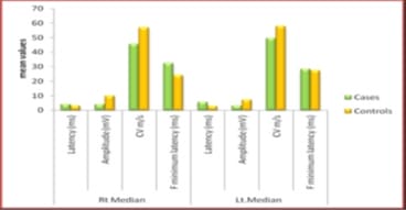
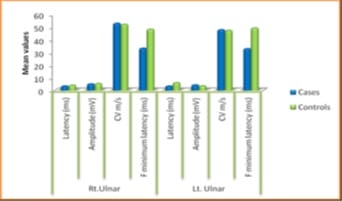
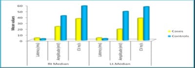
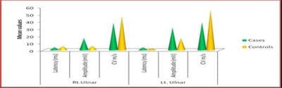
Background: Diabetes Mellitus affects the peripheral nervous system, and in uncontrolled diabetes, symptomatic neuropathy is one of the commonest incapacitating complications. Diabetic neuropathy can be detected at an early stage using Electrodiagnostic tests. Nerve Conduction Study (NCS) is the most sensitive, reliable, non-invasive and objective means of investigations of Diabetic Polyneuropathy. If detected, the proper treatment can be instituted in the early stages with a good outcome. As the peripheral nerve can regenerate, a line of treatment can be planned accordingly. Based on this background, the present study aimed to evaluate the comparative peripheral nerve conduction study of median and ulnar nerves in people with type-2 diabetes with polyneuropathy. Methods: A total of 58 subjects were included in the present study, 29 subjects with Diabetic Neuropathy and 29 age and sex-matched healthy subjects. Motor and Sensory nerve conduction studies were conducted in both upper and lower limbs nerves using bipolar surface electrodes with RMS EMG EP MARK-II machine. Statistical analysis was done using SPSS 20.0. Results: A definite decrease in amplitude and nerve conduction velocities of both the sensory and motor components, axonal and demyelinating type, significantly correlating with higher HbA1c levels. F minimum latencies of Median nerves were increased, possibly more of demyelinating type of polyneuropathy. Conclusion: NCS being a simple, harmless, non-invasive and objective technique along with the easy interpretation of results, can be used routinely to evaluate the status of nerves in patients with Type 2 Diabetes Mellitus to prevent a more disabling state the earliest.

Keywords: Diabetic Polyneuropathy, Nerve Conduction Study, HbA1c

Corresponding Author How to Cite this Article To Browse
Bhagya V, Professor, Department of Physiology, JJM Medical College, Davanagere, Karnataka, India.
Email:
Bhagya V, Brid SV, Comparative Peripheral Nerve Conduction Study of Median and Ulnar nerves in Type-2 Diabetics with Age and Sex Matched Normal Subjects.. Trop J Pathol Microbiol. 2021;7(5):219-225.
Available From
https://pathology.medresearch.in/index.php/jopm/article/view/574

Manuscript Received Review Round 1 Review Round 2 Review Round 3 Accepted
2021-08-30 2021-09-15 2021-09-28 2021-10-15 2021-10-18
Conflict of Interest Funding Ethical Approval Plagiarism X-checker Note
No Nil Yes 6%

© 2021 by Bhagya V, Brid SV and Published by Siddharth Health Research and Social Welfare Society. This is an Open Access article licensed under a Creative Commons Attribution 4.0 International License https://creativecommons.org/licenses/by/4.0/ unported [CC BY 4.0].

Introduction

Diabetes mellitus (DM) is a syndrome of disorders characterized by hyperglycemia caused due to a relative or absolute deficiency of insulin, decreased glucose utilization, increased gluconeogenesis [1]. Diabetes mellitus is a global health problem, affecting more than 170 million individuals and all age groups worldwide. In more developed societies, the prevalence of diabetes mellitus has reached about 6 % [2]. Diabetes is defined as a state of chronic hyperglycemia and is classified into two major types, type 1 diabetes mellitus (T1DM) and type 2 diabetes mellitus (T2DM). Type 1 diabetes mellitus is due to total failure of the pancreas to secrete insulin, autoimmune destruction of insulin-secreting cells in the islets of Langerhans and is also known as Insulin Dependent Diabetes mellitus (IDDM).

Type 2 DM or Non-Insulin Dependent Diabetes mellitus (NIDDM) is a heterogeneous, multifactorial, polygenic disease characterized by insulin secretion defects. It is associated with insulin resistance and qualitative and quantitative insulin deficiency caused by alterations in several gene products [3-4]. The presence of DM predisposes to increased risk of many complications such as cardiovascular diseases, peripheral vascular diseases, stroke, neuropathy, renal failure, retinopathy and blindness. Complications of Diabetes may be classified into macrovascular (coronary heart disease, cerebrovascular disease and peripheral vascular disease) and microvascular (retinopathy, nephropathy and neuropathy). The physiological properties of nerves and muscles are usually modified due to pathophysiological changes occurring from many diseases like diabetes.

Diabetes is fast becoming the epidemic of the 21st century [5-6]. The physiological properties of nerves and muscles are usually modified due to pathophysiological changes resulting from many diseases like diabetes [7]. The number of people with diabetes has risen from 108 million in 1980 to 422 million in 2014. The global prevalence of diabetes among adults over 18 years has been increased from 4.7% in 1980 to 8.5% in 2014. Diabetes prevalence has been rising more rapidly in the middle- and low-income countries [8-10]. A healthy diet, regular physical activity, maintaining average body weight and avoiding tobacco use prevent or delay the onset of type 2 diabetes.

Diabetes can be treated and its consequences avoided or delayed with diet, physical activity, medication and regular screening and treatment for complications[11]. Diabetes Mellitus is one of the diseases that affect the peripheral nervous system, and symptomatic neuropathy in uncontrolled diabetes is one of the commonest long-term incapacitating complications [12]. A widely accepted definition of diabetic peripheral neuropathy is the presence of symptoms and signs of peripheral nerve dysfunction in people with diabetes after exclusion of other causes. Many previous studies have reported a wide range of prevalence of Neuropathy in Diabetics, which estimates from 3% to 32% [13].

Diabetic neuropathy is identified by the presence of peripheral nerve dysfunction in patients with diabetes. The risk of developing peripheral neuropathy is directly proportional to the magnitude and duration of hyperglycemia [14]. Although the precise nature of the injury to the peripheral nerves from hyperglycemia is not known, it is likely related to mechanisms such as polyol accumulation, damage from advanced glycation end products, and oxidative stress. In diabetes, peripheral neuropathy manifests in several forms, including sensory, focal/multifunctional, autonomic neuropathies. Diabetic autonomic neuropathy is a cause of significant morbidity and mortality in patients with diabetes [15].

Nerve problems in Diabetes mellitus can develop at any time, often occurring about ten years after the diagnosis of diabetes. The longer the duration of diabetes and irregular being the treatment, with fluctuating blood sugar levels, the higher the risk of developing diabetic neuropathy. [4]. Electroneurographyis considered the gold standard for measuring peripheral nerve function and has been previously validated for measuring both sensory and motor peripheral nerve function [16-17]. Sensory Neuropathy (SNAP) are better appreciated by nerve conduction studies than conventional vibration tests [18]. Nerve conduction studies (NCS) and electromyography (EMG) can document the characteristics of the neuropathy (e.g., axonal, demyelinating) and the localization (e.g. mononeuropathy versus radiculopathy or distal neuropathy) and, possibly, the severity and even prognosis for morbidity. Multiple consensus panels recommend the inclusion of electrophysiological testing in the evaluation of diabetic neuropathy. These same panels recommend the use of NCS/EMG procedures in clinical research studies.


NCS/EMG establishes diagnosis quite early than other diagnostic procedures because of its sensitivity to detect the slowing of conduction of action potential in a nerve, which is an early indicator of peripheral neuropathy. Neurophysiologic studies supplemented the clinical examination by precisely localizing the lesion and characterizing the conduction abnormalities providing additional information, details and objectivity. They delineate a variety of conditions that may otherwise escape detection [19]. Primarily NCS determination is used compared to EMG to study diabetic neuropathy since it is non-invasive, can easily be reproduced, and is sensitive, which gives a clear recording with minimal disturbances. Early functional impairment of nerve in asymptomatic cases can be studied, i.e. disorder affecting the nerves is insufficient to produce clinical abnormality may be detected as a decrease in Sensory nerve conduction velocity. It is helpful as a diagnostic tool, prognostic and follows up the study of neuropathy [20]. Based on this background, we aimed to evaluate the comparative peripheral nerve conduction study of median and ulnar nerves in people with type-2 diabetes with symptoms of polyneuropathy with age and sex-matched normal subjects.

Materials and Methods

Settings: This study was conducted in the Department of Physiology and Medicine at JJM Medical college and Hospital, Davanagere, Karnataka.

Duration and Type of the Study: This is a case-control study conducted from April 2015-Nov 2019.

Sampling Methods: A total of 29 patients diagnosed with type 2 diabetes mellitus as per American Diabetes Association (ADA) criteria [21] attended to general medicine OPD atJJMMedicalcollege and hospital, Davanagere, Karnataka and also we included29 healthy volunteers (age and sex-matched) as controls.

Sample Size: Total 58 Subjects, 29 Cases and 29 Controls.

Inclusion Criteria: All the subjects aged between 30 to 70 years and patients diagnosed with type 2 Diabetes Mellitus with symptoms of polyneuropathy were included in the present study.

Exclusion Criteria: subjects whoever suffering from Type1 Diabetes Mellitus, Nephropathy, Kidney, Liver and Thyroid diseases, Urinary Tract Infections,

Cardiovascular Diseases, Cerebrovascular and Peripheral Vascular diseases, Active Inflammatory Diseases, Individuals on Thiazolidinediones, Anti- Inflammatory drugs and whoever not willing to participate in these study were excluded.

Data Collection and Procedures: Motor and sensory nerve conduction studies were conducted in both upper limb (Median and Ulnar) nerves using bipolar surface electrodes in 29 Type-2 Diabetics with neuropathy symptoms referred to our neurophysiology lab for NCS. NCS was performed with the RMS EMG EP MARK II machine, and the lab temperature was maintained at 21-23ºC. Latency, amplitude, conduction velocity and F-wave studies were done by placing the active electrode over the motor point and reference electrode on the tendon with stimulation proximally and distally. Both functional and reference electrodes are placed on the nerve in sensory nerve studies with a Ground electrode between stimulating and recording electrode.

Values are taken as reference from normal values of motor and sensory nerve conduction from the survey by Misra and Kalita. Motor nerve conduction studies were carried out by placing an active electrode over the motor point and a reference electrode on the tendon by stimulating proximally and distally. Sensory nerve conduction studies were carried out with both functional and a reference electrode placed on the nerve. The ground electrode is positioned between the stimulating and recording electrode.

Motor nerve conduction studies

Median motor nerve conduction: Recording electrode is placed close to the motor point of abductor pollicis brevis and the reference electrode 3 cm distal at the first metacarpophalangeal joint. A supramaximal stimulation is given at the wrist (3 cm proximal to the distal crease) and elbow (near the volar crease of the brachial pulse)

Ulnar motor nerve conduction: Recording is usually done from Abductor digitiminimi employing surface electrodes with stimulation at wrist and elbow. Ulnar nerve slides back and forth on extension and flexion of the elbow. Therefore a standard position of the elbow with 90º or 135º flexion during stimulation and measurement of distance is recommended. (Harding and Haler, 1983).

Sensory nerve conduction studies


Median sensory nerve conduction: The recording electrode for orthodromic conduction is placed 3 cm proximal to the distal crease, and reference is placed at a distance of 3 cm proximally. For stimulation, ring electrodes are placed at the second or third digits. The cathode is placed at the first interphalangeal joint and anode 3 cm distal.

Ulnar sensory nerve conduction: The orthodromic conduction was done by stimulating the digital nerve by ring electrodes at the interphalangeal joint of the fifth digit.

Ethical Consideration and Permission: All the subjects were included after taking informed consent and approved by Institutional Ethics Committee (IEC).

Statistical analysis: The data were analyzed for the difference in the right and left median, and Ulnar Nerves Latency, Amplitude, CV and F minimum latency between cases and controls was analyzed using unpaired students T-Test. Statistical analysis was performed by using Microsoft Excel Spread Sheets. A p-Value <0.05 was considered statistically significant.

Results

patho_574_01.jpg

Figure 1- Shows the Mean values of Rt. & Lt Median motor nerve conduction.

Figure 1: Shows the right and left median motor nerve conduction in study subjects. There was a reduced amplitude, CV, and increased latency, and minimum latency was observed in cases compared to controls.

Figure 2: Shows the right and left ulnar motor nerve conduction in between cases and controls. A mild increase in motor latency was observed in both right and left ulnar nerve motor conduction in patients with type 2 diabetes mellitus with polyneuropathy compared to healthy controls. There is no change in Amplitude and CV in both right and left ulnar nerve conduction.

patho_574_02.jpg

Figure 2- Shows the Mean values of Rt. &Lt. Ulnar motor nerve conduction.

patho_574_03.jpg

Figure 3: Shows the right and left sensory nerve conduction of median nerves between cases and controls. There was a significantly decreased amplitude, CV, and increased Latency were observed in both right and left sensory nerve conduction of median nerves in patients with type 2 diabetes mellitus with polyneuropathy when compared to healthy controls.

patho_574_04.jpg

Figure 4: Shows the right and left sensory nerve conduction of ulnar nerves between cases and controls. There is a decreased CV and no change in latency in both right and left sensory nerve conduction of ulnar nerves in both subjects.

Discussion

In the present study, there is a definite decrease in amplitude and nerve conduction velocities of both the sensory and motor components, axonal and demyelinating type significantly correlating with higher HBA1c levels and F minimum latencies of Median nerves were increased possibly more of demyelinating type of polyneuropathy. Conduction abnormalities develop diffusely along the entire nerve length but more in distal than the proximal segment [22].


The NCV is gradually diminished by Diabetic Neuropathy, with an estimate of a loss of approximately 0.5m/s/year. A simple rule is that a 1% decrease in HbA1c improves the conduction velocity of about 1.3m/s (Arezzo,1997). Hyperglycemia correction in patients results in improvement of symptoms and nerve conduction abnormalities (Judzewitsch et al.,1983). Elizabeth et al. studied the neuropathy assessment in diabetic patients by analyzing the conduction velocity within the motor and sensory fibres. They concluded that the longer the duration of diabetes, certainty of changes in the motor and sensory responses of the individual is more. These studies were consistent with a previous similar survey done by Muthuselvi K et al. where the amplitude values of both sensory and motor nerve conduction studies were found to be significantly reduced in diabetic individuals than the non-diabetic control group with a significant' p' value. It was also seen that there was an inverse relationship on correlating the blood sugar values, with increasing blood sugar values, sensory nerve conduction velocities are correspondingly decreasing, and motor nerve conduction velocities reducing. However, there are some fluctuations [23-24]. But, in our study, the correlation of cases with blood sugars was not considered, although all the patients in our study mostly presented with blood sugar and higher HbA1c levels. These studies findings were also consistent with the previous study done by Olney, Richard K. The present study was comparable to the study done by Verma et al., who showed that amplitude and conduction velocity range is significantly decreased in diabetic patients in comparison to normal healthy volunteers, [25]. and the most affected nerve in the upper limb is the Median nerve, and the lower limb is a sural nerve which is also comparable with a study done by Zahed Ali et.al.[26]. The values of the F-wave parameters, which were recorded from the median and the ulnar nerves, were following those reported [27-29]. A study done by Xuan Kong et al. demonstrated that NCS did use computer-based electrodiagnostic equipment was a better tool for the diagnosis of Diabetic Polyneuropathy [30].

Conclusion

From our study, we conclude that increased distal motor latencies, the reduced amplitude of CMAP and motor conduction velocities in the Median nerve indicate involvement more of the median nerve in the cases referred compared to the age and sex-matched normal subjects.

What new this study adds to existing knowledge

Neuropathic symptoms may take years to appear, but even before, diabetic neuropathy can be diagnosed by NCS, and further damage to the nerves can be curtailed. The NCS, however, is more powerful in detecting neuropathy which shows that NCS helps see subclinical neuropathies also. Diabetic neuropathy is curable, and hence if detected, the proper treatment can be instituted in the early stages, which can give rise to a good outcome. The drawbacks of the nerve conduction study are that: only the status of distal nerves can be evaluated, and only the velocities in the large fibre can be measured. These drawbacks do not pose any serious problem since, in NIDDM, distal nerves with the most significant myelinated 'A' fibres are chiefly affected.

Authors Contribution

Dr Bhagya. V, Data Collection, Literature Review, Manuscript Preparation, Manuscript Editing and Final Approval. Dr Brid SV, Patients Collection, Data Analysis and Statistical Analysis.

Reference

01. Stumvoll M, Goldstein BJ, van Haeften TW. Type 2 diabetes: principles of pathogenesis and therapy. Lancet. 2005 Apr 9-15;365:1333-46. [Crossref][PubMed][Google Scholar]

02. King H, Aubert RE, Herman WH. Global burden of Diabetes, 1995-2025: prevalence, numerical estimates, and projections. Diabetes Care. 1998 Sep;21(9):1414-3. [Crossref][PubMed][Google Scholar]

03. World Health Organization. Global report on diabetes. Geneva: World Health Organization. 2016. [Crossref][PubMed][Google Scholar]

04. Fowler, Michael J. "Microvascular and macrovascular complications of diabetes. Clinical diabetes. 2008: 77-82. [Crossref][PubMed][Google Scholar]

05. Erratum. American Diabetes Association, Classification and diagnosis of diabetes, In Standards of Medical Care in Diabetes-2016. Diabetes Care. 2016;39(1):S13-22. [Crossref][PubMed][Google Scholar]


06. Frier BM. Diabetes mellitus, 20 th ed, In: Boon A College R, Davidson's Principles and practice of medicine. Churchill Living stone. 2006:805-48. [Crossref][PubMed][Google Scholar]

07. Alzahrani. "Determination of Serum Adiponectin Levels in Type 2 Diabetes Patients of the Saudi Population in the Al-Jouf Region". Romanian Journal of Diabetes Nutrition and Metabolic Diseases. 2020: 257-262. [Crossref][PubMed][Google Scholar]

08. Salam RS, Laikangbam S, Tina D, Wahengbam DD, Bishnupriya P, Purnima MD. Adiponectin and Body Mass Index in Type 2 Diabetes Mellitus. International Journal of Contemporary Medical Research. 2020;04:1-4. [Crossref][PubMed][Google Scholar]

09. Nagarathna R, Bali P, Anand A, Srivastava V, Patil S, Sharma G, et al. Prevalence of Diabetes and Its Determinants in the Young Adults Indian Population-Call for Yoga Intervention. Front Endocrinol. 2020 Dec 11;11:507064. [Crossref][PubMed][Google Scholar]

10. Su N, Zhao N, Wang G, Wang L, Zhang Y, Li R, et al. The Effects of Adiponectin and Adiponectin Receptor 1 Levels on Macrovascular Complications Among Patients with Type 2 Diabetes Mellitus. Cell Physiol Biochem. 2019;52(2):225-231. [Crossref][PubMed][Google Scholar]

11. Krarup C. Nerve conduction studies in selected peripheral nerve disorders. Curr Opin Neurol. 200215(5):579-93. [Crossref][PubMed][Google Scholar]

12. WHO. "Global Report on Diabetes. World Health Organization. 2016;1:1-6. [Crossref][PubMed][Google Scholar]

13. Ziegler D, Gries FA, Spüler M, Lessmann F. The epidemiology of diabetic neuropathy, Dia CAN Multicenter Study Group. Diabet Med. 1993;10: 2:82-86. [Crossref][PubMed][Google Scholar]

14. Muthuselvi K, Shanthi M, Ethiya N. Comparison of Nerve Conduction Studies in Geriatric Normal and Diabetic Subjects. . 2015;4(4):2013–5. [Crossref][PubMed][Google Scholar]

15. Magladery JW, Mcdougal DB. Electrophysiological studies of nerve and reflex activity in normal man I, Identification of certain reflexes in the electromyogram and the conduction velocity of peripheral nerve fibers. Bull Johns Hopkins Hosp. 1950;86(5):265-90. [Crossref][PubMed][Google Scholar]

16. Lambert EH. "Diagnostic value of electrical stimulation of motor nerves". Electroencephalogr Clin Neurophysiol. 1962;22: 9-16. [Crossref][PubMed][Google Scholar]

17. Gilliatt RW, Sears TA. Sensory nerve action potentials in patients with peripheral nerve lesions. J Neurol Neurosurg Psychiatry. 1958;21(2):109-18. [Crossref][PubMed][Google Scholar]

18. Kakrani AL, Gokhale VS, Vohra KV, Chaudhary N. Clinical and nerve conduction study correlation in patients of diabetic neuropathy. J Assoc Physicians India. 2014;62(1):24-7. [Crossref][PubMed][Google Scholar]

19. Kimura J. Long and short of nerve conduction measures: reproducibility for sequential assessments. J Neurol Neurosurg Psychiatry. 2001;71(4):427-30. [Crossref][PubMed][Google Scholar]

20. WagmanIh, Lesse H. Maximum conduction velocities of motor fibers of ulnar nerve in human subjects of various ages and sizes. J Neurophysiol. 1952;15(3):235-44. [Crossref][PubMed][Google Scholar]

21. American Diabetes Association. "Standards of medical care in diabetes". Diabetes Care. 2017;40 (1): S1–35. [Crossref][PubMed][Google Scholar]

22. Misra UK, Kalita J. Clinical neurophysiology: nerve conduction, electromyography, evoked potentials. Elsevier Health Sciences. 2019:14-19. [Crossref][PubMed][Google Scholar]

23. Verma, Anita, Swati M, Prashant K. "Sensory nerve conduction studies in non-insulin dependent diabetes mellitus (NIDDM) patients without symptoms of peripheral neuropathy and healthy volunteers: a comparative study". Int J Basic ApplPhysiol. 2015: 158-62. [Crossref][PubMed][Google Scholar]

24. Ali, Zahed. "Role of electrodiagnostic tests in early detection of diabetic neuropathy". Bangladesh Journal of Neuroscience. 2008: 34-44. [Crossref][PubMed][Google Scholar]

25. Kong X, Lesser EA, Potts FA, Gozani SN. Utilization of nerve conduction studies for the diagnosis of polyneuropathy in patients with diabetes: a retrospective analysis of a large patient series. J Diabetes Sci Technol. 2008;2(2):268-74. [Crossref][PubMed][Google Scholar]


26. Kimura, Jun. "The blink reflex" Electrodiagnosis in disease of nerve and muscle. Principles and practice. 2001;1-9. [Crossref][PubMed][Google Scholar]

27. Hamdan, Farqad B. "Nerve conduction studies in healthy Iraqis: Normative data". Iraqi J Med Sci. 2009: 75-92. [Crossref][PubMed][Google Scholar]

28. Buschbacher RM. Median nerve F-wave latencies recorded from the abductor pollicis brevis. Am J Phys Med Rehabil. 1999 Nov-Dec;78(6):S32-7. [Crossref][PubMed][Google Scholar]

29. Budak F, Efendi H, Apaydin R, Bilen N, Komsuoglu S. The F response parameters in Behçet's disease. Electromyogr Clin Neurophysiol. 2000 Jan-Feb;40(1):45-8. [Crossref][PubMed][Google Scholar]

30. Asad A, Hameed MA, Khan UA, Butt MU, Ahmed N, Nadeem A. Comparison of nerve conduction studies with diabetic neuropathy symptom score and diabetic neuropathy examination score in type-2 diabetics for detection of sensorimotor polyneuropathy. J Pak Med Assoc. 2009;59(9):594-8. [Crossref][PubMed][Google Scholar]