E-ISSN:2456-1487
P-ISSN:2456-9887
RNI:MPENG/2017/70771

Case Report

Cardiac Death

Tropical Journal of Pathology and Microbiology

2021 Volume 7 Number 6 November December
Publisherwww.medresearch.in

A Rare Case of Sudden Cardiac Death

Mulkalwar A.1*, Jadhav T.2, Savla J.3, Vaideeswar P.4
DOI: https://doi.org/10.17511/jopm.2021.i06.10

1* Alhad Mulkalwar, Intern, Department of Pathology, Seth Gordhandas Sunderdas Medical College and King Edward Memorial Hospital, Mumbai, Maharashtra, India.

2 T Jadhav, Intern, Department of Pathology, Seth Gordhandas Sunderdas Medical College and King Edward Memorial Hospital, Mumbai, Maharashtra, India.

3 J Savla, Intern, Department of Pathology, Seth Gordhandas Sunderdas Medical College and King Edward Memorial Hospital, Mumbai, Maharashtra, India.

4 P Vaideeswar, Professor (Additional), Department of Pathology (Cardiovascular & Thoracic Division), Seth Gordhandas Sunderdas Medical College and King Edward Memorial Hospital, Mumbai, Maharashtra, India.

While sudden deaths fall within the purview of forensic experts, they are often found to be caused by cardiovascular pathologies (to the extent of 85%), which may be evident or occult [1]. A sudden cardiac death is an unexpected death owing to a cardiac cause which generally occurs within an hour of onset of symptoms [2, 3]. Most sudden cardiac deaths are due to coronary artery diseases [4, 5]. The remainder is caused by a heterogeneous group of non-ischemic disorders, most of which result in structural cardiac abnormalities [6]. Herein we describe, at autopsy, a case of a sudden cardiac death secondary to an arrhythmogenic cardiomyopathy.

Keywords: Arrhythmogenic cardiomyopathy, Papillary fibroelastoma, Coronary ostitis

Corresponding Author How to Cite this Article To Browse
Alhad Mulkalwar, Intern, Department of Pathology, Seth Gordhandas Sunderdas Medical College and King Edward Memorial Hospital, Mumbai, Maharashtra, India.
Email:
Alhad Mulkalwar, T Jadhav, J Savla, P Vaideeswar, A Rare Case of Sudden Cardiac Death. Trop J Pathol Microbiol. 2021;7(6):329-331.
Available From
https://pathology.medresearch.in/index.php/jopm/article/view/551

Manuscript Received Review Round 1 Review Round 2 Review Round 3 Accepted
2021-11-10 2021-11-12 2021-11-19 2021-11-26 2021-12-03
Conflict of Interest Funding Ethical Approval Plagiarism X-checker Note
Nil Nil Yes 16%

© 2021by Alhad Mulkalwar, T Jadhav, J Savla, P Vaideeswarand Published by Siddharth Health Research and Social Welfare Society. This is an Open Access article licensed under a Creative Commons Attribution 4.0 International License https://creativecommons.org/licenses/by/4.0/ unported [CC BY 4.0].

Case description

On 31st March 2019, a 40-year-old female fell unconscious following an alleged history of chest pain. She was rushed to the casualty of King Edward Memorial Hospital, Mumbai but was declared dead on arrival. Autopsy revealed mildly enlarged ventricles of the heart. There was mild, diffuse adipose tissue infiltration all over, especially over the posterior wall of the left ventricle. A 0.3 cm x 0.3 cm papillary fibroelastoma at the rim of the left coronary ostium was observed [Fig-1]. Histopathological examination of the right ventricle revealed increased epicardial adipose tissue infiltrating the superficial myocardium but without associated fibrosis. Fibers of the myocardium and the endocardium of the right ventricle were completely normal. Myocardium of the left ventricular wall, apex and septum showed mild hypertrophy of fibers with interstitial and perivascular adipose tissue and interstitial fibrosis. These findings were suggestive of a left ventricular dominant arrhythmogenic cardiomyopathy. Histopathology also revealed a focus of thrombus throwing into papillae on the luminal aspect of the origin of the left main coronary artery with extensive adventitial fibrosis and prominent lymphoid aggregate [Fig-2]. The cause of death was concluded to be ostitis of the left main coronary artery with mural thrombosis and associated with arrhythmogenic cardiomyopathy, an extremely rare incidence.

patho_551_01.JPG
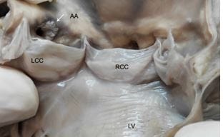
Fig-1: Interior of the heart showing a 0.3cm x 0.3cm papillary fibroelastoma at the rim of the left coronary ostium.

patho_551_02.JPG
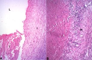
Fig-2: Sections from the left coronary artery - A. Lumen L with thrombus T. Note extreme intimal I fibrocellular thickening (H&E x 250); B. There is marked attenuation of the media M due to transmural destruction by inflammatory cells, lymphocytes and histiocytes and a vague collection of epithelioid cells. (H&E x 250).

Reference

01. Vaideeswar P, Tyagi S, Singaravel S, Marathe SP. Sudden cardiac deaths: Role of non ischemic myocardial disorders—Part II. Indian J Pathol Microbiol 2021;64: 231-7. [Crossref][PubMed][Google Scholar]

02. Engelstein ED, Zipes DP. Sudden cardiac death. In: Alexander RW, Schlant RC, Fuster V, eds. The Heart, Arteries and Veins. New York, NY: McGraw-Hill; 1998:1081–1112 [Crossref][PubMed][Google Scholar]

03. Myerburg RJ, Castellanos A. Cardiac arrest and sudden death. In: Braunwald E, ed. Heart Disease: A Textbook of Cardiovascular Medicine. Philadelphia, Pa: WB Saunders; 1997:742–779 [Crossref][PubMed][Google Scholar]

04. Goldstein S, Landis JR, Leighton R, Ritter G, Vasu CM, Lantis A, Serokman R. Characteristics of the resuscitated out-of-hospital cardiac arrest victim with coronary heart disease. Circulation. 1981; 64:977-984. [Crossref][PubMed][Google Scholar]

05. Goldstein S, Medendorp SV, Landis JR, Wolfe RA, Leighton R, Ritter G, Vasu CM, Acheson A. Analysis of cardiac symptoms preceding cardiac arrest. Am J Cardiol. 1986; 58:1195-1198. [Crossref][PubMed][Google Scholar]

06. Vaideeswar P, Tyagi S, Singaravel S, Marathe SP. Sudden cardiac deaths:


Role of nonischemic myocardial disorders—Part I. Indian J Pathol Microbiol 2021;64: 14-21. [Crossref][PubMed][Google Scholar]