E-ISSN:2456-1487
P-ISSN:2456-9887
RNI:MPENG/2017/70771

Research Article

Cytology

Tropical Journal of Pathology and Microbiology

2021 Volume 7 Number 3 May-June
Publisherwww.medresearch.in

Role of Fine Needle Aspiration Cytology in Effective Management of Patients with Granulomatous Inflammation in Neoplastic and Non-Neoplastic Lesions

Patel D.1*, Shukla A.2, Jivani T.3
DOI: https://doi.org/10.17511/jopm.2021.i03.06

1* Drashti Patel, Senior Resident, Department of Pathology, Surat Municipal Institute of Medical Education and Research, Surat, Gujarat, India.

2 Ashwini Shukla, Professor (Additional), Department of Pathology, Surat Municipal Institute of Medical Education and Research, Surat, Gujrat, India.

3 Toral Jivani, Assistant Professor, Department of Pathology, Surat Municipal Institute of Medical Education and Research, Surat, India.

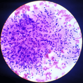
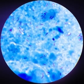
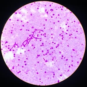
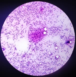
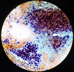
Background: Granulomatous inflammation is a common diagnosis given in fine-needle aspiration cytology (FNAC) and it is a type of chronic inflammation in which tissue reaction appears following cell injury. Aims: To assess the usefulness of the cytological study in the diagnosis of granulomatous lesions. Materials and Methods: An observational study is done based on cytomorphological evaluation of 156 cases of granulomatous inflammation diagnosed at the Department of Pathology, in Surat Municipal Institute of Medical Education & Research over 18 months from February 2018 to July 2019. Results: Out of the total of 156 cases of granulomatous inflammation, tuberculosis was the most common finding in 111 cases (71.15%), followed by fungal infection (05 cases). The male to female ratio was 1.0 to 1.4. The most commonly affected age group was 21 to 30 years. Conclusions: All the cases of granulomatous inflammations are analysed for definitive diagnosis to provide early and proper treatment of underlying diseases, malignancies or lymphoma.

Keywords: Fine needle aspiration cytology, Granuloma, PAS stain, Tuberculosis, ZN Stain

Corresponding Author How to Cite this Article To Browse
Drashti Patel, Senior Resident, Department of Pathology, Surat Municipal Institute of Medical Education and Research, Surat, Gujarat, India.
Email:
Patel D, Shukla A, Jivani T. Role of Fine Needle Aspiration Cytology in Effective Management of Patients with Granulomatous Inflammation in Neoplastic and Non-Neoplastic Lesions. Trop J Pathol Microbiol. 2021;7(3):128-134.
Available From
https://pathology.medresearch.in/index.php/jopm/article/view/530

Manuscript Received Review Round 1 Review Round 2 Review Round 3 Accepted
2021-05-20 2021-05-30 2021-06-03 2021-06-06 2021-06-09
Conflict of Interest Funding Ethical Approval Plagiarism X-checker Note
No Nil Yes 5%

© 2021 by Drashti Patel, Ashwini Shukla, Toral Jivani and Published by Siddharth Health Research and Social Welfare Society. This is an Open Access article licensed under a Creative Commons Attribution 4.0 International License https://creativecommons.org/licenses/by/4.0/ unported [CC BY 4.0].

Introduction

Granulomatous inflammation is a distinctive form of chronic inflammation produced in response to various infectious, autoimmune, toxic, allergic, and neoplastic conditions. [1]. Infectious causes most notably presenting with granuloma are tuberculosis and fungal infections. It is characterized by the formation of distinct granulomas composed of aggregates of epithelioid histiocytes, with a peripheral cuff of lymphocytes and plasma cells, and occasionally a necrotic centre. The clinical value of FNAC is not only in neoplastic conditions but also important in the diagnosis of non-neoplastic conditions. Tuberculosis is the foremost infectious cause of mortality worldwide with 2-3 million deaths being reported each year and accounts for over a quarter of all avoidable deaths globally. [2].

The diagnosis of tuberculosis remains a challenge. [3]. History, clinical examination and many diagnostic tests are helpful. Every test has its sensitivity, specificity and limitations. The commonly performed tests include examination of sputum for Acid Fast Bacilli, Cultures for Mycobacterium tuberculosis, Fine Needle Aspiration Cytology (FNAC), Biopsy, and PCR. [4]. FNAC plays an important role in the diagnosis of tuberculous inflammation and prevents unnecessary surgery. [5]. Cytodiagnosis of tuberculosis depends on the demonstration of epithelioid cell granuloma and caseous necrosis with or without Langhans type of giant cells. Bacteriological confirmation is required by Ziehl Neelsen (ZN) stain/culture for acid-fast bacilli (AFB). Treatment of tuberculosis can be straightway started after FNAC diagnosis by correlation with clinical findings and other investigations.

Granulomatous inflammation is not diagnostic of TB. Others causes must be ruled out before giving anti-tuberculous treatment (AKT). Good clinical history, a close cytological examination and a clinicopathological correlation are essential in making a final diagnosis. Some solid tumours and malignant lymphomas which are associated with epithelioid granulomas are widely observed in FNAC cytosmear and they are underdiagnosed sometimes. Granulomatous inflammation found in lymph nodes draining carcinomas is a recognised phenomenon. [6,7]. These cases with granulomas can lead to diagnostic difficulties. So, the cytological differential diagnosis of a granulomatous lesion should include malignant neoplasms also.

Materials and methods

The present study was carried at the department of Pathology at tertiary care hospital over 18 months from February 2018 to July 2019.

  • Selection of cases:
    • Inclusion criteria:
      • Cases diagnosed as granulomatous inflammation on FNAC
    • Exclusion criteria:
      • Acellular smears/smears with crushed morphology or poorly stained slides will be excluded.
      • Previously diagnosed cases and cases already taking AKT.

Patients fulfilling inclusion criteria will be selected from fine needle aspiration cytology specimens after written consent. A routine stain like Hematoxylin and eosin (H&E), Papanicolaou (PAP) and Giemsa stain will be done. Two extra unstained slides will be smeared from aspiration material. One slide will be stained by Ziehl Nelson's (ZN) stain. The second slide will be stained with Periodic Acid Schiff stain (PAS).

Results

Total 156 patients with granulomatous inflammation diagnosed on FNAC were taken. Granulomas were described as comprising of pale staining epithelioid cells which were round to oval to spindle against an eosinophilic background.

Table-1: Sex distribution of cases

Sex Number of cases % of cases
Female 92 58.97%
Male 64 41.03%
Grand Total 156 100.00%

As shown in Table-1, a slight female preponderance was observed. The male to female ratio was 1.0 to 1.4.

In this study the age of cases ranged from 3 months to 77 years.

As per Table-2, the maximum incidence was in the age group of 21 to 30 years (41 cases).

Table-2: Age wise distribution of cases

Age group Number of cases % of cases
0-1 05 3.21%
1-10 09 5.77%
11-20 27 17.31%

21-30 41 26.28%
31-40 25 16.03%
41-50 19 12.18%
51-60 21 13.46%
61-70 03 1.92%
71-80 06 3.85%
Total 156 100%

In the present study of 156 cases,35 cases had a history of smoking, 4 cases had a history of tobacco and 3 cases had a history of both tobacco and smoking. While 114 cases are devoid of any habits. Out of the total of 38 cases with smoking, 34(89.47%) cases were of tuberculosis, 1 case of fungal infection, 1 case of SCC, 1 case of metastatic SCC and 1 case was labelled as granulomatous inflammation.

Among 156 cases, single swelling was present in 124 cases and multiple swellings were present in 32 cases. Out of which cervical was the commonest site. On FNAC, the nature of aspirate was blood mixed in 104 cases followed by aspiration of pus and cheesy material in 35 and 17 cases respectively.

Table-3: Cytomorphological categorization of cause of granulomatous inflammation

Cytological Diagnosis Number of cases % of cases
HL 01 0.64%
Leprosy 01 0.64%
Micro Filaria 01 0.64%
NHL 01 0.64%
SCC 02 1.28%
De Quervain’s thyroiditis 01 0.64%
Granulomatous Inflammation 29 18.59%
Tuberculosis 111 71.15%
Granulomatous sialadenitis 01 0.64%
Metastatic Carcinoma 02 1.28%
Fungal Infection 05 3.21%
Sarcoidosis 01 0.64%
Total 156 100.00%

Table-3: dictates Cytomorphological diagnosis in the present study of 156 cases of granulomatous inflammation on FNAC, tuberculosis was the most common finding in 111 cases (71.15%).

Table-4: Result on ZN stain

ZN Stain Number of cases % of cases
Negative 45 28.85%
Positive 111 71.15%
Total 156 100.00%

As shown in Table-4, the ZN stain for acid-fast bacilli was positive in 111 cases and negative in 45 cases. Granulomatous inflammation with positive ZN stain was diagnosed as tuberculosis. PAS stain for fungus was positive in 5 cases.

Out of a total of 156 cases, caseation present in 102 cases and absent in 54 cases. Total 102 cases with caseation, 99 cases were of tuberculosis and 1 case of fungal infection. In the remaining 2 cases of granulomatous inflammation, the cause of caseation was not detected. There was an association between AFB positivity and caseation necrosis. We have found 99 cases (89.18%) with caseation out of a total of 111 AFB positive cases, while only 3 cases were found without caseation. Multinucleated giant cells were present in the smear of 51 (32.69%); cases. (Table- 5)

Table-5: Correlation of caseation with ZN stain

  Caseation
ZN stain Absent Present Total
Negative 42 3 45
Positive 12 99 111
Total 54 102 156

Discussion

The granulomatous lesion is seen in a large number of diseases. It is seen in various types of infections, foreign body reactions, aberrant immune reactions and secondary responses in lymph nodes draining carcinoma. [8]. It is also seen in some cases of lymphomas. [9]. Granulomas are most commonly found in routine practice which is characterized by the formation of distinct granulomas composed of aggregates of epithelioid histiocytes, with a peripheral cuff of lymphocytes and plasma cells, and occasionally a necrotic centre. To treat these lesions, demonstration of the etiological agent for definitive diagnosis is essential, which will help inpatient management and outcome. [10].

In developing countries like India, tuberculosis is the most common cause of granulomatous inflammation. Early diagnosis and effective treatment are important for TB control. The diagnosis of TB is confirmed by isolating M. tuberculosis or by finding specific DNA sequences in aspirates. Fine needle aspiration cytology is an inexpensive, less invasive procedure for early diagnosis of such tuberculosis and timely initiation of a specific therapy. [11]. FNAC plays an important role in the diagnosis of tuberculous inflammation and prevents unnecessary surgery.


Samples can also be used for microbiological and biochemical analysis in addition to cytological preparations. Cytodiagnosis of tuberculosis depends on the demonstration of epithelioid cell granuloma and caseous necrosis with or without Langhans type of giant cells. Bacteriological confirmation is required by Ziehl Neelsen (ZN) stain/culture for acid-fast bacilli (AFB). Treatment of tuberculosis can be straightway /started after FNAC diagnosis by correlation with clinical findings and other investigations.

Most of the patients were in the age group of 21 to 30 comprising 41 cases (26.28%). The least affected age groups were between 61 to 70 years, comprising 3 cases (1.92 %). Ergete and Bekele. [12]. observed a similar finding in their study. Similar results were found in the study of Permi HS et al and Pawale JS et al. [13,14]. An individual is energetically most active during this period and therefore the chances of exposure are also more. The present study found that females were more commonly affected than males (male to female ratio was 1:1.4).

The higher incidence of disease among females may be due to the low immunity of Indian females, particularly those belonging to low socio-economic strata and those in the reproductive age group. Paliwal et al and Khajuria et al also noted the female preponderance in their studies. [15,16]. The commonest groups of swellings aspirated in the present study were the cervical group. This study was also consistent with other studies in terms of cervical involvement (69.96%) as the most common anatomic site of granulomatous inflammation. [17].

patho_530_01.png

Figure-1: Well-formed epithelioid cell granuloma in the background of caseation in tuberculosis (H& E stain-40x)

The most common cause of granuloma in this study was tuberculosis (111 cases) (Figure-1&2), which is in concordance with findings of another study of Permi HS et al and Pawale JS et al. [13,14]. However, the second most common infectious cause was fungal infection (Figure-3) in this study.

Figure-2: Acid fast bacilli in background of caseous necrosis (ZN stain-100x)

patho_530_02.jpg

Figure-3: Fungal septate hyphae (PAS stain-40x)

patho_530_03.jpg

A similar finding was observed in the study of


Adhikari RC et al and Mudassar et al. [18,19]. Other infectious causes of granulomatous inflammation found in this study were leprosy, granulomatous sialadenitis, microfilaria and De Quervain’s thyroiditis. [20-23] Non-infectious causes of granuloma found in the present study are Hodgkin lymphoma, non-Hodgkin lymphoma (Figure-4), squamous cell carcinoma and metastatic carcinoma (Figure-5). Sehgal et al observed in their study that granulomas can be seen in draining lymph nodes of patients with malignancy and in both Hodgkin’s disease and non-Hodgkin’s lymphoma. [24]. Koo et al also described the formation of granuloma in lymph nodes containing metastatic carcinoma. [17].

Figure-4: Granuloma in case of non-Hodgkin lymphoma (H&E stain-10x)

patho_530_04.jpg

Figure-5: Granuloma along with malignant cells in lymph node draining metastatic carcinoma (PAP stain-40x)

patho_530_05.jpg

The clinical features of peripheral lymphadenopathy caused by tuberculosis and malignancies are similar. In both conditions, patients have features like painless swelling, weight loss and decreased appetite. Sometimes, cytological findings of granulomas in carcinoma and lymphoma obscure primary malignancy and may favour the underdiagnosis of such cases as infectious etiologies. For appropriate and timely management of these cases, proper identification of the cause of granulomatous inflammation is mandatory. Many mechanisms have been described for the formation of granulomas in malignancies. Antigens, most likely derived from tumour cells, could elicit a hypersensitivity reaction mediated by T-helper cells and thereby stimulate the activation of monocytes to form epithelioid histiocytes. [25].

The insignificant number of cases (21.15%), the cause of granuloma was not known in this study, this finding is similar to the study of Adhikari RC et al. [18]. In the present study of 156 cases of granulomatous inflammation, 111 cases showed positivity for acid-fast bacilli. This finding is agreed with Krishnaswamy H et al. [21]. Whereas it is 44% in the study of Mudassar et al, 45.8% in the study of Das et al and 59.4% in the study of Bezabih et al. [19,27,28]. AFB positivity was notably and more commonly found in granulomatous inflammation with caseous necrosis. 99 out of 111 AFB positive cases associated with caseation necrosis (89.18%), in the current study. This finding is consistently seen in the study of Bezabih et al and Mudassar et al. [28,19].

Conclusion

This study concludes that even after highly effective anti-tubercular treatment, tuberculosis remains the leading cause of granulomatous lesions. FNAC is still a good diagnostic tool in the evaluation of granulomatous lesions. Patients who are not responding to empirical AKT should be considered for other causes of granulomatous inflammation other than TB, and proper workup should be done. This study confirmed the diagnosis of Hodgkin’s lymphoma and few cases of malignancies with the granulomatous reaction that almost masked the malignant component. This reveals out the value of a detailed cytological study in all granulomatous reactions, because of highlighting the malignancy if present. There is a subtle but definite association between malignancy and benign granulomatous inflammation.


There are multiple etiological factors responsible for the co-existence of the two pathologies consist of immunological response to tumour antigen. Scrutiny of such lesions with granuloma is mandatory to avoid underdiagnosis of metastatic disease.

Author contribution

Dr. Drashti Patel: Study design, data analysis, manuscript preparation. Dr. Ashwini Shukla: Concept, data analysis. Dr. Toral Jivani: Literature search

What does this study add to existing knowledge?

This study will provide basic ideas about different causes of granuloma including benign and malignant, by which proper workup of causative agent will be done and underdiagnosis of malignant causes can be prevented.

Reference

01. Shah KK, Pritt BS, Alexander MP. Histopathologic review of granulomatous inflammation. J Clin Tuberc Other Mycobact Dis. 2017 Feb 10;7;1-12. doi: 10.1016/j.jctube. 2017.02.001 [Crossref][PubMed][Google Scholar]

02. World Health Organization (WHO). “Tuberculosis Fact Global and Regional Incidence”. WHO. 1992;Sheet No. 104-2006 [Crossref][PubMed][Google Scholar]

03. Singh S, Dey B, Sachdeva KS, Kabra SK, Chopra KK, Chaudhary VK, Sharma P, Katoch VM. Challenges in tuberculosis diagnosis and management- recommendations of the expert panel. J Lab Physicians. 2015 Jan-Jun;7(1)1-3. doi: 10.4103/0974-2727.154778 [Crossref][PubMed][Google Scholar]

04. Mittal P, Handa U, Mohan H, Gupta V. Comparative evaluation of fine needle aspiration cytology, culture, and PCR in diagnosis of tuberculous lymphadenitis. Diagn Cytopathol. 2011 Nov;39(11)822-6. doi: 10.1002/dc.21472 [Crossref][PubMed][Google Scholar]

05. Nur, Tamanna E, Afroz Shirin, and Moni Mohan Saha. "Diagnostic Accuracy of Fine Needle Aspiration Cytology in Diagnosis of Tuberculous Lymphadenitis. " J Enam Medi Col. 2019;9(1)30-33. [Crossref][PubMed][Google Scholar]

06. Gregorie hb, othersen hb, moore mp. The significance of sarcoid-like lesions in association with malignant neoplasms. Am J Surg. 1962 Oct;104;577-86. doi: 10.1016/0002-9610(62)90399-9. PMID: 13901642 [Crossref][PubMed][Google Scholar]

07. Oberman HA. Invasive carcinoma of the breast with granulomatous response. Am J Clin Pathol. 1987 Dec;88(6)718-21. doi: 10.1093/ajcp/88.6.718 [Crossref][PubMed][Google Scholar]

08. Gorton g, linell f. Malignant tumours and sarcoid reactions in regional lymph nodes. Acta radiol. 1957 May;47(5)381-92. doi: 10.3109/00016925709170911 [Crossref][PubMed][Google Scholar]

09. Hollingsworth HC, Longo DL, Jaffe ES. Small noncleaved cell lymphoma associated with florid epithelioid granulomatous response- A clinicopathologic study of seven patients. Am J Surg Pathol. 1993 Jan;17(1)51-9. doi: 10.1097/00000478-199301000-00006 [Crossref][PubMed][Google Scholar]

10. National tuberculosis centre. Annual report of national tuberculosis control progrmme. Ministry of Health, Kathmandu, Nepal. 2015/2016. [Crossref][PubMed][Google Scholar]

11. Narayana Reddy RA, Narayana SM, Shariff S. Role of fine-needle aspiration cytology and fluid cytology in extra-pulmonary tuberculosis. Diagn Cytopathol. 2013 May;41(5)392-8. doi: 10.1002/dc.22827 [Crossref][PubMed][Google Scholar]

12. Ergete, Wondwossen, and Alemayehu Bekele. "Acid fast bacilli in aspiration smears from tuberculous patients. " Ethiopian Journal of Health Development. 2000;14(1)99-104. [Crossref][PubMed][Google Scholar]

13. Permi H S, Padma S K, Teerthanath S, Mathias M, Kumar S, HL K P. A histopathological study of granulomatous inflammation. Journal of Health and Allied Sciences NU. 2012;2(01)15-19. [Crossref][PubMed][Google Scholar]

14. Pawale, Jayashree, Rekha Puranik, M H Kulkarni. "A histopathological study of granulomatous inflammations with an attempt to find the aetiology. " J Clin Diagn Res. 2011;5(2)301-6. [Crossref][PubMed][Google Scholar]


15. Nidhi P, Sapna T, Shalini M, Kumud G. FNAC in tuberculous lymphadenitis- experience from a tertiary level referral centre. Indian J Tuberc. 2011 Jul;58(3)102-7. [Crossref][PubMed][Google Scholar]

16. Khajuria, R, Goswami K C, Singh K, Dubey V K. Pattern of lymphadenopathy on fine needle aspiration cytology in Jammu. JK Sci. 2006;8(3)157-159. [Crossref][PubMed][Google Scholar]

17. Koo V, Lioe TF, Spence RA. Fine needle aspiration cytology (FNAC) in the diagnosis of granulomatous lymphadenitis. Ulster Med J. 2006 Jan;75(1)59-64. [Crossref][PubMed][Google Scholar]

18. Adhikari R C, K B Shrestha, G Sayami. "Granulomatous inflammation: a histopathological study". Journal of Pathology of Nepal. 2013;3(6)464-468. [Crossref][PubMed][Google Scholar]

19. Majeed MM, Bukhari MH. Evaluation for granulomatous inflammation on fine needle aspiration cytology using special stains. Patholog Res Int. 2011;851524. doi: 10.4061/2011/851524 [Crossref][PubMed][Google Scholar]

20. Sandhu VK, Sharma U, Singh N, Puri A. Cytological spectrum of salivary gland lesions and their correlation with epidemiological parameters. J Oral Maxillofac Pathol. 2017 May-Aug;21(2)203-210. doi: 10.4103/jomfp.JOMFP_61_17 [Crossref][PubMed][Google Scholar]

21. Panicker N K, Buch AC, Vimal S, Dharwadkar AP. Cytological diagnosis of microfilariae in subcutaneous nodule. Medical Journal of Dr DY Patil University. 2012;5(1)71. [Crossref][PubMed][Google Scholar]

22. Dayal, Achaleshwar, and Karthikeyan Selvaraju. "Filariasis of the breast. " (2010). [Crossref][PubMed][Google Scholar]

23. Kushwah, Akanksha, Narendra Bhattarai, Ajay Koirala. "A histopathological study of granulomatous lesions. " Journal of Pathology of Nepal. 2018;8 (2)1341-1345. [Crossref][PubMed][Google Scholar]

24. Sehgal S, Goyal P, Ghosh S, Mittal D, Singh S. Malignancy and granulomatosis: causality or coincidence? Narrative systematic review. Iranian Journal of Pathology. 2014;9(4)237-244. [Crossref][PubMed][Google Scholar]

25. Khurana KK, Stanley MW, Powers CN, Pitman MB. Aspiration cytology of malignant neoplasms associated with granulomas and granuloma-like features- diagnostic dilemmas. Cancer. 1998 Apr 25;84(2)84-91. doi: 10.1002/(sici)1097-0142(19980425)84:2<84::aid-cncr3>3.0.co;2-d [Crossref][PubMed][Google Scholar]

26. Krishnaswamy H, C K Job. "The role of Ziehl Neelsen and Fluorescent stains in tissue sections in the diagnosis of tuberculosis". Indian Journal of Tuberculosis. 1974;21(10)18-21. [Crossref][PubMed][Google Scholar]

27. Das DK, Pant CS, Pant JN, Sodhani P. Transthoracic (percutaneous) fine needle aspiration cytology diagnosis of pulmonary tuberculosis. Tuber Lung Dis. 1995 Feb;76(1)84-9. doi: 10.1016/0962-8479(95)90586-3 [Crossref][PubMed][Google Scholar]

28. Bezabih M, Mariam DW, Selassie SG. Fine needle aspiration cytology of suspected tuberculous lymphadenitis. Cytopathology. 2002 Oct;13(5)284-90. doi: 10.1046/j.1365-2303.2002.00418.x [Crossref][PubMed][Google Scholar]