E-ISSN:2456-1487
P-ISSN:2456-9887
RNI:MPENG/2017/70771

Research Article

Thyroid

Tropical Journal of Pathology and Microbiology

2021 Volume 7 Number 2 March-April
Publisherwww.medresearch.in

Fine needle aspiration cytology of thyroid lesions: A single-centre experience

Sheikh S.1, Abbas F.2, Beigh A.3, Farooq S.4*, Latief M.5
DOI: https://doi.org/10.17511/jopm.2021.i02.04

1 Sheema Sheikh, Associate Professor, Department of Pathology, Government Medical College, Srinagar, Jammu and Kashmir, India.

2 Farhat Abbas, Senior Resident, Department of Pathology, Government Medical College, Srinagar, Jammu and Kashmir, India.

3 Ambreen Beigh, Medical officer, Department of pathology, Government Medical College, Srinagar, Jammu and Kashmir, India.

4* Summiya Farooq, Assistant Professor, Department of Pathology, Government Medical College, Srinagar, Jammu and Kashmir, India.

5 Muzamil Latief, Senior Resident, Department of Nephrology, Government Medical College, Srinagar, Jammu and Kashmir, India.

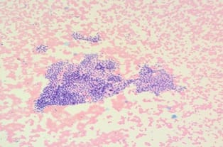
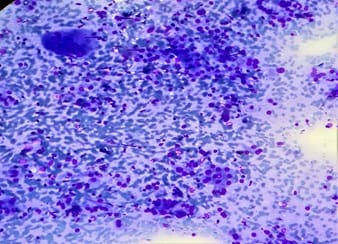
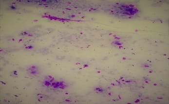
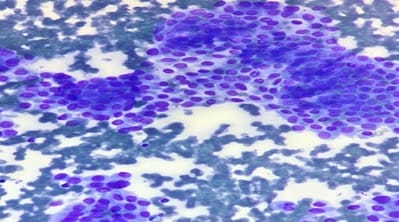
Background FNAC (Fine needle aspiration cytology) is an easy and cost-effective method for the diagnosis of thyroid lesions. The use of the Bethesda system is largely helpful in eliciting the risk of malignancy and helps in further management of patients with thyroid lesions. In this study we studied thyroid lesions and reported them according to the BETHESDA System of Classification based on cytological features on FNAC. Materials and Methods: In this prospective single-centre study conducted over 2 years we included 642 patients with thyroid lesions who underwent FNAC. These lesions were then classified according to the Bethesda system based on cytomorphological features. Results: Out of 642 patients included in our study, 100 were males and 542 were females with a male: female ratio of 1:5.42. The age of the patient population in our study ranged from 4-94 years. Among 642 cases in our study most (61.38%) cases were benign whereas 9.96% cases were found to be malignant and 2.60% cases were suspicious of malignancy. Conclusion: Bethesda System of reporting FNAC of thyroid lesions is a standardized system and helps in deciding diagnostic approach and appropriate management of patients.

Keywords: FNAC, Thyroid, Bethesda system, Cytology

Corresponding Author How to Cite this Article To Browse
Summiya Farooq, Assistant Professor, Department of Pathology, Government Medical College, Srinagar, Jammu and Kashmir, India.
Email:
Sheikh S, Abbas F, Beigh A, Farooq S, Latief M. Fine needle aspiration cytology of thyroid lesions: A single-centre experience. Trop J Pathol Microbiol. 2021;7(2):85-92.
Available From
https://pathology.medresearch.in/index.php/jopm/article/view/524

Manuscript Received Review Round 1 Review Round 2 Review Round 3 Accepted
2021-03-18 2021-03-28 2021-04-07 2021-04-13
Conflict of Interest Funding Ethical Approval Plagiarism X-checker Note
No Nil Yes 8%

© 2021 by Sheema Sheikh, Farhat Abbas, Ambreen Beigh, Summiya Farooq, Muzamil Latief and Published by Siddharth Health Research and Social Welfare Society. This is an Open Access article licensed under a Creative Commons Attribution 4.0 International License https://creativecommons.org/licenses/by/4.0/ unported [CC BY 4.0].

Introduction

Thyroid gland abnormalities are one of the commonly seen pathologies in surgical practice. However accurate diagnosis of thyroid nodules is a challenge for physicians managing patients with thyroid pathologies. Palpable thyroid nodules are more common in women compared to men [1,2]. Thyroid nodules may present with hypothyroidism, hyperthyroidism, cosmetic issues, and problems in other organs including compression of surrounding structures. Some of these lesions have the potential for malignancy [3].

Clinical presentation, hormone levels and imaging are important however, cytological analysis of fine-needle aspiration (FNA) material is the primary modality for initial evaluation of thyroid lesions (1,4-6). Benign lesions require to follow up, while malignant lesions require surgical treatment. FNAC is considered to be the “gold standard” in the selection of patients for surgery [7]. FNAC does not require any previous preparation. It can be performed without local anaesthesia. In medical centres with longstanding experience, diagnostic (adequate) biopsies obtained from solid nodules range from 90–97% [8, 9]. In most clinical settings, the cytology of thyroid nodules is classified into one of six Bethesda categories. [10]

Bethesda categorization is important for cancer risk assessment [11,12] and the preoperative likelihood of low-risk- versus high-risk cancer phenotypes. Furthermore, Bethesda classification may predict high-risk histologic features such as LNM (lymph node metastasis), ETE (extra-thyroidal extension), or lymphovascular invasion (LVI). However, further data are needed to validate such findings [13]. The most common result in thyroid lesions on FNAC is benign cytology which accurately predicts a benign nodule. Three indeterminate groups on cytology show an adequate yet morphologically abnormal sample in which the risk of malignancy is increased though not confirmed.

Such samples are classified as atypia of undetermined significance (AUS), Follicular lesion of undetermined significance (FLUS), suspicious for a follicular neoplasm (FN), or suspicious for malignancy (SUSP). The risk of malignancy is 5–15%, 15–30%, and 60–75% for AUS, FN and SUSP for malignancy respectively [14]. Malignant (M) cytology suggests a highly predictive 97–99% risk of cancer, and thyroidectomy is generally indicated in such cases.

FNAC has undoubtedly improved preoperative risk assessment and provides some information on prognostic indicators according to some studies [15-18].

Materials and Methods

Place of study: GMC Srinagar Kashmir

Type of study: a prospective, observational study

Inclusion criteria: All ages and gender with thyroid nodules or swellings.

Sampling method: fine-needle aspiration cytology(FNAC)

Classification: Bethesda system

This prospective single centre study of thyroid lesions was carried out at the Department of Pathology, Government Medical College, Srinagar which is a tertiary care centre. The study was carried for 2-years from January 2018 to December 2019.

A total of 642 cases of thyroid lesions were included in the study. done during this period. Consent was taken from all patients for the FNAC procedure after informing them about the complications of the procedure. FNAC was performed in all patients presenting with thyroid swellings either blindly or under US(ultrasound) guidance.

Ultrasound guidance was used in cases in which the lesions were either deep-seated, non-palpable or subcentimetric. All cases were diagnosed and classified based on the Bethesda system of classification. In patients with multiple nodules in the same gland, Bethesda classification and analysis were done according to the highest risk thyroid Bethesda system (TBS), as this was most likely to help clinicians to make appropriate decisions.

All FNA was done by the pathologist as the outpatient procedure and ultrasound-guided FNA was done in cases where lesions were non-palpable and deep-seated lesions. Smears prepared were air-dried and stained with May–Grunwald–Giemsa (MGG) and wet smears were stained with Papanicolaou and haematoxylin and eosin stain (H&E).

Stained slides were examined under a light microscope. The results were categorised into Bethesda classification and further subcategorised based on cytological features.


Results

Table 1: Gender distribution of cases

No of males 100
No of females 542
Total 642

In our study the total number of patients included was 642. Out of these 100 were males and 542 were females as shown in the table above

Male: female ratio was 1:5.42.

The age of patients ranged from 4-94 years. Most of the patients were in the age group of 21 -50 years.

All patients were categorised on the basis of Bethesda system as given in table below

Table 2: Bethesda categorization of patients

BETHESDA CATEGORY NO. OF PATIENTS %age
CATEGORY I NON DIAGNOSTIC 118 18.38
CATEGORY II BENIGN 393 61.21
CATEGORY III AUS 9 1.40
CATEGORY IV FN/SUSP FN 40 6.23
CATEGORY V SUSP OF MALIGNANCY 18 2.80
CATEGORY VI MALIGNANT 64 9.96
AUS; Atypia of undetermined significance; FN Follicular neoplasm; SUSP Suspicious of malignancy; M Malignant

Most of the cases belonged to benign category II (61.21%). Atypia of undetermined significance (category III) was seen in 1.40% of cases.

6.23% cases belonged to Category IV designated as Follicular neoplasm or suspicious of follicular neoplasm. 2.80% cases belonged to category V or were suspicious for malignancy and malignant lesions accounted for 9.96% of all cases.

Bethesda category III which included benign lesions was further subcategorized into colloid /adenomatoid goitre, lymphocytic or Hashimoto's thyroiditis and granulomatous thyroiditis.18.38%cases were non-diagnostic.


Table-3: Age distribution of thyroid lesions

AGE COLLOID GOITRE/ ADENO- MATOID GOITRE LYMPH- OCYTIC THYR-OIDITIS GRANULO- MATOUS THYROIDITIS FOLLICULAR NEOPLASM HURTHLE CELL NEOPLASM PAPILLARY CARCINOMA MEDU- LLARY CA ANAPLASTIC CA METASTASIS
</=10 4 0 0 0 0 0 0    
11-20 15 6 0 3 0 7 0    
21-30 56 29 0 4 0 30 2    
31-40 82 19 4 7 4 12 0    
41-50 70 8 3 4 3 7 0   1
51-60 44 9 2 5 2 3 0    
61-70 28 0 0 4 2 0 1 1  
71-80 10 1 0 0   1 1    
81-90 2 0 0 0          
>90 1 0 0 0          
total 312 72 9 27 13 58 4 1 1

Colloid goitre was seen predominantly in the 31-40 years age group. 43 of 312 patients of colloid goitre were males and 269 patients were females. Lymphocytic thyroiditis was seen in a predominantly 21-30years age group among whom 5 of 72 patients were males and 67 were females. Most patients with granulomatous thyroiditis were in the age group of 31-40. The most common age group for follicular/ hurtle cell neoplasms was 41-50 years among these 10 out of 40 patients were males and 30 were females. Malignant lesions were most predominant in the 31-40 years age group among these 19 of 64 patients were males and 45 were females.

patho_524_01.jpg


Figure 1: Showing monolayered sheet of follicular cells forming honeycomb in case of a colloid goitre on haematoxylin and eosin staining

patho_524_02.jpg

Figure 2: Hashimoto’s thyroiditis showing hurthle cells in a lymphoid background on MGG staining

patho_524_03.jpg

Figure 3: Epitheloid cell collection in case of granulomatous thyroiditis on MGG staining

Follicular neoplasms were also subcategorized into follicular neoplasms and hurthle cell neoplasms. Also malignant tumors were categorized into subcategories based on cytomorphological features. Sub categorization of various thyroid lesions is shown in table 2 along with age distribution.

patho_524_04.jpg

Figure 4: Papillary carcinoma thyroid: showing nuclear overlapping grooving and intranuclear inclusions.

patho_524_05.jpg

Figure 5: Medullary carcinoma showing dyscohesive cell cluster with plasmacytoid cells on MGG staining

patho_524_06.jpg

Figure 6: Highly pleomorphic cells and mitotic figures in a case of anaplastic carcinoma

Discussion

The Bethesda system has standardized thyroid nodule terminology in cytology. Using this system the prevalence of malignancy could be effectively conveyed to both the patient and the practitioner, for better decision making. Also it has become easy for the pathologist to interpret the cases where a specific pathology cannot be elicited. Therefore, it remains the gold standard for the initial evaluation of thyroid lesions. In our study 642 cases of thyroid lesions were categorised into the Bethesda system and were further subcategorised based on cytological features of FNA Samples.

The age of patients ranged from 4-94 years with most cases in the age range of 31 to 40 (28.34%) followed by 21-30 years (4.92%). Comparable results were found in other studies done previously.


(19,20,21,22,23) In our study 542 patients were females with a male to female ratio of 1:5.42. The results were similar to a study done by Bhagat et al (20) in which the ratio was 1:5.67 and Kumar et al where the ratio was1:4.4 (24).

Female preponderance in thyroid lesions was seen in other studies done by yang J et al, Bagga PK et al, Rabaglia JL et al and others. (25-32,14)

Majority of patients presented with diffuse swelling similar to the study done by Vasudha et al. (33)

Out of 642 cases 18.38% were non-diagnostic, 61.21% were benign, 1.40% cases showed AUS, 6.23% cases showed follicular neoplasm/FLUS, 2.80% were suspicious of malignancy and 9.96% were malignant as per the Bethesda system. Comparison of patients as per Bethesda category is shown below


Bethesda Category (%) Current Study Prathima et al [34] Bhagat et al [20] Sinna et al [35] Mondak SK al [36]
N (Total Cases) 642 178 172 296 1020
Non diagnostic 18.38 11.7 5.6 7.1 1.2
Benign 61.21 77.5 87.5 33.1 87.5
Atypia of undetermined significance 1.40 1.12 15 13.5 1
Follicular neoplasm /suspicious of follicular neoplasm 6.23 3.9 3.1 16.5 4.2
Suspicious of malignancy 2.80 2.2 0.6 10.1 1.4
Malignant 9.96 3.3 3.1 19.5 4.7

In our study the number of cases in the non-diagnostic category (category I) was more compared to other studies. This could be because of a large number of total cases in our study.

Also the percentage of follicular neoplasms, malignant cases and cases suspicious for malignancy was higher in our study compared to other studies. These differences could be due to a large number of overall cases in our study. In our study the benign lesions which belong to Bethesda category II, were further classified into colloid/adenomatoid goitre, lymphocytic thyroiditis and granulomatous thyroiditis.

Among this most common entity was colloid/adenomatoid goitre accounting for 79.38% cases, followed by lymphocytic thyroiditis accounting for 18.38% and then granulomatous thyroiditis accounted for 2.29% cases of benign lesions.

This distinction of category II cases helped the clinicians to treat these entities better. Similar results were found by Agarwal et al where the commonest pathology was colloid or adenomatoid goitre followed by lymphocytic thyroiditis followed by Hashimoto's thyroiditis, followed by granulomatous thyroiditis. (37) Colloid/adenomatoid goitre was the common disease overall 312/642(48.59%) cases. This is comparable to a study done by Lakshmi et al where nodular goitre was the commonest disease accounting for almost 50.59% of cases. [38]

In our study only 1.40% of cases belonged to category III, due to strict adherence to diagnostic criteria. Similar results were found by Savita S et al. (39) In our study among category IV 27/40(67.5%) cases belonged to follicular neoplasms and 13/40 (32.5%) cases belonged to hurthle cell neoplasms. Malignant cases accounted for 9.96% of our study and cases suspicious for malignancy were 2.80%. This was unlike the studies done by Nandekar et al where malignant cases were 1.98 %. (40) and Mehra et al where the malignant cases were only 2.2%. (41) The commonest malignancy was papillary thyroid carcinoma 58/64(90.625%), followed by medullary carcinoma 4/64(6.25%). We had a single case of anaplastic carcinoma and metastasis to the thyroid each. This was similar to the study done by Lakshmi et al where the commonest malignancy was papillary carcinoma thyroid. [38]

Conclusion

Bethesda system is a reliable system for classifying thyroid lesions. It also provides prognostic information about cancer type, variant, and risk of recurrence. It also gives clear cut guidelines for the surgical management of patients. This study was done to show the distribution of various thyroid lesions in Kashmir valley and the increased prevalence of malignant lesions of thyroid in this part of the world compared to others. Neoplastic lesions of the thyroid designated by category IV V and VI were more common in our study.


What does this study add to existing knowledge?

Bethesda system provides prognostic information about cancer type, variant, and risk of recurrence. It also gives clear cut guidelines for the surgical management of patients.

Author contributions

SS, FA and SF collected the data and conducted this study. FA and AB did data analysis. ML and FA did manuscript drafting. All authors were involved in revising and approved the final version of the manuscript.

Reference

  1. Cooper DS, Doherty GM, Haugen BR, et al. American Thyroid Association (ATA) Guidelines Taskforce on Thyroid Nodules and Differentiated Thyroid Cancer- Revised American Thyroid Association management guidelines for patients with thyroid nodules and differentiated thyroid cancer. Thyroid. 2009;19(11)1167–214.
  2. Tan GH, Gharib H. Thyroid incidentalomas- management approaches to nonpalpable nodules discovered incidentally on thyroid imaging. Ann Intern Med. 1997 Feb 1;126(3)226-31.
  3. Roman SA. Endocrine tumors- evaluation of the thyroid nodule. Curr Opin Oncol. 2003 Jan;15(1)66-70.
  4. Levine RA. Current guidelines for the management of thyroid nodules. Endocr Pract. 2012 Jul-Aug;18(4)596-9.
  5. Gharib H, Papini E, Paschke R, Duick DS, Valcavi R, Hegedüs L, Vitti P. AACE/AME/ETA Task Force on Thyroid Nodules, American Association of Clinical Endocrinologists, Associazione Medici Endocrinologi, and EuropeanThyroid Association Medical Guidelines for Clinical Practice for the Diagnosis and Management of Thyroid Nodules. Endocr Pract. 2010 May-Jun;16 Suppl 1;1-43.
  1. Moon WJ, Baek JH, Jung SL, Kim DW, Kim EK, Kim JY, Kwak JY, Lee JH, Lee JH, Lee YH, Na DG, Park JS, Park SW. Korean Society of Thyroid Radiology (KSThR); Korean Society of Radiology, Ultrasonography and the ultrasound-based management of thyroid nodules- consensus statement and recommendations. Korean J Radiol. 2011 Jan-Feb;12(1)1-14.
  2. Polyzos SA, Kita M, Avramidis A. Thyroid nodules - stepwise diagnosis and management. Hormones (Athens). 2007 Apr-Jun;6(2)101-19.
  3. Wong CK, Wheeler MH. Thyroid nodules- rational management. World J Surg. 2000;24(8)
  4. Landis SH, Murray T, Bolden S, Wingo PA. Cancer statistics, 1998. CA Cancer J Clin. 1998 Jan-Feb;48(1)6-29.
  5. Cibas ES, Ali SZ. The Bethesda System for Reporting Thyroid Cytopathology. Thyroid. 2009 Nov;19(11)1159-65.
  6. VanderLaan PA, Marqusee E, Krane JF. Features associated with locoregional spread of papillary carcinoma correlate with diagnostic category in the Bethesda System for reporting thyroid cytopathology. Cancer Cytopathol. 2012 Aug 25;120(4)245-53.
  7. Kleiman DA, Beninato T, Soni A, Shou Y, Zarnegar R, Fahey TJ 3rd. Does bethesda category predict aggressive features in malignant thyroid nodules?. Ann Surg Oncol. 2013 Oct;20(11)3484-90.
  8. Yassa L, Cibas ES, Benson CB, Frates MC, Doubilet PM, Gawande AA, Moore FD Jr, Kim BW, Nosé V, Marqusee E, Larsen PR, Alexander EK. Long-term assessment of a multidisciplinary approach to thyroid nodule diagnostic evaluation. Cancer. 2007 Dec 25;111(6)508-16.

  1. Rabaglia JL, Kabbani W, Wallace L, Holt S, Watumull L, Pruitt J, Snyder WH, Nwariaku FE. Effect of the Bethesda system for reporting thyroid cytopathology on thyroidectomy rates and malignancy risk in cytologically indeterminate lesions. Surgery. 2010 Dec;148(6)1267-72; discussion 1272-3.
  2. Hirsch D, Robenshtok E, Bachar G, Braslavsky D, Benbassat C. The Implementation of the Bethesda System for Reporting Thyroid Cytopathology Improves Malignancy Detection Despite Lower Rate of Thyroidectomy in Indeterminate Nodules. World J Surg. 2015;39
  3. Yoon JH, Lee HS, Kim EK, Moon HJ, Kwak JY. Thyroid Nodules- Nondiagnostic Cytologic Results according to Thyroid Imaging Reporting and Data System before and after Application of the Bethesda System. Radiology. 2015 Aug;276(2)579-87.
  4. Srbova L, Gabalec F, Ryska A, Cap J. Results of retrospective classification of thyroid FNAs according to the Bethesda system- would this have improved accuracy?. Cytopathology. 2015 Aug;26(4)231-7.
  5. Sullivan PS, Hirschowitz SL, Fung PC, Apple SK. The impact of atypia/follicular lesion of undetermined significance and repeat fine-needle aspiration- 5 years before and after implementation of the Bethesda System. Cancer Cytopathol. 2014 Dec;122(12)866-72.
  6. Ugurluoglu C, Dobur F, Karabagli P, Celik ZE. Fine needle aspiration biopsy of thyroid nodules- cytologic and histopathologic correlation of 1096 patients. Int J Clin Exp Pathol. 2015;8(11)14800
  7. Bhagat DV, Tailor DH, Kaptan DK, et al. Diagnostic role of the Bethesda system for reporting thyroid lesions- Effective tool for managing thyroid lesions. Global J of Medical Research. 2014;14 (1)12-8.
  1. Garg S, Desai NJ, Mehta D, Vaishnav M. To Establish Bethesda System for Diagnosis of Thyroid Nodules on the Basis of Fnac with Histopathological Correlation. J Clin Diagn Res. 2015 Dec;9(12)EC17-21.
  2. Kulkarni DC, Mittal DM, Nema DM, et al. Diagnostic role of the Bethesda system for reporting thyroid cytopathology in an academic institute of central India- One year experience. Indian Journal of Basic and Applied Medical Research. 2016;5;157-66.
  3. Sawarkar PR, Mahore SD, Bothale KA, et al. Cytological evaluation of thyroid lesions based on the Bethesda system. IOSR-JDMS. 2016;15;112-7.
  4. Kumar D, Dwivedi G, Bhargava S. The Bethesda system for reporting thyroid cytopathology. Journal of medical science and clinical research. 2014;2(12)3171-6.
  5. Yang J, Schnadig V, Logrono R, Wasserman PG. Fine-needle aspiration of thyroid nodules- a study of 4703 patients with histologic and clinical correlations. Cancer. 2007 Oct 25;111(5)306-15.
  6. Bagga PK, Mahajan NC. Fine needle aspiration cytology of thyroid swellings- how useful and accurate is it?. Indian J Cancer. 2010 Oct-Dec;47(4)437-42.
  7. Harvey AM, Mody DR, Amrikachi M. Thyroid fine-needle aspiration reporting rates and outcomes before and after Bethesda implementation within a combined academic and community hospital system. Arch Pathol Lab Med. 2013 Nov;137(11)1664-8.
  8. Patel MM, Patel K, Kaptan KR, et al. Fine needle aspiration cytology as a first line investigation in thyroid Lesions. National Journal of Medical Research. 2013;3(02)106-10.

  1. Park JH, Yoon SO, Son EJ, Kim HM, Nahm JH, Hong S. Incidence and malignancy rates of diagnoses in the bethesda system for reporting thyroid aspiration cytology- an institutional experience. Korean J Pathol. 2014 Apr;48(2)133-9.
  2. Naz S, Hashmi AA, Khurshid A, Faridi N, Edhi MM, Kamal A, Khan M. Diagnostic accuracy of Bethesda system for reporting thyroid cytopathology- an institutional perspective. Int Arch Med. 2014 Oct 31;7;46.
  3. Bolland MJ, Eagleton C, Orr-Walker B. Diagnostic category agreement and malignancy rates in clinician-categorised, non-standardised thyroid cytology reports. N Z Med J. 2014 Dec 19;127(1407)49-55.
  4. Arul P, Masilamani S. A correlative study of solitary thyroid nodules using the bethesda system for reporting thyroid cytopathology. J Cancer Res Ther. 2015 Jul-Sep;11(3)617-22.
  5. Vasudha MB, Hemali JT, Kumar BRK. Diagnostic role of Bethesda system of reporting thyroid lesions, Global journals Inc (US). Global journal of medical research- Microbiology and pathology. 2014;14(1)57;87554.
  6. Prathima S, Suresh TN, Kumar MH, et al. Impact of the Bethesda system in reporting thyroid cytopathology. Thyroid fresearch and practice. 2016;13(1)9.
  7. Sinna EA, Ezzat N. Diagnostic accuracy of fine needle aspiration cytology in thyroid lesions. J Egypt Natl Canc Inst. 2012 Jun;24(2)63-70.
  1. Mondal SK, Sinha S, Basak B, Roy DN, Sinha SK. The Bethesda system for reporting thyroid fine needle aspirates- A cytologic study with histologic follow-up. J Cytol. 2013 Apr;30(2)94-9.
  2. Agrawal D, Bhake AS, Rastogi N, et al. Role of Bethesda system for reporting thyroid lesion and its correlation with histopathological diagnosis. Journal of Datta Meghe Institute of Medical Sciences university(DU). 2019;14(2)74-81.
  3. Lakshmi S, Latha B, Nirmala C. Spectrum Of Thyroid Lesions On Fine Needle Aspiration Cytology. Journal Of International Medicine And Dentistry. 2015;2(1)53-62.
  4. Soni S, Deepika Gupta D, Bordia S, et al. Categorization of thyroid lesions FNAC in Bethesda system- An institutional study in southern Rajasthan. International Journal of Clinical and Diagnostic Pathology. 2020;3(2)159-164.
  5. Nandedkar SS, Dixit M, Malukani K, Varma AV, Gambhir S. Evaluation of Thyroid Lesions by Fine-needle Aspiration Cytology According to Bethesda System and its Histopathological Correlation. Int J Appl Basic Med Res. 2018 Apr-Jun;8(2)76-82.
  6. Mehra P, Verma AK. Thyroid cytopathology reporting by the bethesda system- a two-year prospective study in an academic institution. Patholog Res Int. 2015;2015;240505.