E-ISSN:2456-1487
P-ISSN:2456-9887
RNI:MPENG/2017/70771

Case Report

Abscess

Tropical Journal of Pathology and Microbiology

2020 Volume 6 Number 7 September-October
Publisherwww.medresearch.in

Scrotal abscess with xanthogranulomatous epididymo-orchitis: a case report of rare diagnostic entity

Verma R.1, Dalal N.2*, Yadav A.3, Pawar R.4, Singh S.5
DOI: https://doi.org/10.17511/jopm.2020.i07.04

1 Renuka Verma, Assistant Professor, Department of Pathology, Pandit Bhagwat Dayal Sharma Post Graduate Institute of Medical Sciences, Rohtak, Haryana, India.

2* Niti Dalal, Senior Resident, Department of Pathology, Pandit Bhagwat Dayal Sharma Post Graduate Institute of Medical Sciences, Rohtak, Haryana, India.

3 Archana Yadav, Resident, Department of Pathology, Pandit Bhagwat Dayal Sharma Post Graduate Institute of Medical Sciences, Rohtak, Haryana, India.

4 Richa Pawar, Assistant Professor, Department of Pathology, Pandit Bhagwat Dayal Sharma Post Graduate Institute of Medical Sciences, Rohtak, Haryana, India.

5 Sunita Singh, Senior Professor, Department of Pathology, Pandit Bhagwat Dayal Sharma Post Graduate Institute of Medical Sciences, Rohtak, Haryana, India.

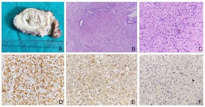
Xanthogranulomatous epididymo-orchitis is an extremely rare diagnostic entity presenting as a scrotal mass with or without pain. Rarely, it can affect prostate, epididymis, and testicle. It can mimic a testicular malignancy both clinically as well as radiologically, hence, careful pathological examination and biomarker study are essential to rule out malignancy. We report a case of 50 years old male with increasing right testicular swelling with a discharging pus. Skin over scrotum was gangrenous. Right orchidectomy was performed. Histopathological diagnosis revealed features of xanthogranulomatous epididymo-orchitis with no evidence of malignancy. IHC markers for tumor were negative.

Keywords: Xanthogranuloma, Epididymitis, Orchitis, Malignancy

Corresponding Author How to Cite this Article To Browse
Niti Dalal, Senior Resident, Department of Pathology, Pandit Bhagwat Dayal Sharma Post Graduate Institute of Medical Sciences, Rohtak, Haryana, India.
Email:
Verma R, Dalal N, Yadav A, Pawar R, Singh S. Scrotal abscess with xanthogranulomatous epididymo-orchitis: a case report of rare diagnostic entity. Trop J Pathol Microbiol. 2020;6(7):443-445.
Available From
https://pathology.medresearch.in/index.php/jopm/article/view/486

Manuscript Received Review Round 1 Review Round 2 Review Round 3 Accepted
2020-09-29 2020-10-09 2020-10-15 2020-10-20
Conflict of Interest Funding Ethical Approval Plagiarism X-checker Note
No Nil Yes 9%

© 2020 by Renuka Verma, Niti Dalal, Archana Yadav, Richa Pawar, Sunita Singh and Published by Siddharth Health Research and Social Welfare Society. This is an Open Access article licensed under a Creative Commons Attribution 4.0 International License https://creativecommons.org/licenses/by/4.0/ unported [CC BY 4.0].

Introduction

Xanthogranulomatous epididymo-orchitis is a rare inflammatory condition. It is characterized by the destruction of testicular tissue which is replaced by prominent macrophage infiltration, lymphocytes and plasma cells along with giant cells, and necrosis [1]. Kidney and gall bladder are the most commonly affected organs followed by liver, appendix, ovaries, vagina, bones, urinary bladder and rarely affecting prostate, epididymis, and testicle [2].

Exact pathogenesis of xanthogranulomatous inflammation in epididymis and testis remains unknown. Possible risk factors include diabetes, obstruction of the epididymis, ischemic process of the testis and genital tract, and urinary tract infection.[3] We report a case of scrotal abscess in 50 years old male and to the best of our knowledge, only 23 cases of xanthogranulomatous epididymo-orchitis had been reported, so far. [1,2,4,5,6]

Case history

A 50 years old male presented with pain and increasing right testicular swelling with a discharging pus. Skin over scrotum was gangrenous. Approximately 150ml of pus was drained out and a presumptive diagnosis of scrotal abscess was made. USG showed heterogenous mass lesion in right testis. Right orchidectomy was performed and the specimen was received in the department of pathology.

Gross examination showed testis with distorted outlines covered by scrotal skin with a draining sinus measuring 7.5x4x4 cm. On cut open, a cavity measuring 3.5cm with yellowish purulent material was identified. (Figure A) Sections were processed from representative areas. Histopathological examination showed necrotic tissue and neutrophilic exudate surrounded by granulation tissue, fibrosis and chronic inflammatory infiltrate. Numerous histiocytes, giant cells and extensive xanthomatous change replaced the entire testis.

Compressed normal epididymal tissue was seen at periphery. (Figure B,C) Staining for acid fast bacilli using 20% H2SO4 was negative. On PAS staining, no infectious organism could be identified. Immunohistochemical staining revealed CD 68 positivity, Inhibin, Melan A, Calretinin, ER and PR negativity. (Figure D,E,F) A final histopathological diagnosis of scrotal abscess with xanthogranulomatous epididymo-orchitis was given.

patho_486_01.jpg

Figure : A : Gross specimen showed distorted architecture of testis along with a cavity filled with pus ; B : Microscopy showed necrotic tissue, neutrophilic exudate surrounded by granulation tissue, fibrosis and chronic inflammatory infiltrate along with compressed epididymis (H&E, 100X); C : Microscopy revealed cells with abundant eosinophilic cytoplasm with neutrophilic exudate (H&E, 400X); D : CD68 positivity (IHC, 400X); E: Calretinin negativity (IHC, 400X); F : Inhibin negativity (IHC, 400X).

Discussion

Xanthogranulomatous epididymo-orchitis is a non-neoplastic condition with wide age range of distribution viz., 3rd to 7th decade of life [1]. Various theories regarding etiology have been suggested including obstruction of epididymis or urinary obstruction, testicular ischemia secondary to atherosclerosis, diabetes, autoimmune process, and defect in chemotactic activity [7]. Infectious etiology like Escherichia coli, Proteus mirabilis, Pseudomonas aeruginosa has also been postulated [2,6].

Its diagnosis is quite challenging as it can mimic testicular malignancy, malakoplakia, Rosai-Dorfman disease of the testis and lepromatous leprosy [8]. Sometimes, testicular malignancy is associated with normal serum tumor markers [6]. Testicular germ cell tumors and seminoma had been associated with xanthogranulomatous inflammation, therefore, extensive sampling is recommended to rule out malignancy [2].

In such cases, immunohistochemistry like PLAP, OCT3/4 had been proved to be helpful. In order to differentiate non-neoplastic conditions, malakoplakia showed intracytoplasmic inclusion in macrophages known as Michaelis Gutmann bodies. Rosai-Dorfman disease of the testis has a characterstic feature of emperipolesis.


Macrophages in other xanthogranulomatous conditions and lepromatous leprosy are S-100 positive. Bacterial infection usually improves after taking antibiotics therapy [2,8]. In the present case, macrophages are CD68 positive and other immunohistochemical markers for malignancy like inhibin, Melan-A, Calretinin, ER and PR are negative.

The diagnostic utilities of inguinal ultrasound, CT scan, fine needle aspiration cytology, serologic tumor makers and clinical history are very limited [1]. Only histopathology and immunohistochemistry can aid in the diagnosis of xanthogranulomatous epididymo-orchitis. In case of testicular abscess, orchidectomy with hemiscrotectomy is recommended [2].

Conclusion

Xanthogranulomatous epididymo-orchitis is a clinically significant entity due to its close resemblance with malignancy. Both clinicians and pathologist should be aware of this condition. Thorough histopathological examination is the key to the diagnosis.

Acknowledgement

All the authors have contributed to concept, literature search, data acquisition, data analysis, manuscript editing and review.

Reference

  1. Said SHA, Abdalla RY, Aghaways I, Abdullah AM. Xanthogranulomatous orchitis- Review of the published work, and report of one case. Urology Case Reports. 2019;27;100908.
  2. Sharma P, Ramteke P, Satapathy A, Ray MD. Xanthogranulomatous Orchitis Presenting as a Scrotal Mass in an Elderly Male- Malignancy or Mimicker?. Clinical Medicine Insights- Case Reports. 2019;12;1–4.
  3. Demirci D, Ekmekcioglu O, Soyuer I, Emirdogan M. Xanthogranulomatous orchitis with scrotal fistulas. Int J Urol. 2004;11;686– 8.
  4. Gongora e Silva RF, Pinto IC, Constantino EC, Querichelli AF, Spessoto LC, Fácio FN Jr. Xanthogranulomatous orchitis- case report of a rare condition. AME Case Rep. 2019;3;4.
  1. Rami S, Alazab RS, Ghawanmeh HM, Al-Okour RK, Alshammari A, Lafi T, Al-Karasneh AI, Al-Bashir S, Al-Ghazo M. Xanthogranulomatous Orchitis- Rare Case with Brief Literature Review. Urology Case Reports. 2017;13;92-3.
  2. Zoran Persec Z, Bulimbasic S, Persec J, Ljubanovic D, Bartolin Z, Patrlj L, Hrgovic Z. Xanthogranulomatous epididymitis- clinical report and immunohistochemical analysis. Wien Klin Wochenschr. 2008.
  3. Al-Said S, Ali A, Alobaidy AK, Mojeeb E, Al-Naimi A, Shokeir AA. Xanthogranulomatous orchitis- Review of the published work and report of one case. International Journal of Urology. 2007;14;452–4.
  4. Nistal M, Gonzalez-Peramato P, Serrano A, Regadera J. Xanthogranulomatous funiculitis and orchiepididymitis- report of 2 cases with immunohistochemical study and literature review. Arch Pathol Lab Med. 2004;128;911–4.