E-ISSN:2456-1487
P-ISSN:2456-9887
RNI:MPENG/2017/70771

Case Report

Desmoplastic

Tropical Journal of Pathology and Microbiology

2020 Volume 6 Number 7 September-October
Publisherwww.medresearch.in

Desmoplastic Infantile Astrocytoma and Desmoplastic Infantile Ganglioglioma – not so infantile anymore

Sahai J.1*, Sahu S.2
DOI: https://doi.org/10.17511/jopm.2020.i07.05

1* Jyotsna Sahai, Resident, Department of Pathology, MGM Medical College and Hospital, Navi Mumbai, Maharashtra, India.

2 Shilpi Sahu, Professor and HOD, Department of Pathology, MGM Medical College and Hospital, Navi Mumbai, Maharashtra, India.

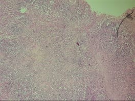
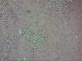
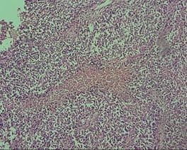
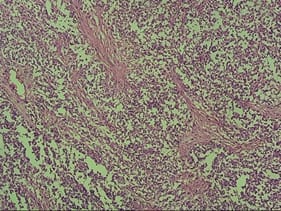
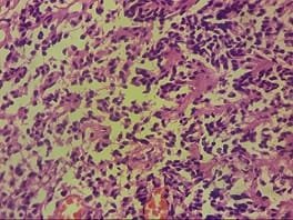
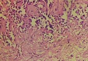
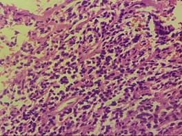
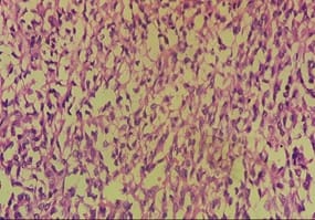
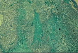
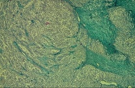
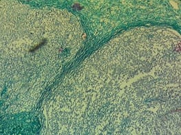
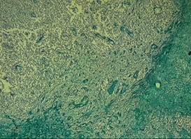
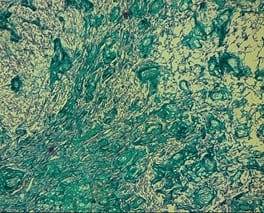
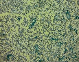
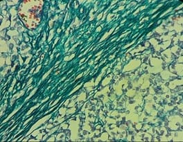
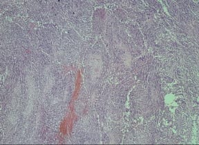
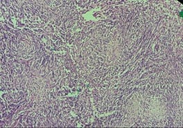
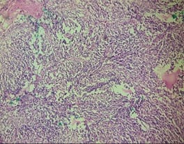
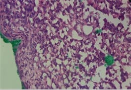
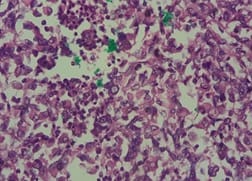
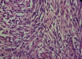
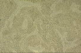
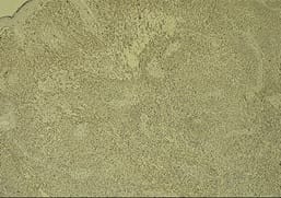
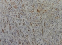
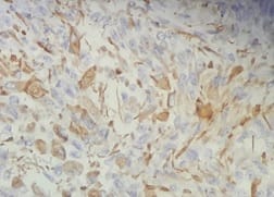
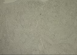
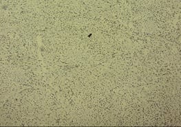
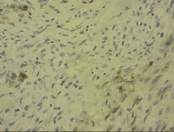
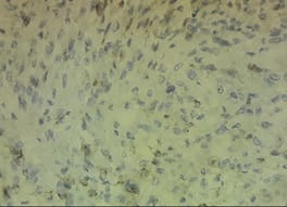
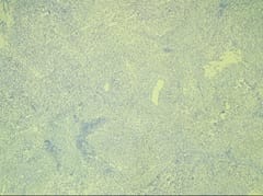
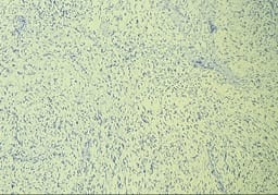
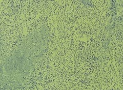
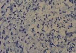
DIA and DIG are rare, infantile, supratentorial neoplasms that usually occur in children before 2 years of age and are exceedingly rare in older age groups. They appear as hypodense, cystic masses with solid components showing dural attachment on neuroimaging. They are characterized by reticulin-rich spindle cell stroma containing connective tissue due to meningeal involvement, microscopically. These tumors have potential for misdiagnosis because they contain varying proportions of neoplastic glial, neuronal and poorly differentiated cells, which causes them to have a “small round blue cell tumor” like appearance, though they have a good prognosis if correctly diagnosed. The current study report two cases diagnosed at our institution that had very late presentation with varying complaints which challenged the normally believed dictum of these tumors being entirely infantile.

Keywords: Astrocytoma, CNS, Desmoplastic, Ganglioglioma, Infantile, Tumor

Corresponding Author How to Cite this Article To Browse
Jyotsna Sahai, Resident, Department of Pathology, MGM Medical College and Hospital, Navi Mumbai, Maharashtra, India.
Email:
Sahai J, Sahu S. Desmoplastic Infantile Astrocytoma and Desmoplastic Infantile Ganglioglioma – not so infantile anymore. Trop J Pathol Microbiol. 2020;6(7):446-454.
Available From
https://pathology.medresearch.in/index.php/jopm/article/view/485

Manuscript Received Review Round 1 Review Round 2 Review Round 3 Accepted
2020-09-28 2020-10-08 2020-10-15 2020-10-20
Conflict of Interest Funding Ethical Approval Plagiarism X-checker Note
No Nil Yes 7%

© 2020 by Jyotsna Sahai, Shilpi Sahu and Published by Siddharth Health Research and Social Welfare Society. This is an Open Access article licensed under a Creative Commons Attribution 4.0 International License https://creativecommons.org/licenses/by/4.0/ unported [CC BY 4.0].

Introduction

The terms desmoplastic infantile astrocytoma (DIA) and desmoplastic infantile ganglioglioma (DIG) were coined by VandenBerg in 1987 based on immunohistochemistry and electron microscopy findings [1].

DIA and DIG are rare, massive, enhancing, supratentorial neoplasms that usually occur in children, before 2 years of age with a median of 6 months [2]. The WHO categorizes DIA / DIG as grade I “neuronal and mixed neuronal-glial” entity in its classification of Central Nervous System (CNS) neoplasms [3].

Before the WHO classification, DIA and DIG were thought to be separate entities. (4) But given their similar clinical, neuroimaging and pathological features, they were grouped together by WHO in 2000 [5,6]. The tumors invariably arise in the supratentorial region and commonly involve more than one lobe, preferentially the temporal and frontal lobes.

Neuroimaging studies reveal these neoplasms as large, hypodense cystic masses with a solid isodense or slightly hyperdense superficial portion along with dural attachment [7]. Grossly, these tumors are large with a solid and cystic component, attached to the dura and are slow-growing with good prognosis after complete resection.

Microscopically, DIG / DIA are characterized by a reticulin–rich spindle cell stroma containing connective tissue due to prominent leptomeningeal involvement [1]. These tumors have potential for misdiagnosis due to the fact that they contain varying proportions of neoplastic glial, neuronal and poorly differentiated cells, the latter lending a “small round blue cell tumor” feature to them.

The primitive appearing element can lead to misdiagnosis of a WHO grade IV embryonal neoplasm. DIA contains only the primitive element plus astrocytic tumor cells, whereas DIG has a predominant astrocytic tumor population along with a neoplastic ganglion cell component too. The latter is usually composed of larger sized ganglion cells [8].

The present study report of two cases with the late occurrence and unusual presentation throwing light on the fact that these neoplasms could also have a non – infantile presentation.

Case Report

CASE ONE

A 6 year old girl presented with headache, projectile vomiting and history of repeated falls since one month. Her mother denied a history of previous similar complaints and gave the normal birth history. On examination, the child was irritable, but all other parameters and systemic examinations were normal.

CNS examination revealed that she had no neurological deficit. On computerized tomographic (CT) scan, a large, solid, enhancing lesion in right frontoparietal lobe with dilatation of lateral, III and IVth ventricles and tentorial herniation was noted. The lesion also caused a midline shift of 1.2 cm towards the left.

HISTOPATHOLOGICAL EXAMINATION

Gross specimen received was fragmented with multiple, grey-brown, soft to firm tissue pieces, the largest measuring 3.5 x 3 x 1.5 cm and the smallest measuring 1 x 1 x 0.5 cm.

Microscopically, a study of H and E stained sections revealed neoplastic cells arranged in fascicles and lobular pattern. The individual cells had eosinophilic cytoplasm with highly pleomorphic vesicular nuclei containing prominent nucleoli along with atypical mitoses.

Also noted were few poorly differentiated neoplastic cells with small, round, deeply basophilic nuclei having minimal cytoplasm. The surrounding stroma showed abundant desmoplasia along with focal hyalinization.

patho_485_01.jpg


patho_485_02.jpg

patho_485_03.jpg

Fig-1: H and E stain, 4X view – Tumor cells arranged in lobules and fascicles with stroma showing desmoplasia.

patho_485_04.jpg

patho_485_05.jpg

patho_485_06.jpg

patho_485_07.jpg

patho_485_08.jpg


patho_485_09.jpg

Fig-2: (10X and 40X views, H and E stain) – Tumor cells having abundant eosinophilic cytoplasm and vesicular nuclei with other groups of neoplastic cells being poorly differentiated with minimal cytoplasm.

patho_485_10.jpg

patho_485_11.jpg

patho_485_12.jpg

patho_485_13.jpg

patho_485_14.jpg

patho_485_15.jpg

Fig-3: (4X and 10X views, Masson Trichrome stain) - Showing positively stained collagen bundles and tumor cells staining negatively

patho_485_16.jpg


Fig-4: (40X view, Masson Trichrome stain) – Showing positively stained collagen bundles and negatively stained tumor cells.

The histomorphological features were suggestive of DIG, which was confirmed on further immunohistochemistry.

The IHC profile performed was as follows –

  1. Glial Fibrillary Acidic Protein (GFAP) – Positive
  2. Smooth Muscle Actin (SMA) – Positive
  3. Synaptophysin - Positive
  4. Vimentin – Positive
  5. ALK – Negative
  6. Calponin – Negative

The immunohistochemical markers confirmed the diagnosis of DIG.

CASE TWO

A 4-year-old girl presented to the surgery OPD with complaints of headache, repeated falls for two months along left-sided weakness for three days following a fall from the bed. CT scan showed a 5.8 x 5.2 x 5.5 cm sized heterodense, predominantly hyperdense lesion in the right frontoparietal lobe causing midline shift of 1.1 cm to left and also causing a mass effect on right lateral ventricle with specs of calcification and necrotic tissue within.

There was evidence of vasogenic edema and thinning of skull vault on the right side (right frontal bone).

patho_485_17.jpg

Fig-5: CT Scan image showing a right-sided lesion in the frontoparietal lobe causing midline shift, mass effect on right ventricle as well as specs of calcification (Arrow pointing to the tumor).

The radiological differential diagnoses were –

  1. Dysembryoplastic Neuroepithelial Tumor (DNET)
  2. Diffuse high-grade astrocytoma
  3. Rhabdoid tumor

HISTOPATHOLOGICAL EXAMINATION

The gross specimen received was fragmented, well encapsulated, soft to firm with the larger piece measuring 7 x 4.5 x 4.3 cm, and the smaller one measuring 4.5 x 2 x 1.5 cm. The cut section showed greyish white fleshy tissue with few areas showing hemorrhage.

A microscopic study of hematoxylin and eosin-stained sections showed a well-circumscribed cellular tumor comprising of neuroepithelial cells and spindly fibroblastic cells. The spindle cells showed a fascicular and storiform arrangement.

The individual tumor cells were round to oval with abundant eosinophilic cytoplasm. Few gemistocytes were also seen. The surrounding stroma was scanty. The tumor showed high vascularity comprising of variable-sized blood vessels. There were focal areas of hemorrhage.

No evidence of atypical mitoses and necrosis or microvascular proliferation was seen in the sections studied.

patho_485_18.jpg

Fig-6: (H and E stain, 4X view) – Highly cellular and fibroblastic tumor.

patho_485_19.jpg


patho_485_20.jpg

patho_485_21.jpg

Fig-7: (H and E stain, 10X view) – Cells arranged in a fascicular and storiform pattern.

patho_485_22.jpg

patho_485_23.jpg

patho_485_24.jpg

Fig-8: (H and E stain, 40X view) – Round to oval tumor cells with abundant eosinophilic cytoplasm arranged in a storiform pattern.

Special stain Reticulin was positive.

The above findings were suggestive of the following differential diagnoses –

  1. Desmoplastic Infantile Ganglioglioma (DIG)/ Desmoplastic Infantile Astrocytoma (DIA)
  2. Fibroblastic Meningioma

Immunohistochemistry was performed to confirm the same. As shown in figures 9 - 11, the IHC profile showed

  1. Glial Fibrillary Acidic Protein (GFAP) – Diffuse Positivity
  2. Epithelial membrane Antigen (EMA) – Focal Positivity; and
  3. Synaptophysin – Negative

patho_485_25.jpg

patho_485_26.jpg


patho_485_27.jpg

patho_485_28.jpg

Fig-9: GFAP - Diffuse positivity.

patho_485_29.jpg

patho_485_30.jpg

patho_485_31.jpg

patho_485_32.jpg

Fig-10: EMA - Focal Positivity.

patho_485_33.jpg

patho_485_34.jpg


patho_485_35.jpg

patho_485_36.jpg

Fig-11 – Synaptophysin – Negative.

The immunohistochemical markers confirmed the diagnosis of DIA.

Discussion

DIA and DIG are rare neuroepithelial, low grade, benign neoplasms usually occurring before the age of 24 months of life with boys being affected more than girls, and exceedingly rare in the older age group. Our study showed the occurrence of both cases in females and at a later age than usually found.

The clinical presentations of DIA / DIG are varied, but usually are those effects due to space-occupying lesions. The most common complaints are increased intracranial pressure, seizures, vomiting, and headaches. Our cases showed the patients presented with complaints of headaches and vomiting which were concordant with those of Samkari et al. Other symptoms could be neck stiffness, hemiparesis, poor verbalization, syncope, nausea, delayed developmental milestones, skull deformities, dysphonia, irritability, apnea, motor and / or sensory loss or weakness [9].

Grossly, the tumors are usually massive as was seen in our cases also and are firmly attached to the dura, which was also seen in our cases. They are composed of two components, solid and cystic. On histopathological examination, they are well-circumscribed and highly desmoplastic tumors.

The cell population is typically glial and / or neuronal. They are characterized by spindle cells arranged in storiform pattern and stroma composed of reticulin fibers surrounding the tumor cells having eosinophilic cytoplasm. Mitotic figures, necrosis, and atypia are all uncommon features in DIA / DIG, indicative of low-grade tumors [9]

The diagnosis is confirmed by immunohistochemistry, as was done in our cases. These tumors stain positively for GFAP (Diffusely) and focally for synaptophysin (neuronal cells). The findings of our cases were similar to these with DIG showing positivity for GFAP, Synaptophysin whereas DIA showed diffuse positivity for GFAP and negative Synaptophysin (as it contained no neuronal elements).

These tumors have an indolent course and usually do not recur after complete resection, suggestive of a favorable prognosis. Our patients were followed up for 6 months and both are hale and hearty with no recurrence of tumors or complaints.

Conclusion

The two cases discussed above show that such cases can present later in life and vary from the usual presentation of DIA / DIG as thought so far and thus should always be kept in mind as a differential diagnosis when reporting CNS neoplasms.

Reference

  1. Vandenberg SR. Desmoplastic infantile ganglioglioma and desmoplastic cerebral astrocytoma of infancy. Brain Pathol. 1993; 3(3)275-81.
  2. Brat D, VandenBerg S, Figarella-Branger D, Reuss D. Desmoplastic infantile astrocytoma and ganglioglioma, In- Louis DN, Ohgaki H, Wiestler OD, et al, editors, WHO Classification of Tumours of the Central Nervous System. Lyon- IARC. 2016,pp;144–146,3.

  1. Louis DN, Ohgaki H, Wiestler OD, Cavenee WK, Figarella-Branger D, Reifenberger G, et al. WHO classification of tumours of the central nervous system. Lyon, France- World Health Organization. 2016.
  2. Naylor RM, Wohl A, Raghunathan A, Eckel LJ, Keating GF, Daniels DJ. Novel suprasellar location of desmoplastic infantile astrocytoma and ganglioglioma- a single institution’s experience. J Neurosurg Pediatr. 2018;22(4)397-403.
  3. Taratuto AL, Vandenberg SR, Rorke LB. Desmoplastic infantile astrocytoma and ganglioglioma, In- Kleihues P, Cavenee WK, editors, WHO Classification of Tumors, Pathology and Genetics- Tumors of the Nervous System. Lyon- IARC. 2000;pp- 99–102.
  4. Brat D, VandenBerg S, Figarella-Branger D, Taratuto A. Desmoplastic infantile astrocytoma and ganglioglioma, In- Louis D, Ohgaki H, Wiestler O, Cavenee W, editors, WHO Classification of Tumours of the Central Nervous System. Lyon- IARC. 2007;pp- 96–98.
  1. Gelabert-Gonzalez M, Serramito-García R, Arcos-Algaba A. Desmoplastic infantile and non-infantile ganglioglioma- Review of the literature. Neurosurg Rev. 2011;34(2)151-158.
  2. Greer A, Foreman NK, Donson A, Davies KD, Kleinschmidt‐DeMasters BK. Desmoplastic infantile astrocytoma/ganglioglioma with rare BRAF V600D mutation. Pediatr Blood Cancer. 2017;64(6)e26350.
  3. Samkari A, Alzahrani F, Almehdar A, Algahtani H. Desmoplastic infantile astrocytoma and ganglioglioma- case report and review of the literature. Clin Neuropathol. 2017;36(1)31.