E-ISSN:2456-1487
P-ISSN:2456-9887
RNI:MPENG/2017/70771

Research Article

Tonsillar

Tropical Journal of Pathology and Microbiology

2020 Volume 6 Number 8 November-December
Publisherwww.medresearch.in

The histomorphological pattern of various tonsillar lesions. A 3-year study in the tertiary care center.

Sharma R.1, Bansal M.2*, Kumar N.3, Kumar G.4
DOI: https://doi.org/10.17511/jopm.2020.i08.05

1 Ritu Sharma, Associate Professor, Department of Pathology, T.S. Misra Medical College and Hospital, Lucknow, Uttar Pradesh, India.

2* Megha Bansal, Associate Professor, Department of Pathology, T.S. Misra Medical College and Hospital, Lucknow, Uttar Pradesh, India.

3 (Brig.) Nikhilesh Kumar, Professor and Head of Department, Department of Pathology, T.S. Misra Medical College and Hospital, Lucknow, Uttar Pradesh, India.

4 Gaurav Kumar, Associate professor, Department of E.N.T, T.S. Misra Medical College and Hospital, Lucknow, Uttar Pradesh, India.

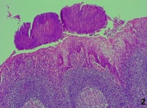
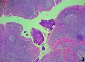
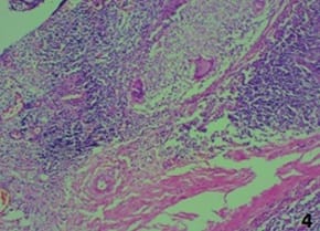
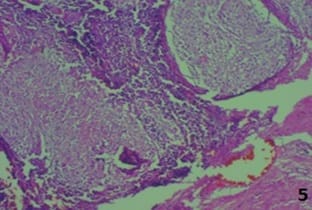
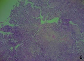
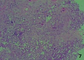
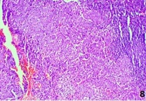
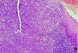
Introduction: Palatine tonsils are paired masses of lymphoid tissue, which act as an immunologic barrier against the entry of pathogenic agents into the respiratory and digestive tracts. Tonsillitis is a common disease, especially among children. Chronic tonsillitis is a disease with repeated attacks of acute tonsillitis or a sub-clinic form of a resistant or poorly treated infection. Aim: To determine the clinicopathological findings in various lesions of tonsillectomy in the palatine tonsil. Materials and methods: This is a retrospective study where 105 (Unilateral-20, Bilateral) cases of histopathologically identified tonsillectomy specimens as well as 4 tonsillar biopsies were included. The available data for all the patients as regards age, sex, and clinical symptoms were collected. Sections were taken from tonsillectomy specimens from representative areas while tonsillar biopsies were processed completely. Routine hematoxylin and eosin staining was done along with this special stain- PAS (periodic acid- Schiff) for actinomycosis and ZN stain for AFB. Results: Amongst the cases, 78 cases showed chronic tonsillitis. 11 cases were chronic tonsillitis with actinomycosis, acute chronic tonsillitis in 07 cases, granulomatous tonsillitis in 03 cases, acute ulcerative tonsillitis with microabscesses in 01 case, and reactive lymphoid hyperplasia in 02 cases. Three malignancies were observed – Two cases of squamous cell carcinoma and one case of undifferentiated carcinoma/lymphoma. Conclusion: Chronic tonsillitis is a common problem facing in all age groups, Histopathology plays a significant role in diagnosing both benign and malignant lesions of the tonsil.

Keywords: Chronic Tonsillitis, Tonsillectomy, Histopathology

Corresponding Author How to Cite this Article To Browse
Megha Bansal, Associate Professor, Department of Pathology, T.S. Misra Medical College and Hospital, Lucknow, Uttar Pradesh, India.
Email:
Sharma R, Bansal M, Kumar N, Kumar G. The histomorphological pattern of various tonsillar lesions. A 3-year study in the tertiary care center.. Trop J Pathol Microbiol. 2020;6(8):486-492.
Available From
https://pathology.medresearch.in/index.php/jopm/article/view/472

Manuscript Received Review Round 1 Review Round 2 Review Round 3 Accepted
2020-07-17 2020-08-25 2020-09-10 2020-11-21
Conflict of Interest Funding Ethical Approval Plagiarism X-checker Note
No Nil Yes 6%

© 2020 by Ritu Sharma, Megha Bansal, (Brig.) Nikhilesh Kumar, Gaurav Kumar and Published by Siddharth Health Research and Social Welfare Society. This is an Open Access article licensed under a Creative Commons Attribution 4.0 International License https://creativecommons.org/licenses/by/4.0/ unported [CC BY 4.0].

Introduction

Tonsillar and adenoid diseases are the most common health-related issues in ear, nose, and throat (ENT) problems in children and adults [1]. The palatine tonsils are paired nodular masses of lymphoid tissue situated on either side of the oropharynx having an extremely remarkable role in the antimicrobial defense of the body [2].

They are covered by non-keratinized stratified squamous epithelium along with deep crypts that invaginate into the parenchyma, in which B–lymphocytes are found [3]. Tonsillitis is one of the commonest infectious diseases seen commonly in the young age group. Various organisms including viruses like Reovirus, Adenovirus, Influenza virus, and Echovirus, and bacteria like beta-hemolytic Streptococcus are implicated in the causation of tonsillitis.

Rarely, it can be caused by Fungi or Parasites [4]. Chronic tonsillitis is still the commonest indication for tonsillectomy [5]. One of the definite indications is asymmetric tonsil for histopathological evaluation to rule out malignancy [6,7].

Tonsillectomy is generally indicated when there are frequent attacks of acute tonsillitis. Other indications of tonsillectomy include obstructive sleep apnea, quinsy, tonsilloliths, tonsillar cysts, and suspicion of malignancy, Adeno-tonsillectomy is performed in children with obstructive sleep apnea [8]. The current study report 105 cases of enlarged tonsils, out of which 101 were undergone for tonsillectomy.

Material and Methods

This is a retrospective study conducted in the Department of Pathology, T.S. Misra Medical College and Hospital, Lucknow (U.P.) from June 2018 to June 2020.

Study duration: Total two-year duration study.

Necessary approvals were taken from the ethical committee of the institution. The available data for all the patients as regards age, sex, clinical symptoms was collected from the medical records section and hospital information system (HIS).

Histopathological reports of the patients who underwent surgical procedure tonsillectomy were also retrieved.

The preoperative clinical history that included clinical presentation, clinical course, and complete medical history were gleaned from the record and clarified with the attending surgeons [Ear Nose and Throat (ENT) dept.] wherever necessary. Total 105 cases were received, out of which 101 were tonsillectomy specimens [Unilateral-20, Bilateral-81] and 04 were tonsillar biopsies. The specimens were fixed in 10% formalin. Sections were taken from tonsillectomy specimens from representative areas while tonsillar biopsies were submitted entirely to make paraffin blocks. The sections were cut at 3-4 micron thickness and stained with Hematoxylin and Eosin. Special stains like PAS (periodic acid- Schiff) for actinomycosis and ZN stain for AFB was also done. Microscopic examination of all the stained slides was done.

Inclusion criteria

All tonsillar tissue (punch/ excision biopsies/ tonsillectomy tissue) received in the histopathology department.

Exclusion Criteria

Inadequate biopsies (Very superficial biopsy/ tonsillar anterior pillar excision biopsy without tonsil tissue)

Result

Total 105 cases were received, out of which 101 were tonsillectomy specimens and 04 were tonsillar biopsies. There were 46 males and 59 females, ranging in age from 1-70 years. The female mean age was of 30 years and the male mean age 34 years. A slight predominance of females (56.2%) was noted (Figure 1).

patho_472_01.jpg

Fig-1: Percentage of distribution of cases in males and females.

The most common age group affected is 21-30 years (Table 2). The most common clinical presentation was recurrent intermittent episodes of throat pain and odynophagia (Table 3). The most common clinical sign is congestion of tonsils, anterior pillars, a peritonsillar region seen in 47.6% of cases (Table 4).


Jugulodigastric lymph nodes were palpable in cases of chronic tonsillitis. Two malignant cases were reported had unilateral enlargement of the tonsil along with ulcero-proliferative growth respectively (Table 1). Amongst 105 cases, histopathological examination showed 78 cases showed chronic tonsillitis only and 11 cases of chronic tonsillitis showed actinomycotic colonies, however, there was no tissue reaction (Figure 2 and 3). 07 cases were acute on chronic tonsillitis, acute ulcerative tonsillitis with microabscesses in one case, and reactive lymphoid hyperplasia in two cases was seen. Granulomatous tonsillitis was seen in three cases (Figure 4 and 5). Malignancy in the tonsil was observed in 3 cases, out of which two cases reported squamous cell carcinoma (Figure 6 and 7) and one as undifferentiated carcinoma/ lymphoma (Figure 8 and 9).

Table-1: Distribution of cases based on Histopathological findings.

Diagnosis No. of cases Percentage (%)
Chronic tonsillitis 78 74.2
Chronic Tonsillitis with Actinomycosis 11 10.5
Acute on chronic tonsillitis 07 6.7
Granulomatous tonsillitis 03 2.8
Reactive lymphoid hyperplasia 02 1.9
Acute ulcerative tonsillitis with microabscesses 01 0.95
Squamous cell carcinoma 02 1.9
Undifferentiated carcinoma / Lymphoma 01 0.95
Total 105 100

Table-2: Distribution of cases in different age groups.

Age group No. of patients (n) Percentage (%)
1-10 09 8.6
11- 20 19 18.1
21- 30 37 35.2
31- 40 29 27.6
41-50 06 5.7
51- 60 03 2.8
61- 70 02 1.9

Table-3: Most common clinical presentation.

Symptoms Percentage (%)
Throat pain 38.1
Odynophagia 17.1
Recurrent throat infection 14.3
Dysphagia or voice quality changes 12.4
History of fever and cough 8.6
Snoring 5.7
Apneic spells 3.8

Table-4: Most common clinical sign.

Common signs Percentage (%)
Congestion of tonsils, anterior pillars, peritonsillar region 47.6
Enlarged tonsil 24.7
Halitosis 20.0
Cheesy material in crypts of tonsil 1.9
Fibrosis 3.81
Growth and ulceration 1.9

patho_472_02.jpg patho_472_03.jpg

Fig-2 and 3: Photos of Actinomycetes colonies in palatine tonsil (10x and 40x).

patho_472_04.jpg


patho_472_05.jpg

Fig-4 and 5: Granulomatous tonsillitis in palatine tonsil (10x and 40x).

patho_472_06.jpg patho_472_07.jpg

Fig-6 and 7: Squamous cell carcinoma in palatine tonsil (40x and 10x).

patho-472_08.jpg

patho_472_09.jpg

Fig-8 and 9: Undifferentiated carcinoma/ lymphoma in palatine tonsil (40x).

Discussion

There has been a debate on the necessity of histopathological examination of routine tonsillectomy or adenotonsillectomy specimens for decades. Tonsils are important components of the immune system and their infections are very frequent. Tonsils are immunologically more active in the first years of life [9].

The pathogenesis of infectious/inflammatory disease in the tonsils most likely has its basis in their anatomic location and their inherent function as an organ of immunity, processing infectious material, and other antigens and then becoming, paradoxically, a focus of infection/ inflammation [9]. During aging, whereas lymphoid tissue regresses, subepithelial tissue changes into fibrotic tissue, and crypts alter into cavities filled with keratin.

In case of infection, bacteria that inhabit the crypts spread into the tonsil and leave their toxins and other products in it, eventually leading to polymorphonuclear leukocyte infiltration, swelling, necrosis, and surface ulceration in tonsils [10]. Chronic tonsillitis most often affects children, but can be seen in adults, probably due to a local dysfunction of the epithelium.

The recurrent nature of acute tonsillitis is attributed to the bacteria surviving intracellularly, thus avoiding antibiotic killing and causing re-infection. Repeated attacks of tonsillitis can lead to tonsillar hypertrophy causing airway obstruction, thus leading to excision [11].

In the present study patients, age group ranged from 1-70 years, majority of the patients belong to the age group of 21-30 years not coincide with a study done by Irmeen Manzoor et al [12].


However the patients with tonsillar malignancy were of higher age group conceding with a study done by Ugras et al [13] lying in the age group 51-70 years.

Ugras et al [13] investigated eight histopathological criteria in all palatine tonsils: 1- Presence of slight-moderate lymphocyte infiltration in the surface epithelium, 2- Presence of abscess leading to the defect in the surface epithelium (Ugras’s abscess), 3- Presence of diffuse lymphocyte infiltration leading to the defect in the surface epithelium, 4- Presence of polymorphonuclear leukocytes in the surface epithelium and the subepithelial area, 5- Presence of lymphoid hyperplasia, 6- Increase in the plasma cells number in the subepithelial area and the interfollicular area, 7- Presence of fibrosis and 8- Presence of atrophy.

Seven out of eight criteria they studied were more closely associated with chronic tonsillitis, only one criterion (the presence of lymphoid hyperplasia) was higher in chronic tonsillar hypertrophy compared to chronic tonsillitis. In the present study the presence of slight-moderate lymphocyte infiltration with areas of mild fibrosis in the surface epithelium seen in 84 (80.0%) cases. Only two cases are of purely Reactive lymphoid hyperplasia and 01 cases of acute tonsillitis with microabscess formation are also noted.

Actinomycetes are filamentous branched bacteria and live as commensal organisms within the oral cavity. When present in tonsillar tissue, they may present with recurrent tonsillitis along with complaints of sore throat, fever. In the present study, eleven cases of chronic tonsillitis showed actinomycotic colonies which are similar to the study done by Irmeen Manzoor et al [12], Sujatha N et al [14].

Histopathological evaluation is not simply important to rule out malignancy but it is equally important for suspicious cases of TB. The present study encountered 03 cases of TB who had preoperative risk factors of family history of TB, high ESR, unilateral tonsillar enlargement which is similar to the study done by Ozbay et al [15] and Sujatha N et al [14].

Cancer of the palatine tonsils was the most common tumor of the oropharynx. Carcinoma arising from these sites usually is squamous in origin and related strongly to smoking, HPV infection, and a lesser degree alcohol ingestion [16].

However, during the past 2 decades, numerous studies have shown that human papillomaviruses (HPV) are a risk factor for the development of oropharyngeal carcinoma [17]. Squamous cell carcinoma (SCC) is the most common malignancy followed by Non-Hodgkin’s lymphomas (NHL) in the tonsil. NHL of the oral cavity and oropharynx usually accounts for 13% of all primary extranodal NHL with approximately 70% occurring in the tonsils [18].

Risk factors for malignancy as old age patient with a history of smoking, chewing paan leaf/ betel nut, history of cancer and constitutional symptoms; associated with examination findings such as tonsil asymmetry, tonsil lesion and neck mass similar to the findings of Agoda et al [19].

In the present study, it was observed two cases of poorly differentiated SCC and one case of NHL were noted which is quite similar to the study done by Agoda et al [19] and Sushna Maharjan et al [20]. Early biopsies of Tonsillar growth will give the key to diagnosis for the pathology to decide the proper management of the disease

Conclusion

Chronic tonsillitis is a common problem facing all age groups including from pediatrics to Geriatrics age groups. In children and young adults, the most common pathology is repeated bacterial and viral infection while in adults of 30-50 years of age and Geriatrics having habits of tobacco chewing and smoking having early symptoms of growth and irritation in the throat. Early biopsy of that growth gives better results of the outcome of the disease process.

After every surgery of Tonsillectomy in any groups and biopsy of any growth in the Oropharynx, Histopathology plays a key role in diagnosing both benign and malignant lesions of the tonsil.

What does the study adds to the existing knowledge?

In children and young adults, the most common cause of chronic tonsillitis is repeated bacterial and viral infection while in adults of 30-50 years of age and Geriatrics having habits of tobacco chewing and smoking have early symptoms of growth and irritation in the throat. Early biopsy of that growth gives better results of the outcome of the disease process.


After every surgery of tonsillectomy in any groups and biopsy of any growth in the Oropharynx, histopathology plays a key role in diagnosing both benign and malignant lesions of the tonsil.

Author’s contribution

Dr. Ritu Sharma: Concept, Dr. Megha Bansal: Study design and Manuscript preparation, Dr. (Brig.) Nikhilesh Kumar: Manuscript preparation, Dr. Gaurav Kumar: Data acquisition

Reference

  1. Guerra MM, Garcia E, de Mendonça Pilan RR, Rapoport PB, Campanholo CB, Martinelli EO. Antibiotic use in post-adenotonsillectomy morbidity- a randomized prospective study. Brazil J Otorhinolaryngol. 2008;74(3)337-341.
  2. Sulhyan KR, Deshmukh BD, Wattamwar RP. Histopathological spectrum of lesions of tonsil- A 2 year experience from tertiary care hospital of Maharashtra, India. Int J Med Res Rev. 2016;4(12)2164-2169.
  3. Singh I. Lymphatics and Lymphatic Tissue Textbook of human histology. 4th ed, Jaypee Brothers Medical Publishers, India. 2002;191.
  4. Alnori HA, Mahmod KA, Mohammed AM. Bacteriological, Serological and Histopathological Study on Tonsillectomy Specimens. Iraqi Postgraduate Med J. 2014;13(2)219-225.
  5. Zhang PC, Pang YT, Loh KS, Wang DY. Comparison of histology between recurrent tonsillitis and tonsillar hypertrophy. Clinical Otolaryngol Allied Sci. 2003;28(3)235-239.
  6. Dolev Y, Daniel SJ. The presence of unilateral tonsillar enlargement in patients diagnosed with palatine tonsil lymphoma- experience at a tertiary care pediatric hospital. Int J Pediatr Otorhinolaryngol. 2008;72(1)9-12.
  1. Oluwasanmi AF, Wood SJ, Baldwin DL, Sipaul F. Malignancy in asymmetrical but otherwise normal palatine tonsils. Ear, Nose Throat J. 2006;85(10)661-663.
  2. Shintani T, Asakura K, Kataura A. The effect of adenotonsillectomy in children with OSA. Int J Pediatr Otorhinolaryngol. 1998;44(1)51-58.
  3. Uppal K, Bais AS. Tonsillar microflora superficial surface versus deep. J Laryngol Otol. 1989;103(2)175-177.
  4. Discolo CM, Darrow DH, Koltai PJ. Infectious Indications for tonsillectomy. Pediatr Clin North America. 2003;50(2)445-458.
  5. Erwin S. Unsuspected sarcoidosis of the tonsil. Otolaryngol Head Neck Surg. 1989;100(3)245-247.
  6. Manzoor I, Khandeparker S, Kulkarni M, Andrabi D. Histopathological Spectrum of Lesions of Palatine Tonsil - A 3 Year Study. Int J Sci Res. 2018;7(9)649-653.
  7. Uğraş S and Kutluhan A. Chronic tonsillitis can be diagnosed with histopathologic findings. Europe J Gen Med. 2008;5(2)95-103.
  8. Sujatha N, Manimaran M, Rao RN, Hussain AK, Jothi SS. Histopathological Features of Tonsils & Significance of Actinomycosis in Chronic Tonsillitis. J Dent Med Sci. 2015;14(4)105-109.
  9. İ Özbay, M Gençoğlu, HH Balıkçı, C Kucur, F Oğhan. Histopathological analysis of tonsillectomy specimens- a report from Southeastern Anatolia. Kulak burunbogazihtisderg. 2014;24(5)254- 258.

  1. Johansen LV, Overgaard J, Overgaard M, Birkler N, Fisker A. Squamous cell carcinoma of the oropharynx- an analysis of 213 consecutive patients scheduled for primary radiotherapy. Laryngoscope. 1990;100(9)985-990.
  2. Näsman A, Attner P, Hammarstedt L, Du J, Eriksson M, Giraud G, et al. Incidence of human papillomavirus (HPV) positive tonsillar carcinoma in Stockholm, Sweden- An epidemic of viral-induced carcinoma. Int J Cancer. 2009;125(2)362-366.
  3. Mohamad I, Hassan S, Salim R. The routine histopathological examination of tonsillectomy specimens at hospital University Sains Malaysia - a retrospective study and its implication. Malays J Med Sci. 2007;14(1)53-55.
  1. Adoga AS, Maan DN, Nuhu SI. Is routine histopathology of tonsil specimen necessary?. Afr J Paediatr Surg. 2011;8(3)283-285.
  2. Maharjan S, Neopane P, Parajuli R. Histopathological Examination of Routine Tonsil or Adenoid Specimens in a Tertiary Care Centre of Nepal- Evaluation of Its Necessity and Cost-Benefit Rate Analysis. Glob J Oto. 2017;8(2)555733.